Development of the GI tract Sanjaya Adikari Dept


- Slides: 31

Development of the GI tract Sanjaya Adikari Dept. of Anatomy


Ampulla of Vater

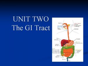
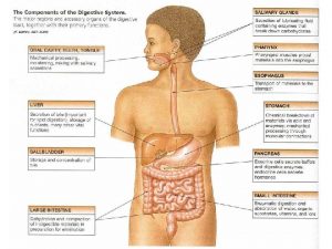
Development of the GUT • Starts at 4 th week IUL due to flexion of embryo • Formed by the endoderm lined yolk sac • Epithelium and secretory components of glands derive from endoderm • Muscles and connective tissues derive from splanchnic mesoderm • Primitive gut consists of four parts -Pharynx -Foregut -Midgut -Hindgut • Foregut, midgut and hindgut, each has its own artery

Foregut Bucco-pharyngeal membrane Midgut Vitelline duct Allantois Cloacal membrane Hindgut


Foregut Coeliac artery Midgut Sup. mesenteric artery Hindgut Inf. mesenteric artery


Foregut • Supplied by Coeliac artery • Extends from the bucco-pharyngeal membrane to a point just distal to hepatic diverticulum • Its proximal part extends up to tracheo-bronchial diverticulum • Its distal part extends from TB diverticulum to HD • Derivatives: Pharynx, Oesophagus, stomach, liver, gall bladder, pancreas and duodenum up to duodenal papilla

Development & rotation of stomach • Tube dilates, posterior wall grows rapidly than the anterior wall: Produce lesser & greater curvatures • Dorsal mesogastrium lengthens rapidly & forms greater omentum • Rotates 90 clock wise: left and right vagus nerves become anterior and posterior

Rotation of stomach 90 rotation

Development of spleen • Develops from the dorsal mesogastrium


Development of duodenum • Develops from distal foregut & proximal midgut • Acquires ‘C’ shape due to stomach rotation and growth of pancreatic buds • Dorsal mesentery gets absorbed into posterior abdominal wall: 2 nd and 3 rd Parts becomes retroperitoneal with pancreas

Development of liver & gall bladder • Liver parenchyma develops from liver bud/hepatic diverticulum • Connective tissue, Kupffer cells and haemopoietic tissue of liver develop from septum transversum • Gall bladder, cystic duct and common bile duct develop from cystic diverticulum

Development of pancreas • Exocrine part develops from the ventral & dorsal pancreatic buds • Endocrine part (Islets of Langerhans) develop from the neural crest cells

Hepatic diverticulum Cystic diverticulum Ventral pancreatic bud Dorsal pancreatic bud

Accessory pancreatic duct Common bile duct Dorsal bud Uncinate process (ventral bud) Gall bladder Main pancreatic duct

Midgut • Supplied by Superior mesenteric artery • Extends from the hepatic diverticulum to the junction of proximal 2/3 and distal 1/3 of the transverse colon • Connected to the yolk sac by vitelline duct through umbilical cord • Undergoes 270 rotation anticlockwise • Derivatives: Part of duodenum, small intestine, caecum, ascending colon and prox. 2/3 of transverse colon

Midgut… • At 6 th week I. U. L, mid gut loop herniates through the umbilical region – Physiological umbilical hernia • This is due to rapid increase in length relative to the size of the abdominal cavity • At 10 th week I. U. L, it returns to the abdominal cavity • Rotates 90 when herniates and 180 when returns

Hindgut • Supplied by Inferior mesenteric artery • Extends from the junction of proximal 2/3 and distal 1/3 of the transverse colon to Cloacal membrane • Derivates: Distal 1/3 of TC, descending colon, sigmoid colon, rectum and upper part of anus

Perineum Coccyx Anal triangle subpubic angle Urogenital triangle

Urorectal septum Cloacal membrane Cloaca Urorectal septum divides the cloaca into urogenital part and an anorectal part. This septum also divides the cloacal membrane into urogenital and anal membranes. The septum itself becomes the perineal body.

Developmental defects - Foregut • Pyloric stenosis: Hypertrophy • Atresia of bile duct: failure of pyloric sphincter muscles to recanalize the cystic diverticulum

Developmental defects - Foregut • Duplication of gall • Annular pancreas: mal fusion of bladder: formation of ventral & dorsal pancreatic buds two cystic diverticula leading to duodenal stenosis

Developmental defects - Midgut • Vitelline fistula: Persistence of vitelline duct • Vitelline cyst: Cyst formation with ligament on either side • Meckels diverticulum: Persistence of small part of vitelline duct connected to gut

Developmental defects - Midgut • Omphalocoele: Persistence of physiological umbilical hernia/ nonreturn of intestinal loops at 10 th week IUL

Developmental defects - Hindgut • Imperforate anus: Nonrupture of anal membrane

Developmental defects - Hindgut • Urorectal fistula: Persistent connection between urinary tract & rectum due to defective formation of urorectal septum

Developmental defects - Hindgut • Congenital megacolon: Absence of parasympathetic ganglia in the bowel wall (aganglionic megacolon or Hirschsprung disease)
 Perforators of lower limb
 Sanjaya adikari
 Pyramidal vs extrapyramidal lesions
 Rubrospinal tract
 Ayling sanjaya
 Ayling sanjaya
 Dr ayling sanjaya
 Frcpath part 1
 Meningomielokel
 Dr ayling
 Dr ayling
 Ayling sanjaya
 Dept nmr spectroscopy
 Florida dept of agriculture and consumer services
 Finance department organizational chart
 Worcester building department
 Dept. name of organization
 Mn dept of education
 Liz welch mississippi
 Dept. name of organization
 Ohio dept of dd
 Poster affiliation
 Vaginal dept
 Gome dept
 Gome dept
 Nyttofunktion
 Gome dept
 Hoe dept
 La city fire interview
 Maine dept of agriculture
 Dept of education
 Florida dept of agriculture and consumer services
