Radiation Hypoplasia Anterior Shoulder Dislocation Thalassemia Major Ewings


 • Findings: – Complete compression of C 3")


 –")


 – Normal")


- Slides: 179
Radiation Hypoplasia
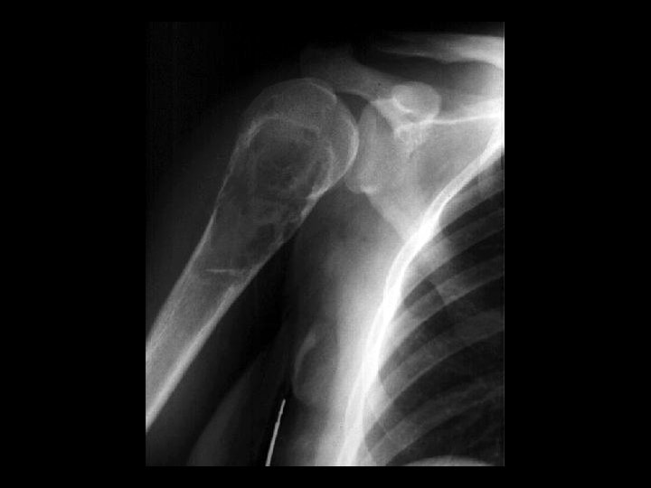
Anterior Shoulder Dislocation
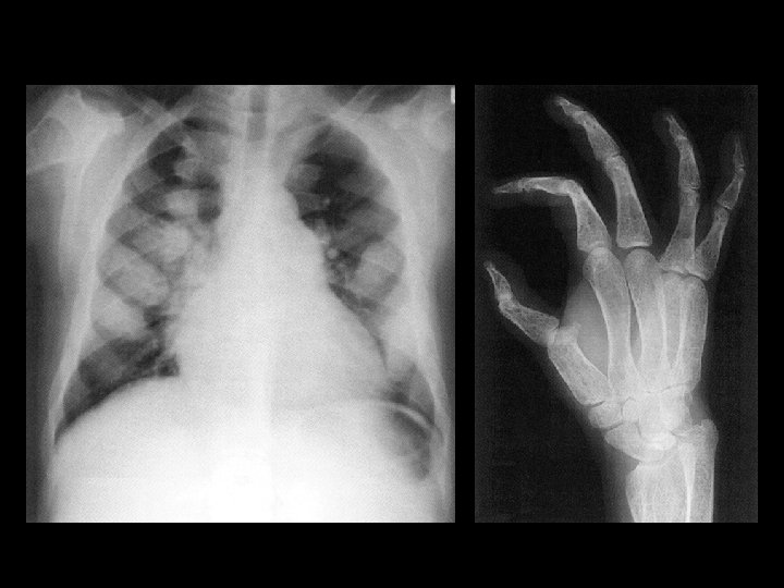
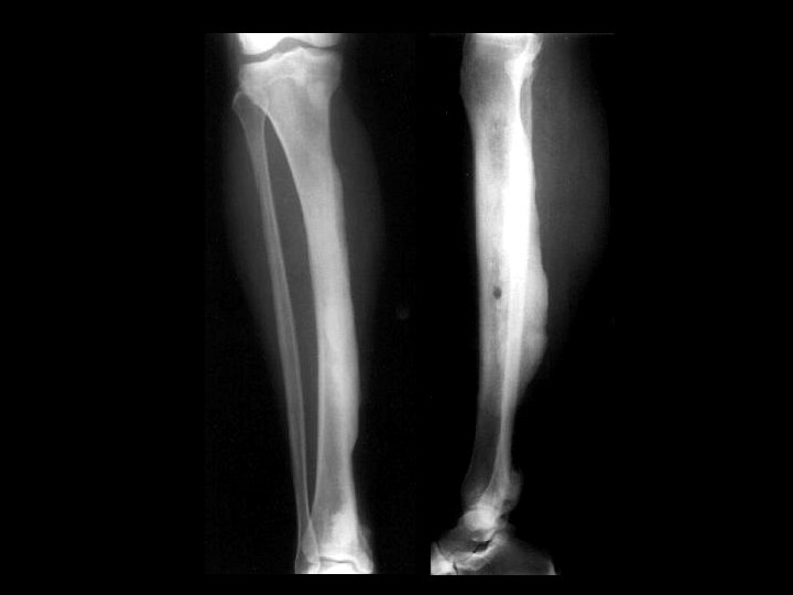
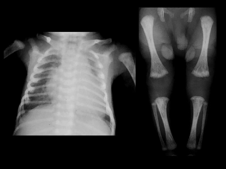
Thalassemia Major
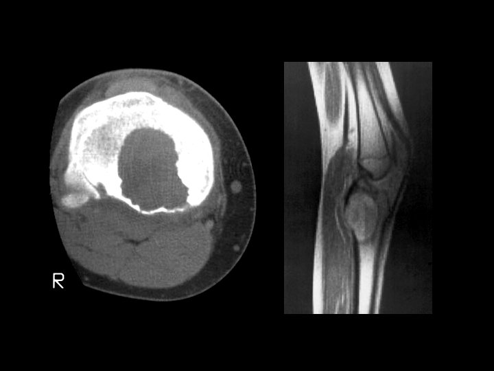
Ewing’s Sarcoma
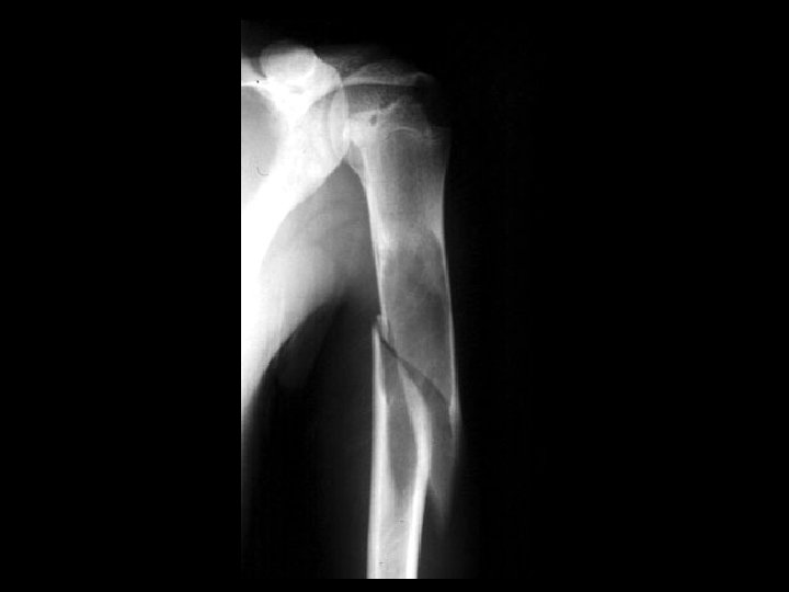
Monteggia fracture
Osteoid Osteoma
Parosteal osteosarcoma
Synovial Osteochondromatosis
UBC w/ path fx
Paget’s Dz of the Spine
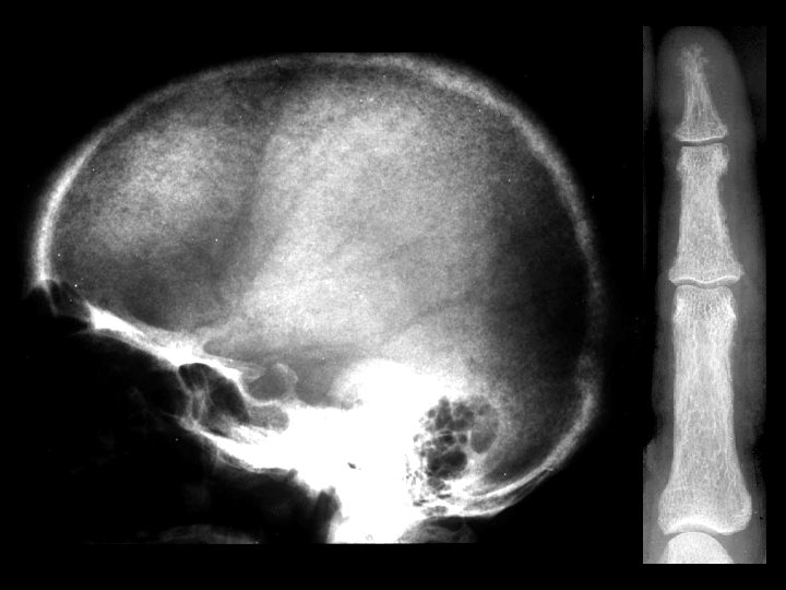
Osteopetrosis • Findings: – generalized increased bone density • rare bone dysplasia • Trabecular --> compact • Anemia w/extramedullary hematopoiesis • ddx: (children) – pyknodysostosis – renal osteodystrophy – vit A, D – Lead – Fluorosis
Paget’s Disease • Findings: – Enlargement of the right femoral head with course and thickened trabecula – Asymmetric lucency of the right superior acetabulum • ddx: – NONE! – This is an Aunt Minnie!
Brown Tumor • Findings: – expansile, lytic rib lesion – lytic lesion of 2 nd PP – subperiosteal bone resorption – arterial Ca 2+ • Usually secondary to CRF but may be primary • check for PT adenoma • if on dialysis, check for amyloid arthropathy • ddx: – multiple myeloma – metastases
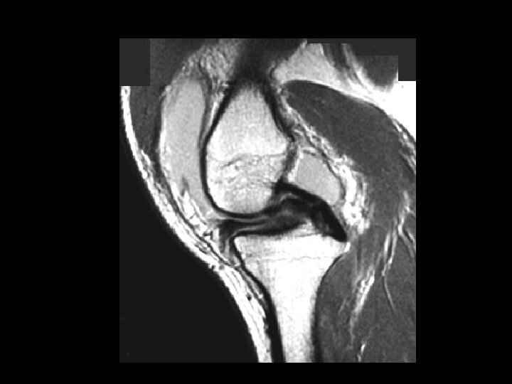
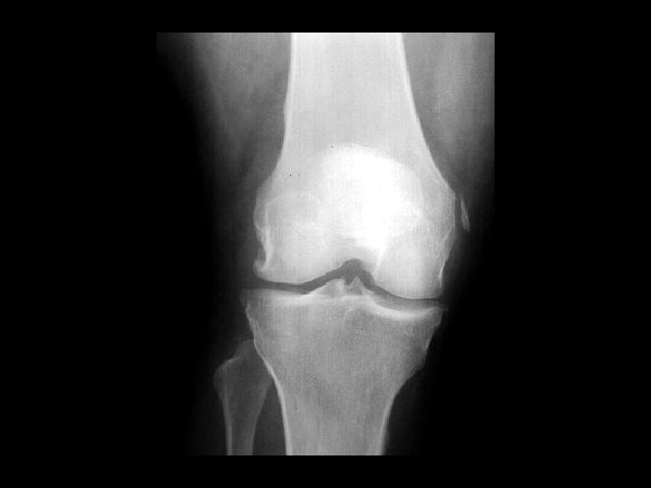
Bucket-Handle Tear • Findings: – “double PCL sign” - torn meniscus BELOW normal PCL on sag view – Above ACL on cor view – Truncation of medial meniscus – Joint effusion • Medial = 3 x lateral • Locked knee • ddx: – torn ACL, PCL – torn meniscus
Ankylosing Spondylitis • Findings: – fused SI joints – right hip erosions – lumbar syndesmophytes • Sero-negative chronic inflammatory disease • Starts in the low back and progesses upward • ddx (sacroilitis) – bilateral • ank spond • IBD – unilateral • Reiter’s • psoriasis
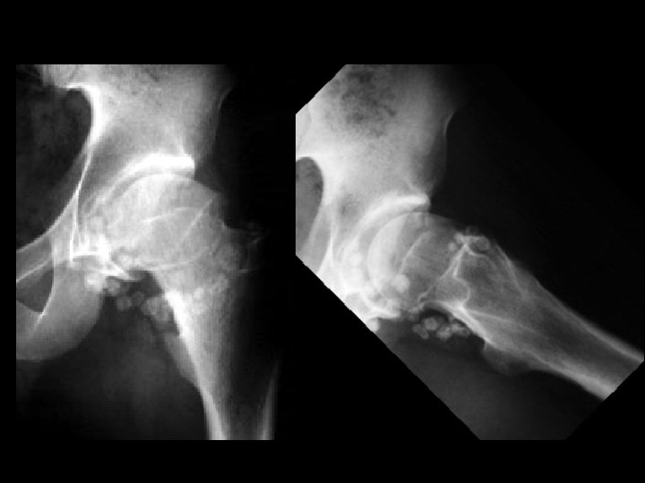
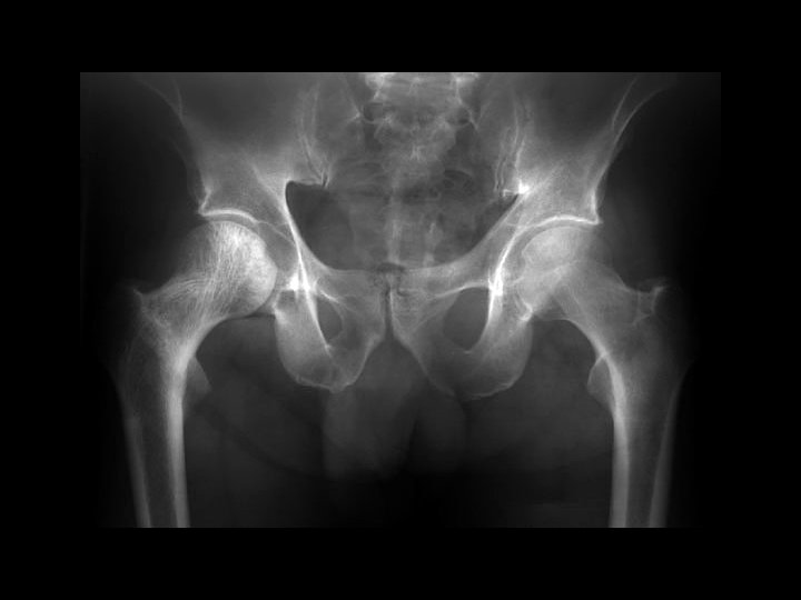
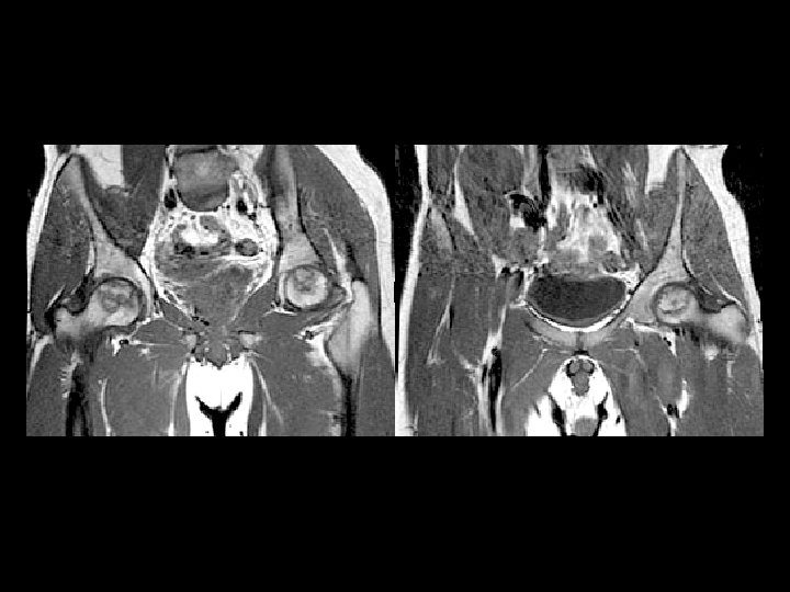
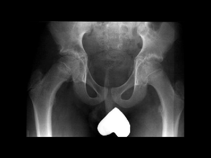
Femoral Head AVN • Findings: – bilateral femoral head AVN w/o collapse – right pelvic renal tx • Ddx: – Pancreatitis – Sickle Cell Dz – Caison’s Dz – etc…
Lateral Patellar Dislocation • Findings: – bone marrow edema of lateral femoral condyle and medial patella – injury of the medial patellar retinaculum – joint effusion • Patella usually dislocates laterally • Look for constellation of findings • Ddx: – NONE! – This is an Aunt Minnie
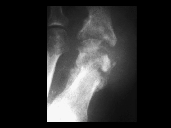
Metastatic Renal Cell • Findings: – expansile lytic lesion of the distal ulnar metaphysis – well-defined margins – internal septations – No periostitis • ddx: – ABC – Giant cell tumor
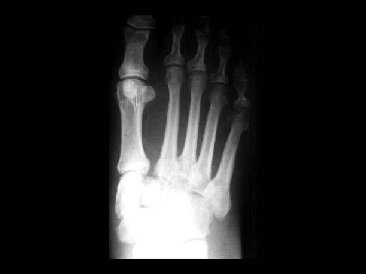
Homolateral Lisfranc farcture/dislocation • Findings – Widening between the base of 1 st and 2 nd metatarsals. – lateral subluxation of the second through fifth metatarsals • dislocation is relative to the cuneiforms: – homolateral – divergent (1 st MT goes medial) • can be due to trauma or in patients with diabetic neuropathy
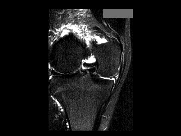
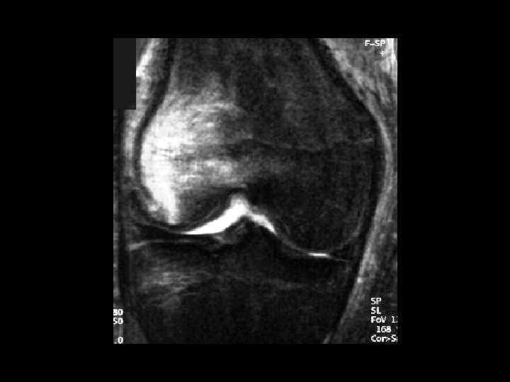
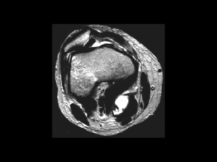
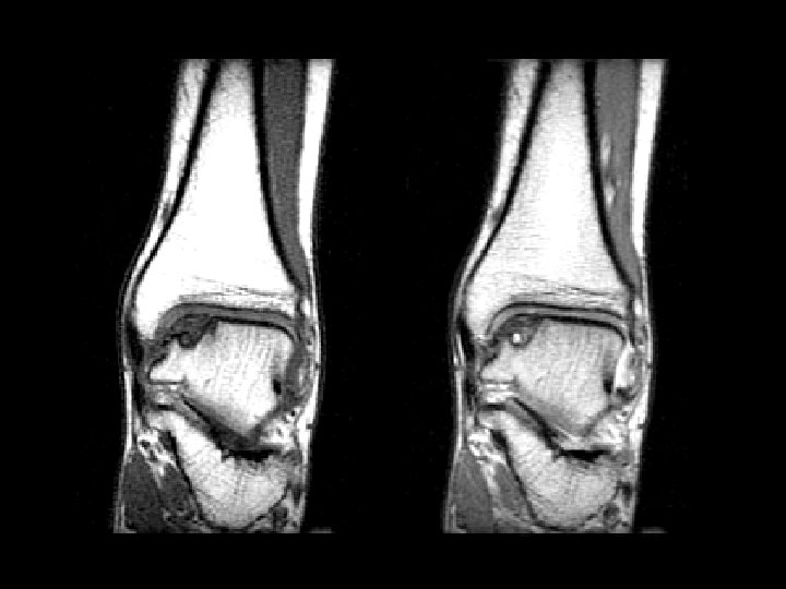
Osteochondritis Dissecans • • • Findings: – defect in the medial talar dome representing an osteochondral fracture Unknown etiology but most common causes are: – trauma – osteonecrosis Do MRI w/ or w/o arthro to look for free fragment
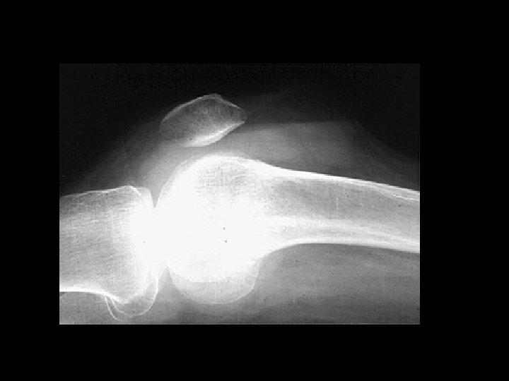
Pellegrini-Stieda disease • • Findings: – Linear ossific density adjacent to the medial femoral condyle Calcification or ossification of the MCL at its insertion site Sequela of previous injury NOT acute, usually not the site of pain
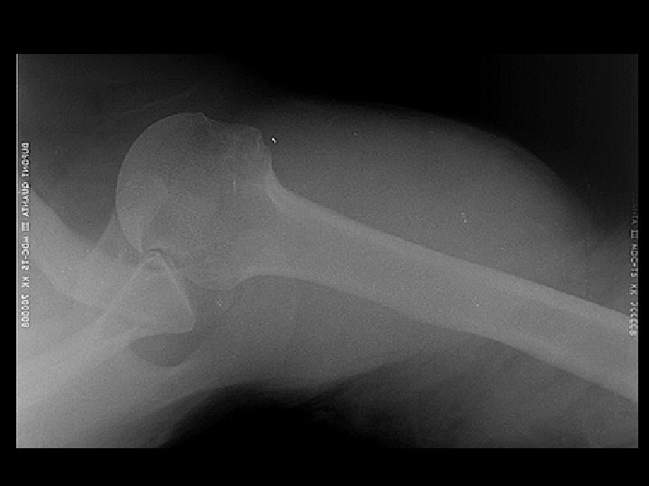
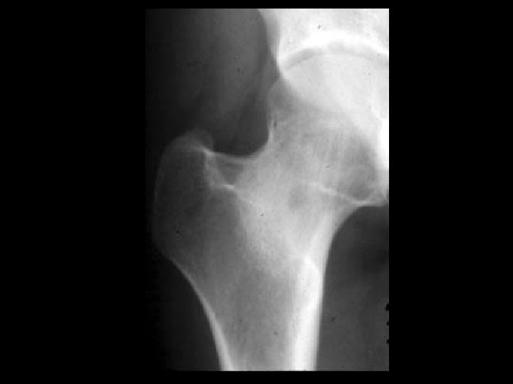
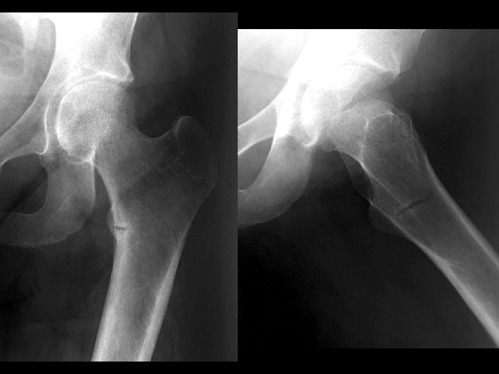
Bilateral SCFE • • Findings: – posteromedial displacement of femoral epiphyses Most common cause of limp in adolescents SH-I shearing fx high incidence in black, overweight pubertal males 75% uni, 25% bi Look for line along lateral femoral neck to transect 1/6 th of epiphysis Must be pinned or fixed
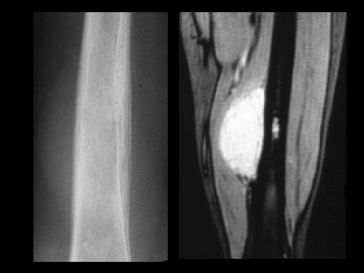
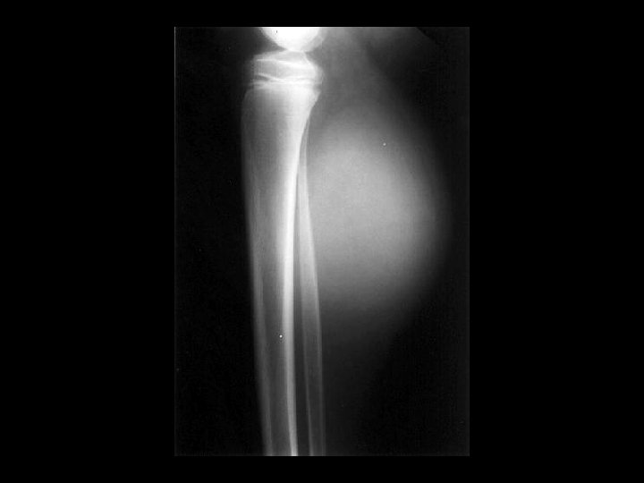
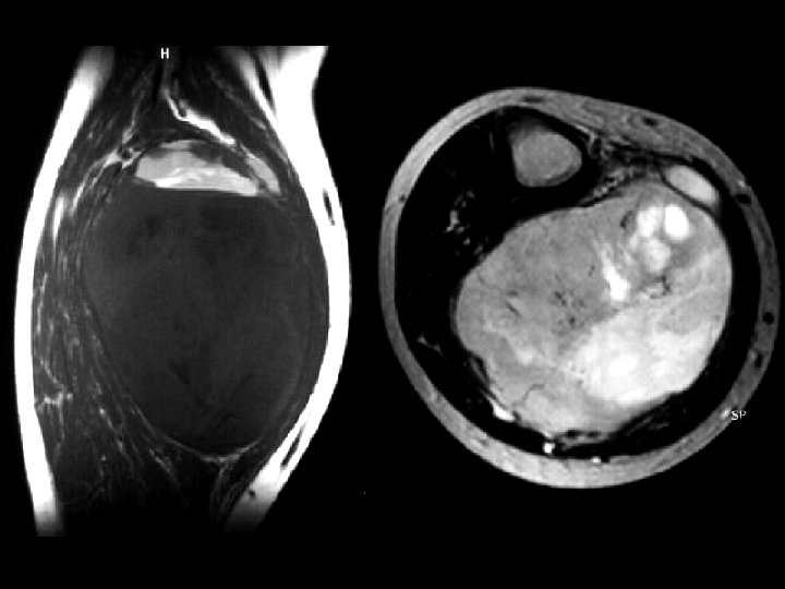
Synovial Sarcoma • • Findings: – Dense soft tissue mass – no bony involvement – heterogeneous T 1 Rare soft tissue tumor of children May show calcification Internal hemorrhage and necrosis are common Found close to joints Prone to metastases ddx: (plain film only) – hematoma
Unicameral Bone Cyst • • • Findings: – lytic, slightly expansile lesion of the proximal metaphysis with a thin sclerotic margin and fracture a. k. a. simple bone cyst fluid-filled cavity common lesion of children, unknown etiology asymptomatic unless fracture (“fallen fragment sign”)
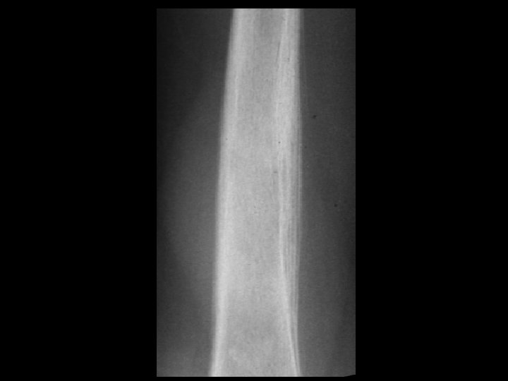
Melorheostosis • • • Findings: – Peripheral hyperostosis of the tibia producing a wavy sclerotic diaphyseal contour Rare bone disorder of childhood “candle wax” dripping down the bone appearance presents with PAIN and joint swelling ddx: – Paget’s – myelofibrosis – renal osteodystrophy – sclerotic mets
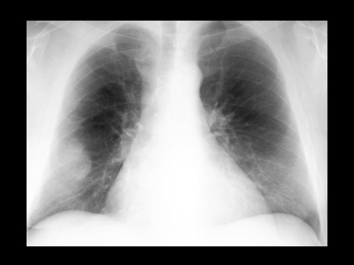
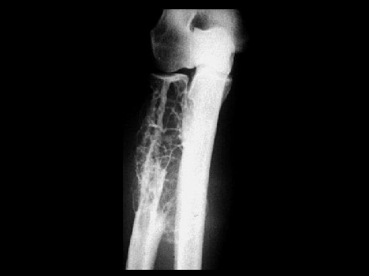
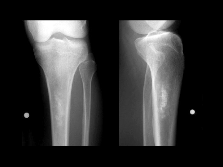
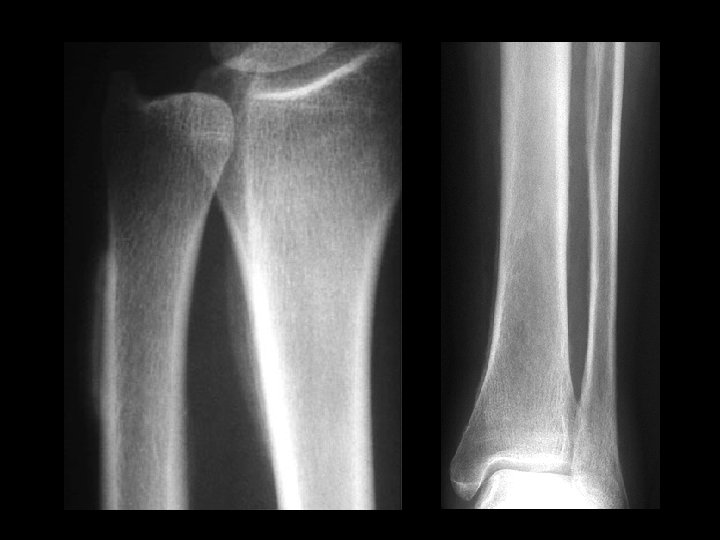
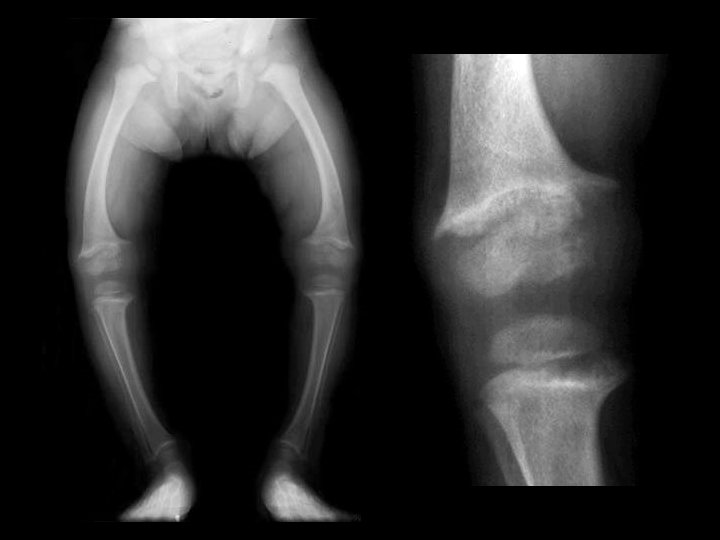
Congenital Reubella w/patent ductus arteriosis • • • Findings: – cardiomegaly, enlarged pulm arteries – longitudinal striations of sclerotic and radiolucent areas at the metaphyses (“celery stalk” appearance) – epiphyseal center not seen – dense, irregular metaphyseal bands most common viral infxn w/bone changes (also CMV) IUGR, TTP, cataracts, sensorineural hearing loss, PDA, pulm art and Ao stenoses
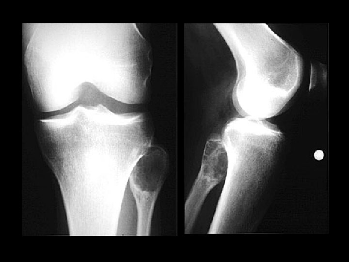
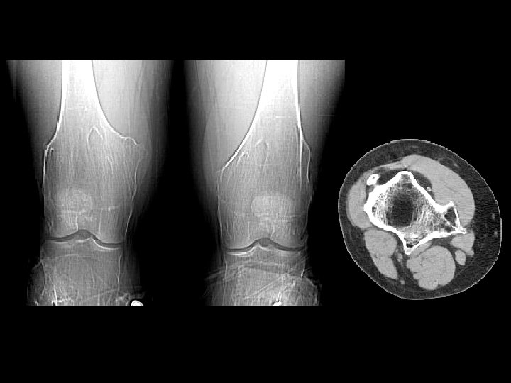
Aneurysmal bone cyst • Findings: – Lucent end of bone lesion in the proximal tibia – Slightly expansile, mild periosteal reaction – Fluid-fluid level on MRI • ddx: – Giant cell tumor – Unicameral bone cyst – Fibrous dysplasia – Chondroblastoma (rare)
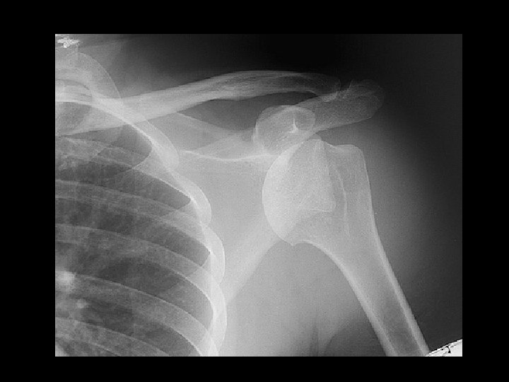
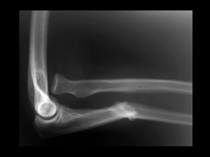
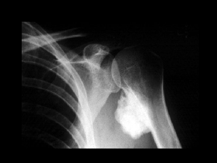
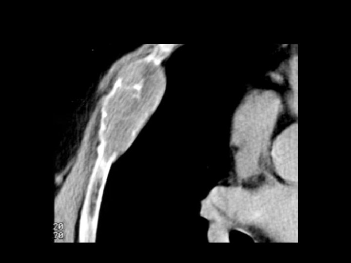
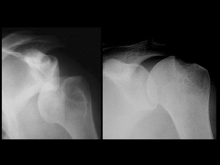
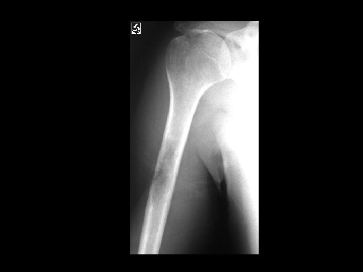
Inferior shoulder dislocation • Findings: – Inferior dislocation of humeral head and a deep cleft in the superior portion • ddx: – Anterior dislocation
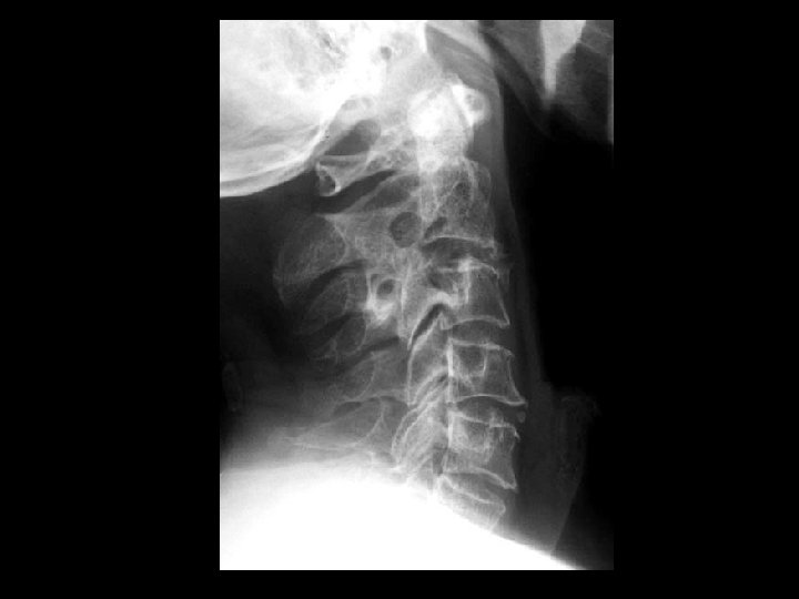
Dens metastasis, pathologic fracture, and C 1 -2 instability • Findings: – 8 mm anterior subluxation of C 1 on C 2 with flexion – Conventional tomogram shows C 2/dens lytic lesion and pathologic fracture • ddx: – Rheumatoid – Dens fracture
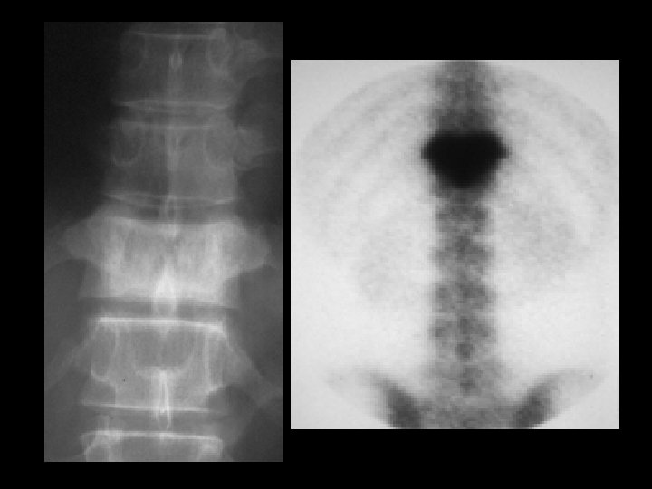
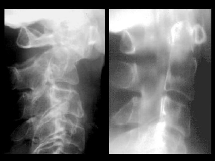
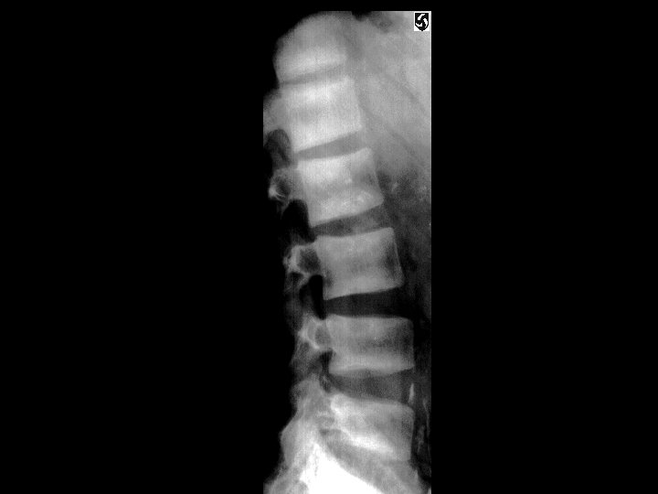
C 3 vertebra plana (multiple myeloma) • Findings: – Complete compression of C 3 (look at the vertebral bodies and their corresponding posterior elements!) • ddx: – Metastasis – Kommel’s disease (if see gas and collapse) – EG
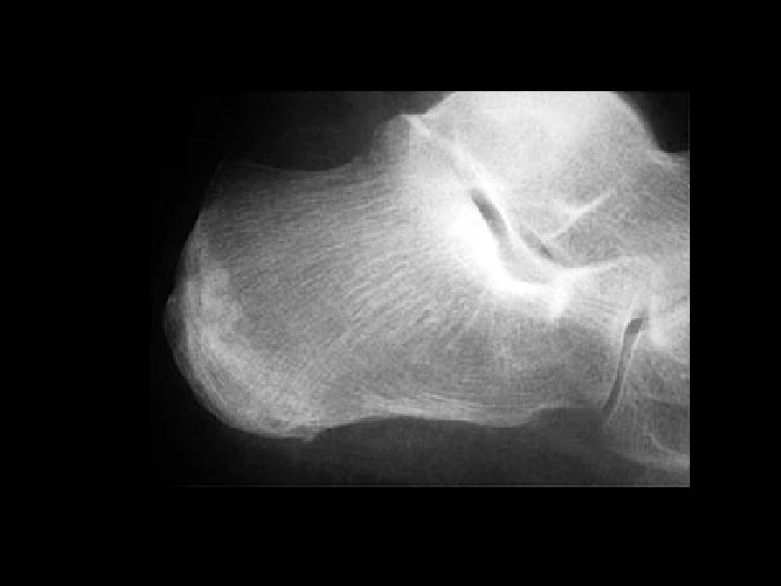
Calcaneal stress fracture • Findings: – Patchy linear and arclike sclerosis in the posterior calcaneous • ddx: – NONE! – This is an Aunt Minnie!
Enchondroma • Findings: – Patchy sclerotic intramedullay lesion in the proximal tibial metadiaphysis – “Rings and arcs” calcification • ddx: – Bone infarct
Cleidocranial dysplasia • Findings: – Absent central portion of clavicle with rudimentary medial and lateral portions – Congenital coxa vara – Widened appearance of pubis (due to delayed ossification) • ddx: – NONE! – This is an Aunt Minnie!
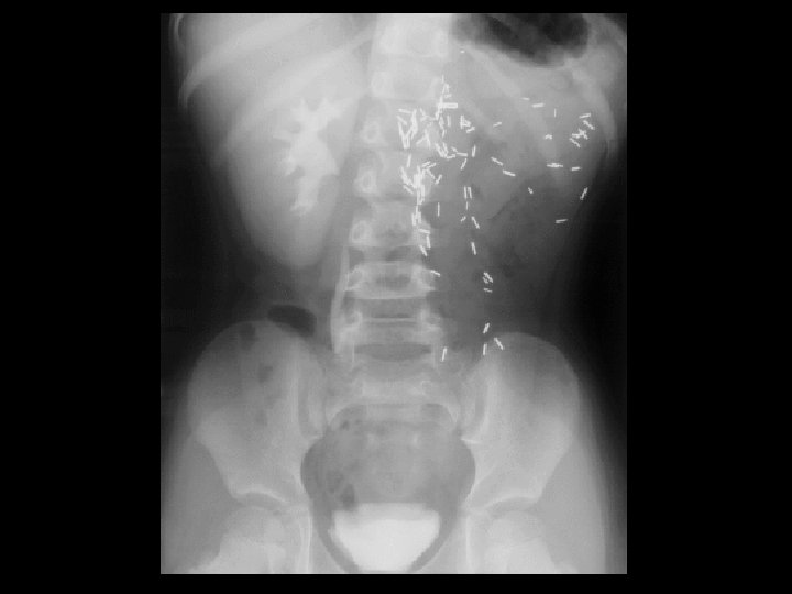
Cystercercosis • Findings: – Innumerable round and oval soft tissue calcifications • ddx: – Diffuse dystrophic or metastatic calcifications
Giant cell tumor • Findings: – Expansile lucent lesion in the fibular head containing multiple thin septa • ddx: – Aneurysmal bone cyst – Fibrous dysplasia – Lytic metastasis – Brown tumor – Chondroblastoma (rare)
Hypertrophic Osteoarthritis • • Findings: – Diffuse symmetrical periosteal reaction of the distal radius, ulna, and tibia ddx: – Adults • Pulmonary • Pachydermoperiostitis • Vascular insufficiency • Thyroid acropachy • Fluorosis – Peds • Caffey disease (up to 6 mo. ) • Leukemia • Rickets • Hypervitaminosis A
Hyperparathyroidism • Findings: – “salt and pepper” skull – Osteopenia with course trabecula – Subperiosteal resorption • ddx: – NONE! – This is an Aunt Minnie!
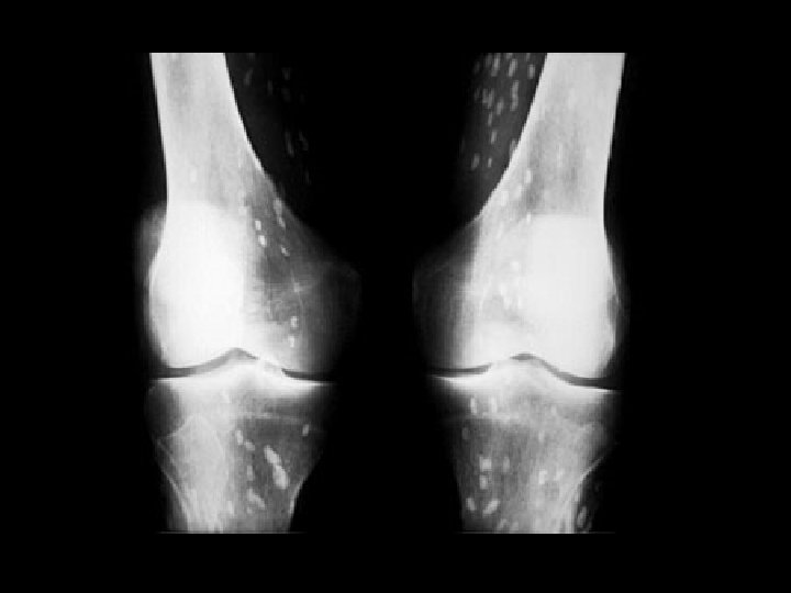
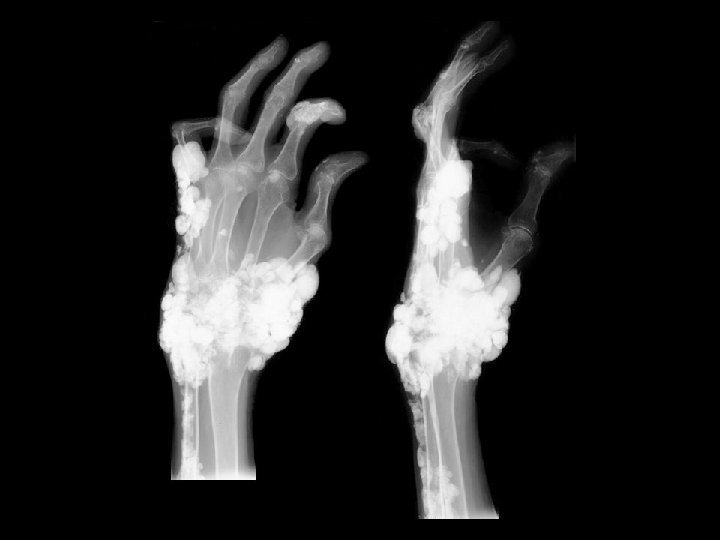
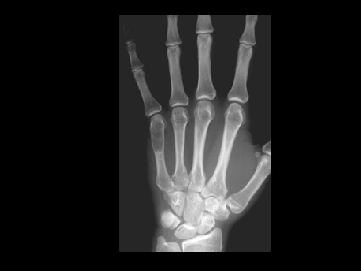
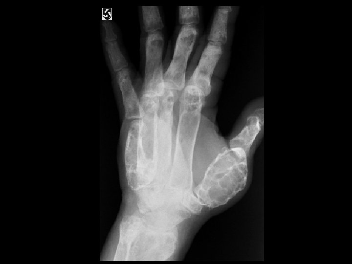
Tumoral Calcinosis • Findings: – Multiple dense calcifications around the joints of the fingers and wrists • ddx: (soft tissue Ca 2+) – Renal osteodystrophy – Heterotopic ossification – Metastatic Ca 2+ – Milk-alkali syndrome – Hypervitaminosis D – Scleroderma – Dermatomyositis
Lymphoma • Findings: – Permeative lytic lesion of the humeral diaphysis – Periosteal reaction and soft tissue swelling • ddx: – Osteomyelitis – Metastasis – Ewing’s Sarcoma – EG
Osteomalacia w/ Looser’s zone • Findings: – Osteopenia – Horizontal linear lucency in the medial femoral metaphysis – Causes: • Renal osteodystrophy • Hyperparathyroidism • Vit D deficiency • ddx: – NONE! – This is an Aunt Minnie!
Acute fracture • Findings: – Lipohemarthrosis – No visible fracture but one is definitely present • ddx: – NONE! – This is an Aunt Minnie!
Osteochondromatosis • Findings: – Flared metaphyses due to multiple bony outgrowths = exostoses – Contiguous with cortex and medulla – Point away from joint – Look for pain (after bony maturation) – check cartilagenous cap = >2 cm, risk of chondrosarcoma • ddx: – NONE! – This is an Aunt Minnie!
Paget’s Disease w/ sarcomatous degeneration • Findings: – Cortical thickening with mixed lytic and sclerotic trabecular pattern and bony enlargement – Large lytic lesion of the right ilium and sacral ala • ddx: – Diffuse sclerotic and lytic mets
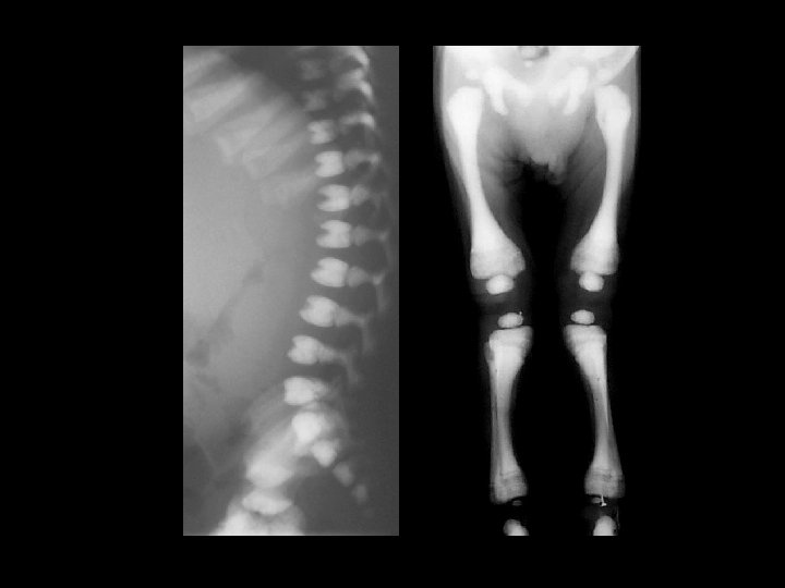
Rickets • Findings: – Cupped and frayed metaphyses – Wide physes (increased osteoid) – Long bone bowing – Decreased bone density • ddx: – NONE! – This is an Aunt Minnie!
Bilateral perched lumbar facets • Findings: – Mild anterolisthesis of L 4 on L 5 – Inferior L 4 facets perched on superior L 5 facets • ddx: – NONE! – This is an Aunt Minnie!
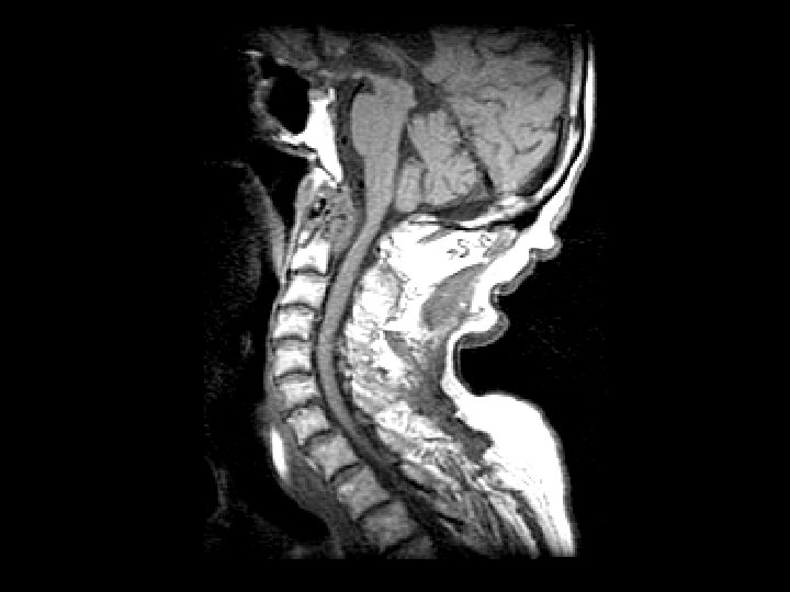
Rheumatoid arthritis • Findings: – Destruction of the dens and the anterior C 1 arch by a soft tissue mass – Impression of the anterior thecal sac with slight deformation of the cervical cord • ddx: – Infection – Metastasis
Septic Arthritis • Findings: – Lytic and sclerotic destruction of the great toe MTP joint – Joint space narrowing – Periosteal reaction – Soft tissue swelling • ddx: – Charcot joint
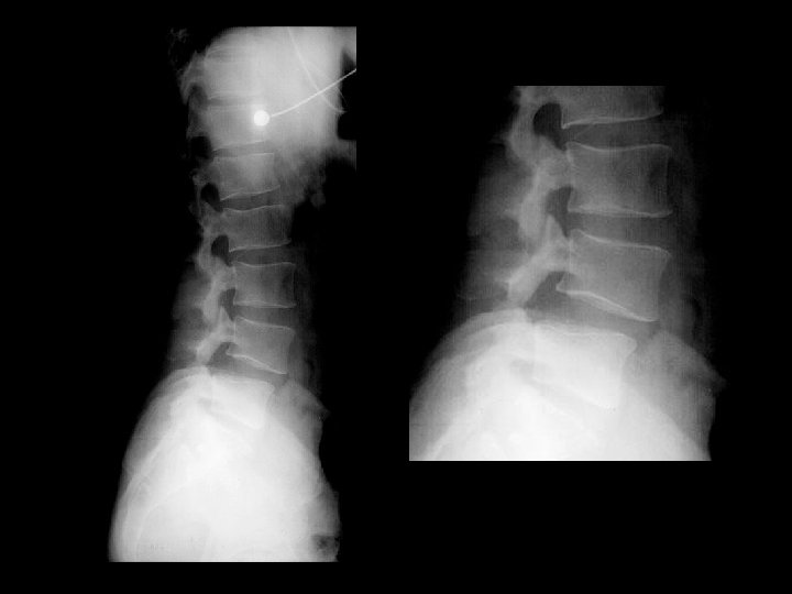
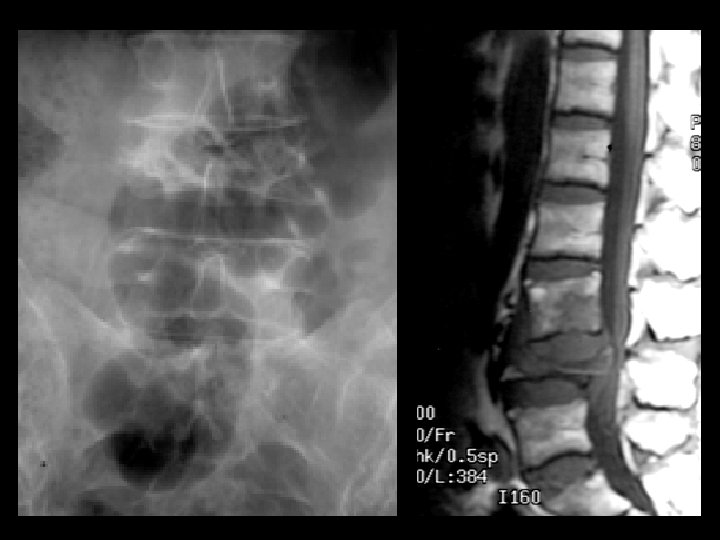
Potts Disease • Findings: – Complete collapse of L 4 and cauda equina compression – Subligamentous soft tissue extension to L 3 body • ddx: – Staph aurius – Gram negatives (IVDA)
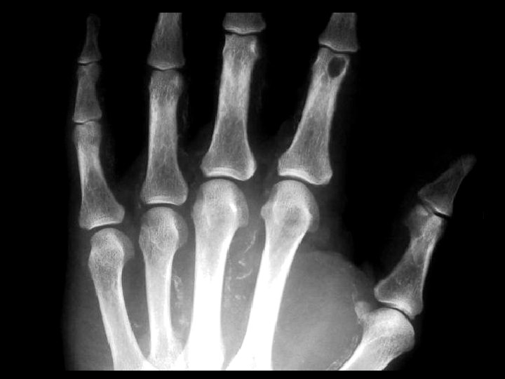
Enchondroma • Findings: – Lucent, slightly expansile lesion of the fifth MC • ddx: – Giant cell tumor – UBC – Fibrous dysplasia
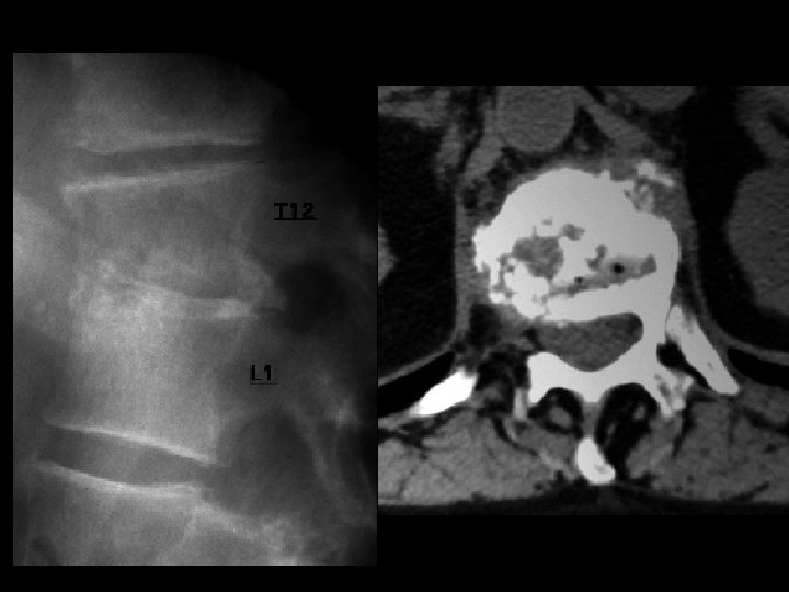
Discitis • Findings: – T 12 -L 1 disc space narrowing with endplate destruction – CT demonstrates presence of gas • ddx: – NONE! – This is an Aunt Minnie!
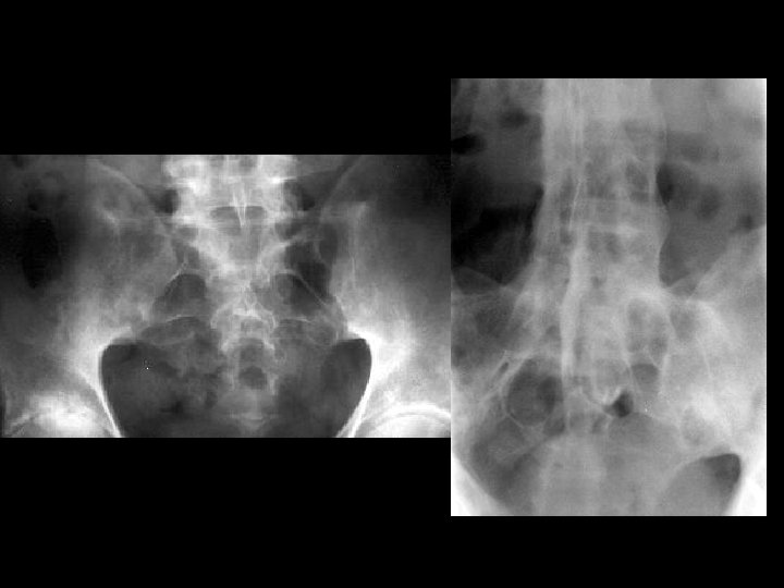
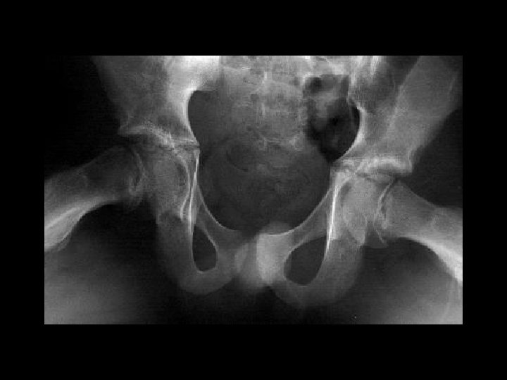
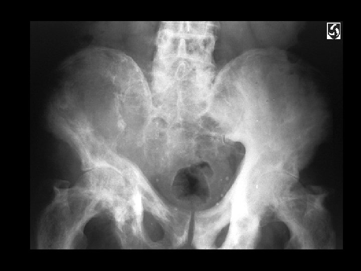
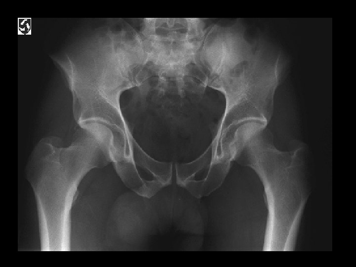
Right hip synovitis • Findings: – Asymmetric teardrop distance (greater on right) – Normal joint space • ddx: – Infection – Rheumatoid
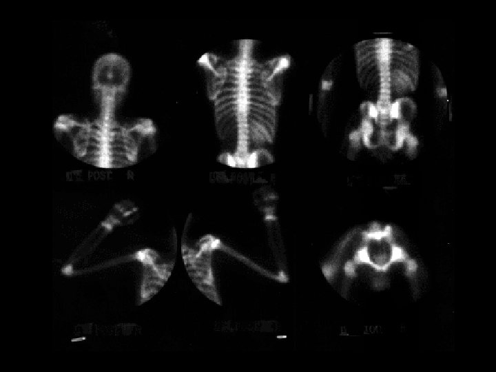
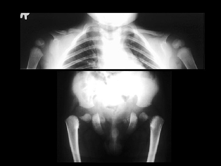
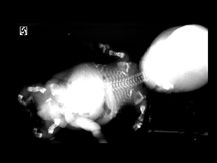
Osteogenesis Imperfecta Type II • Findings: – multiple fractures, bowing and short ribbon-like bones • Type II - lethal - “ribbon bones” and fractures too numerous to count • Disturbances in type I collagen – lack of normal collagen – osteoperosis – fractures – blue sclera • ddx: – NONE! – This is an Aunt Minnie!
Polyostotic fibrous dysplasia • Findings: – Multiple lucent and expansile lesions – “ground glass matrix” – May be seen on precocious puberty – Mc. Cune Albright syndrom • ddx: – Multiple enchondromas
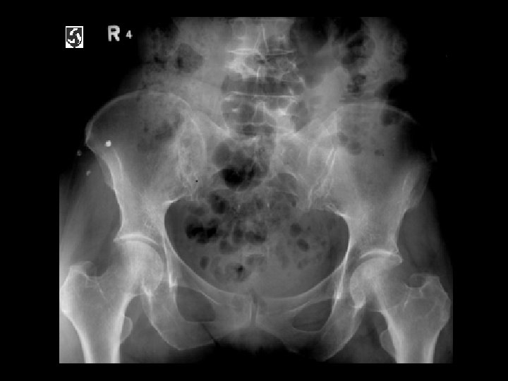
Renal osteodystrophy • Findings: – Osteopenia – Prominent trabecular pattern – “rugger jersey” spine • ddx: – NONE! – This is an Aunt Minnie!
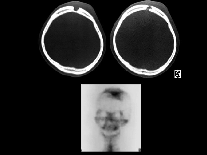
Eosinophilic Granuloma • Findings: – Well-defined lytic lesion in the frontal skull crossing the diploic space – Overlying scalp swelling – Positive bone scan • ddx: – Osteomyelitis – Metastasis
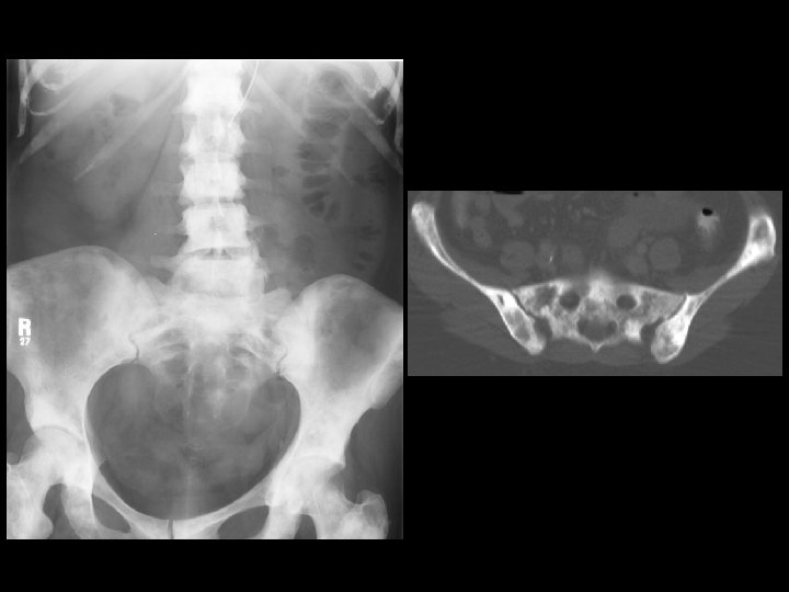
Prostate metastases • Findings: – Diffuse sclerosis • ddx: – Breat, GI mets – myelofibrosis – renal osteodystrophy – osteopetrosis – multiple myeloma