
Human Genetics of Urinary Tract Malformation Ali Gharavi


- Slides: 56
Human Genetics of Urinary Tract Malformation Ali Gharavi, MD Division of Nephrology Columbia University New York, NY ag 2239@columbia. edu
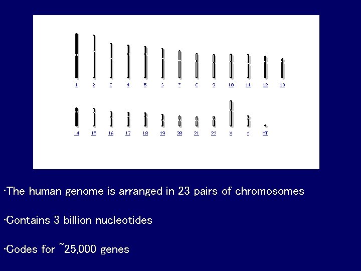
• The human genome is arranged in 23 pairs of chromosomes • Contains 3 billion nucleotides • Codes for ~25, 000 genes
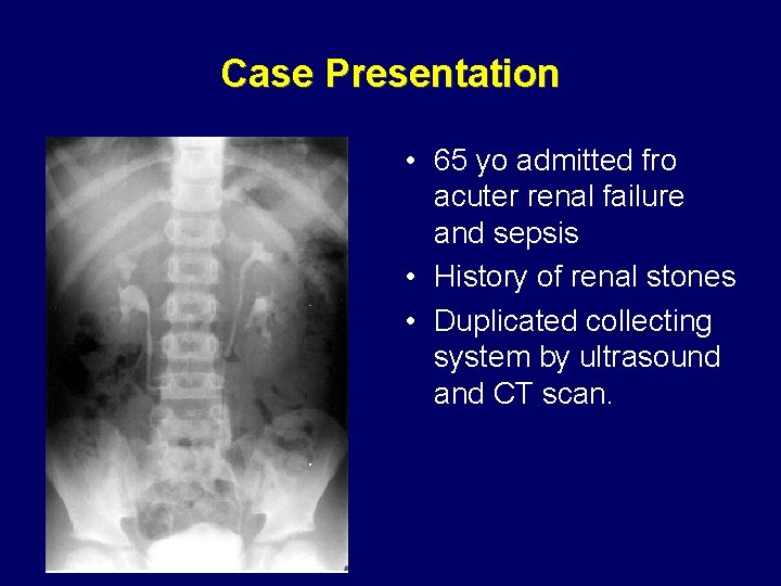
Case Presentation • 65 yo admitted fro acuter renal failure and sepsis • History of renal stones • Duplicated collecting system by ultrasound and CT scan.
Dupl ureter Kidney Problem?
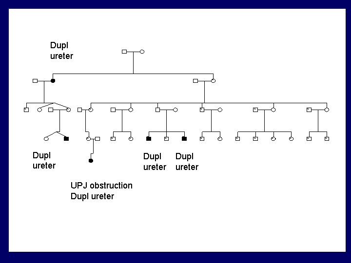
Dupl ureter ? ? ? Dupl ureter ? ? Dupl ureter UPJ obstruction Dupl ureter ? ? ?
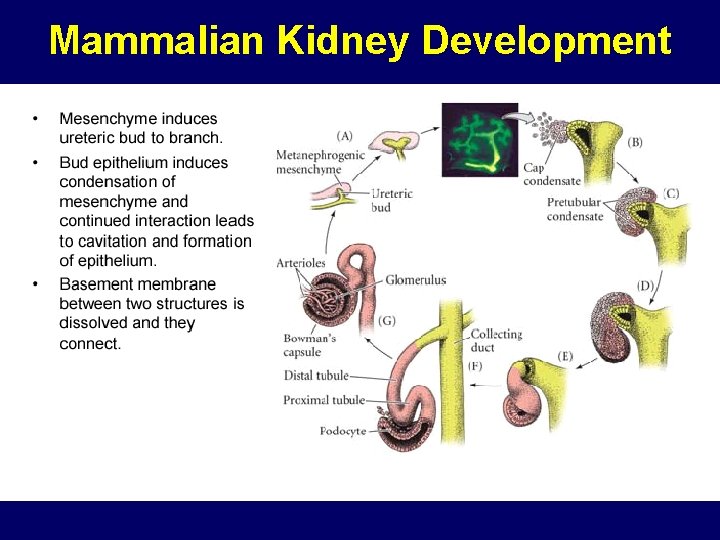
Mammalian Kidney Development
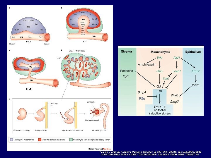
Vainio S and Lin Y. Nature Reviews Genetics 3; 533 -543 (2002); doi: 10. 1038/nrg 842 COORDINATING EARLY KIDNEY DEVELOPMENT: LESSONS FROM GENE TARGETING
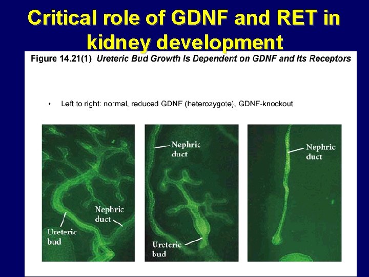
Critical role of GDNF and RET in kidney development
Epidemiology of Urinary tract Abnormalities • Account for one-third of birth defects • 40% of pediatric end-stage renal disease • 10% of adult end-stage renal disease in some countries • Etiology poorly understood because of variable expression and incomplete penetrance
Clinical Features • Due to overlap between developmental pathways, phenotypes are complex, involving anatomic defects in both upper and lower urinary tract • Often asymmetric • Severe phenotypes result in perinatal death due to pulmonary hypoplasia • The majority of cases are nonsyndromic
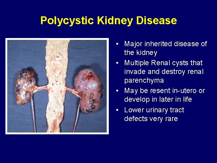
Polycystic Kidney Disease • Major inherited disease of the kidney • Multiple Renal cysts that invade and destroy renal parenchyma • May be resent in-utero or develop in later in life • Lower urinary tract defects very rare
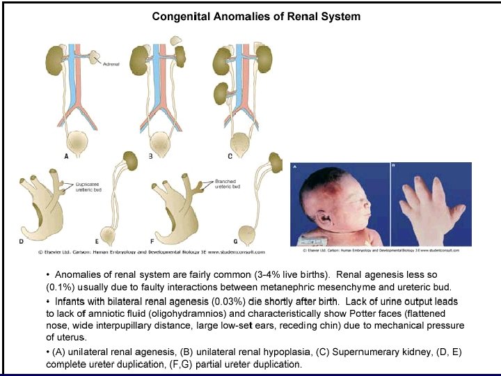
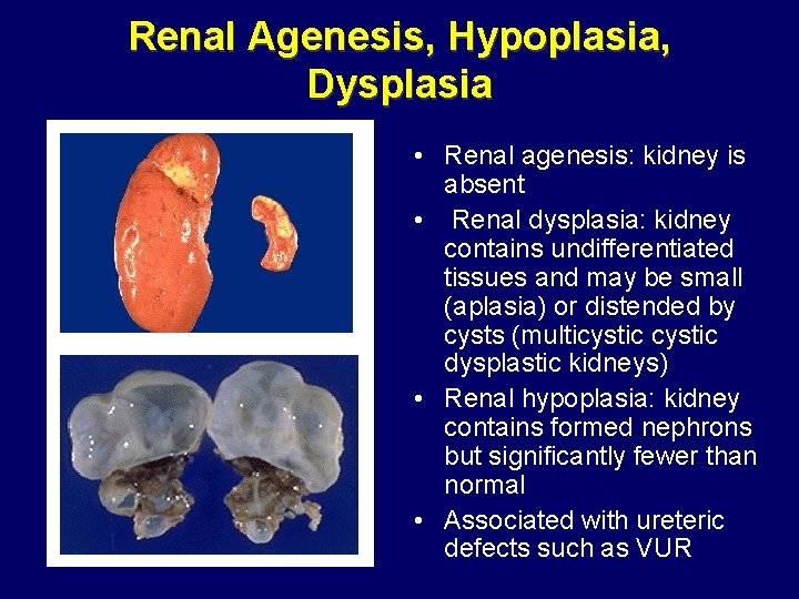
Renal Agenesis, Hypoplasia, Dysplasia • Renal agenesis: kidney is absent • Renal dysplasia: kidney contains undifferentiated tissues and may be small (aplasia) or distended by cysts (multicystic dysplastic kidneys) • Renal hypoplasia: kidney contains formed nephrons but significantly fewer than normal • Associated with ureteric defects such as VUR
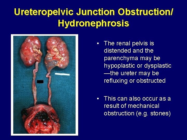
Ureteropelvic Junction Obstruction/ Hydronephrosis • The renal pelvis is distended and the parenchyma may be hypoplastic or dysplastic —the ureter may be refluxing or obstructed • This can also occur as a result of mechanical obstruction (e. g. stones)
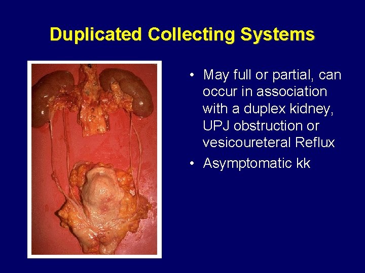
Duplicated Collecting Systems • May full or partial, can occur in association with a duplex kidney, UPJ obstruction or vesicoureteral Reflux • Asymptomatic kk
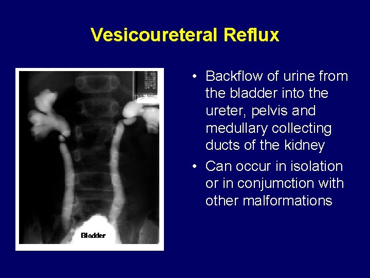
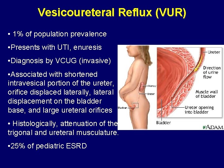
Vesicoureteral Reflux • Backflow of urine from the bladder into the ureter, pelvis and medullary collecting ducts of the kidney • Can occur in isolation or in conjumction with other malformations
Vesicoureteral Reflux (VUR) • 1% of population prevalence • Presents with UTI, enuresis • Diagnosis by VCUG (invasive) • Associated with shortened intravesical portion of the ureter, orifice displaced laterally, lateral displacement on the bladder base, and large ureteral orifices • Histologically, attenuation of the trigonal and ureteral musculature. • 25% of pediatric ESRD
Inheritance of VUR • Prospective screening of 354 siblings of 275 index patients with VUR revealed reflux in 119 (34%) cases • Spontaneous resolution of VUR in patients maintained on antibiotic prophylaxis over 10 years (49 -69%) • Most urologists screen sibs, particularly age<5 • Complex inheritance Noe J Urology, 1992, Greenfeld et al J urology J , Scott et al , Lancet 1997
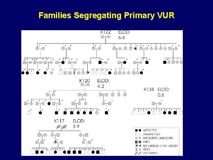
Families Segregating Primary VUR
Chromosomal abnormalities
Syndromic forms • Associated with certain chromosomal abnormalities – Deletion 4 q, 18 q – Duplication 3 q, 10 q • Implicate defects in multiple genes in the development of the trait • Associated with multiple organ defects
10 q deletion syndrome • Cardiac, urogenital, and respiratory complications, orofacial dysmorphism, and psychomotor retardation which vary with different karyotypes. • Urogenital system: Cryptorchidism, genital hypoplasia, and streaked ovaries. Urinary anomalies include kidney aplasia or hypoplasia, hydronephrosis, hydroureter, and cystic disease. • Systematic analysis suggest that deletion of 10 q 26 segment results in this phenotype Ogata et al. Kidney Int , 2000
Single Gene Disorders in Humans
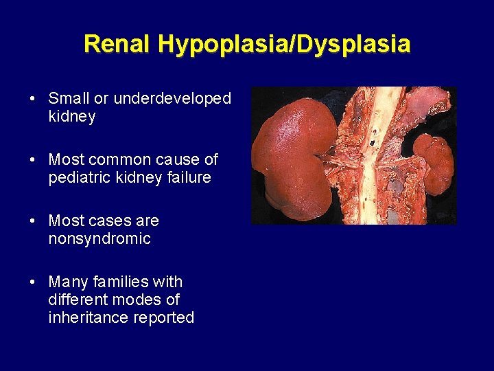
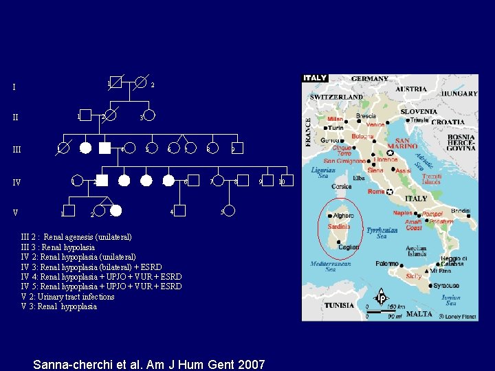
Renal Hypoplasia/Dysplasia • Small or underdeveloped kidney • Most common cause of pediatric kidney failure • Most cases are nonsyndromic • Many families with different modes of inheritance reported
1 I 1 II III 1 V 1 2 2 1 IV 2 3 3 4 2 2 3 3 5 4 6 5 7 6 4 8 9 7 8 9 5 III 2 : Renal agenesis (unilateral) III 3 : Renal hypolasia IV 2: Renal hypoplasia (unilateral) IV 3: Renal hypoplasia (bilateral) + ESRD IV 4: Renal hypoplasia + UPJO + VUR + ESRD IV 5: Renal hypoplasia + UPJO + VUR + ESRD V 2: Urinary tract infections V 3: Renal hypoplasia Sanna-cherchi et al. Am J Hum Gent 2007 10
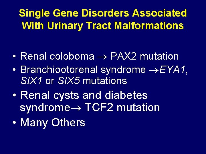
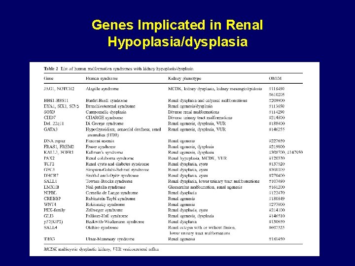
Single Gene Disorders Associated With Urinary Tract Malformations • Renal coloboma PAX 2 mutation • Branchiootorenal syndrome EYA 1, SIX 1 or SIX 5 mutations • Renal cysts and diabetes syndrome TCF 2 mutation • Many Others
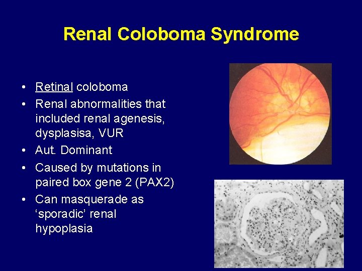
Renal Coloboma Syndrome • Retinal coloboma • Renal abnormalities that included renal agenesis, dysplasisa, VUR • Aut. Dominant • Caused by mutations in paired box gene 2 (PAX 2) • Can masquerade as ‘sporadic’ renal hypoplasia
PAX 2 in Renal Development • Transcription factor • The PAX 2 gene is expressed in primitive cells of the kidney, ureter, eye, ear, and central nervous system • During renal development, expression in nephric duct formation, then in the UB, and finally in proximal elements of the metanephric mesenchyme • Expression absent in adult kidney
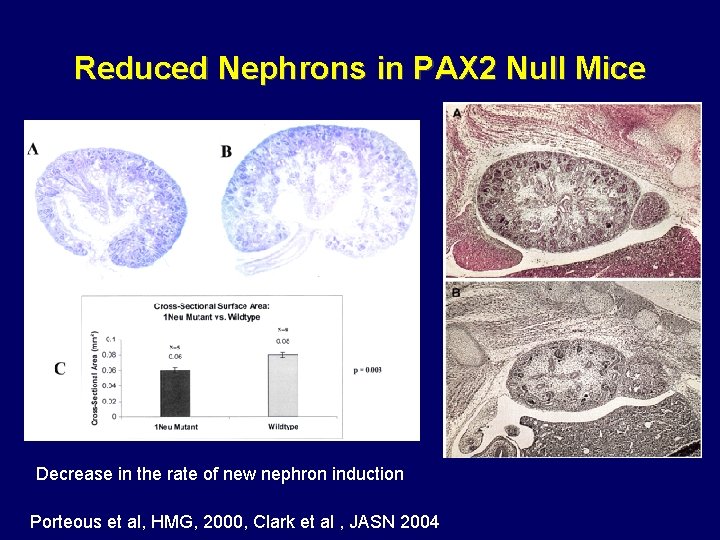
Reduced Nephrons in PAX 2 Null Mice Decrease in the rate of new nephron induction Porteous et al, HMG, 2000, Clark et al , JASN 2004
Genes Implicated in Renal Hypoplasia/dysplasia
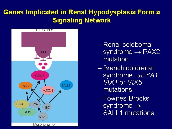
Genes Implicated in Renal Hypodysplasia Form a Signaling Network – Renal coloboma syndrome PAX 2 mutation – Branchiootorenal syndrome EYA 1, SIX 1 or SIX 5 mutations – Townes-Brocks syndrome SALL 1 mutations
Cystic Kidney Disease
Major Subtypes • Autosomal Dominant Polycystic Kidney Disease • Autosomal Recessive Polycystic Kidney Disease • Multicystic Dysplastic kidney (MCDK) – Diabetes and renal cysts syndrome • Medullary Cystic/Nephronophthsis • Bardet-Biedl syndrome • Many Others
ADPKD • Prevalence of 1: 500 to 1: 2000 in the general population • Affects all population worldwide • 7% of cases of end-stage renal disease in USA • Focal and sporadic development of cysts in kidney and other organs
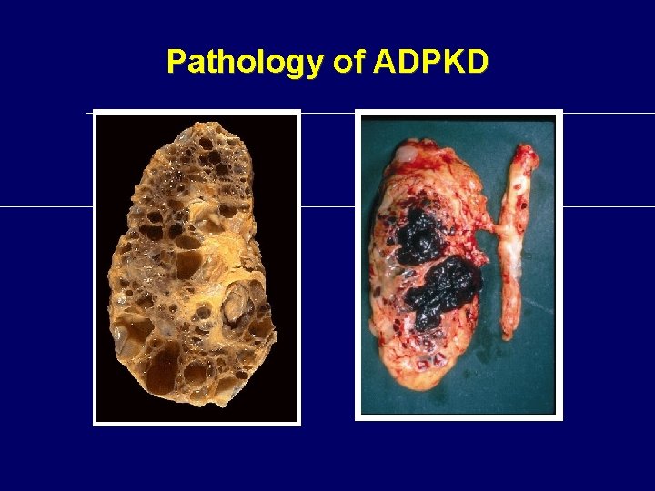
Pathology of ADPKD
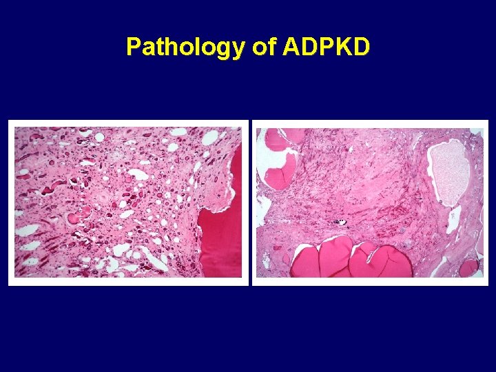
Pathology of ADPKD
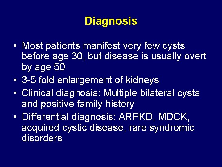
Diagnosis • Most patients manifest very few cysts before age 30, but disease is usually overt by age 50 • 3 -5 fold enlargement of kidneys • Clinical diagnosis: Multiple bilateral cysts and positive family history • Differential diagnosis: ARPKD, MDCK, acquired cystic disease, rare syndromic disorders
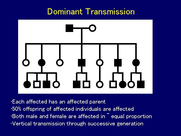
Dominant Transmission • Each affected has an affected parent • 50% offspring of affected individuals are affected • Both male and female are affected in ~ equal proportion • Vertical transmission through successive generation
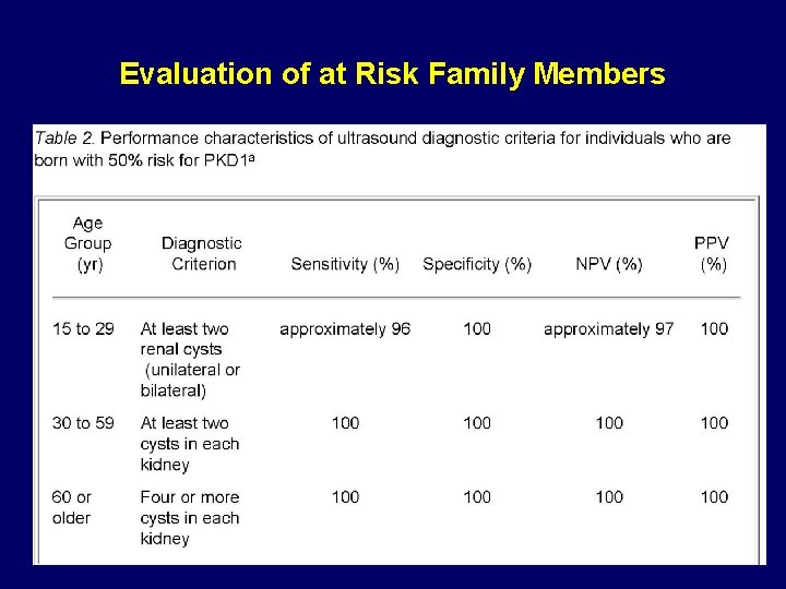
Evaluation of at Risk Family Members
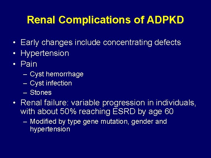
Renal Complications of ADPKD • Early changes include concentrating defects • Hypertension • Pain – Cyst hemorrhage – Cyst infection – Stones • Renal failure: variable progression in individuals, with about 50% reaching ESRD by age 60 – Modified by type gene mutation, gender and hypertension
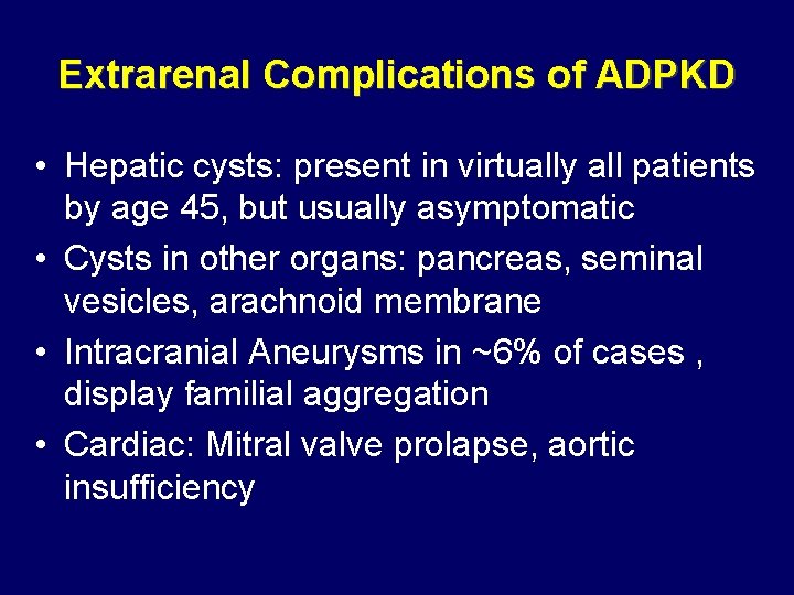
Extrarenal Complications of ADPKD • Hepatic cysts: present in virtually all patients by age 45, but usually asymptomatic • Cysts in other organs: pancreas, seminal vesicles, arachnoid membrane • Intracranial Aneurysms in ~6% of cases , display familial aggregation • Cardiac: Mitral valve prolapse, aortic insufficiency
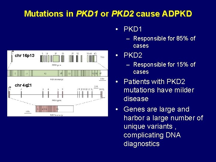
Mutations in PKD 1 or PKD 2 cause ADPKD • PKD 1 – Responsible for 85% of cases chr 16 p 13 • PKD 2 – Responsible for 15% of cases chr 4 q 21 • Patients with PKD 2 mutations have milder disease • Genes are large and harbor a large number of unique variants , complicating DNA diagnostics
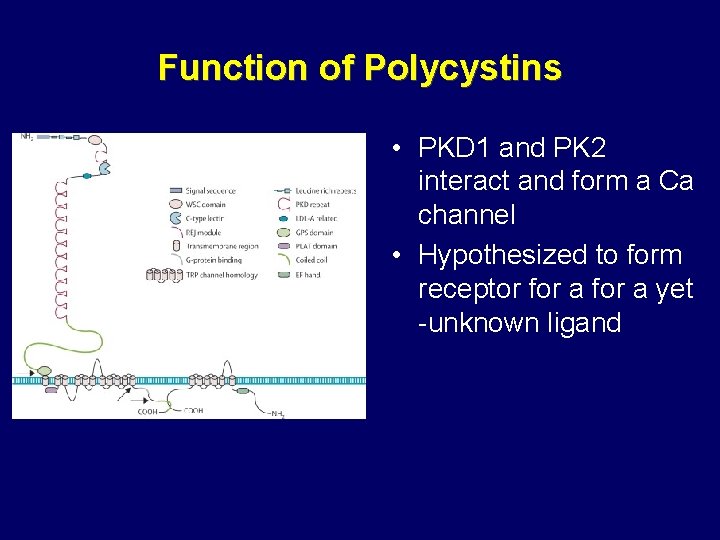
Function of Polycystins • PKD 1 and PK 2 interact and form a Ca channel • Hypothesized to form receptor for a yet -unknown ligand
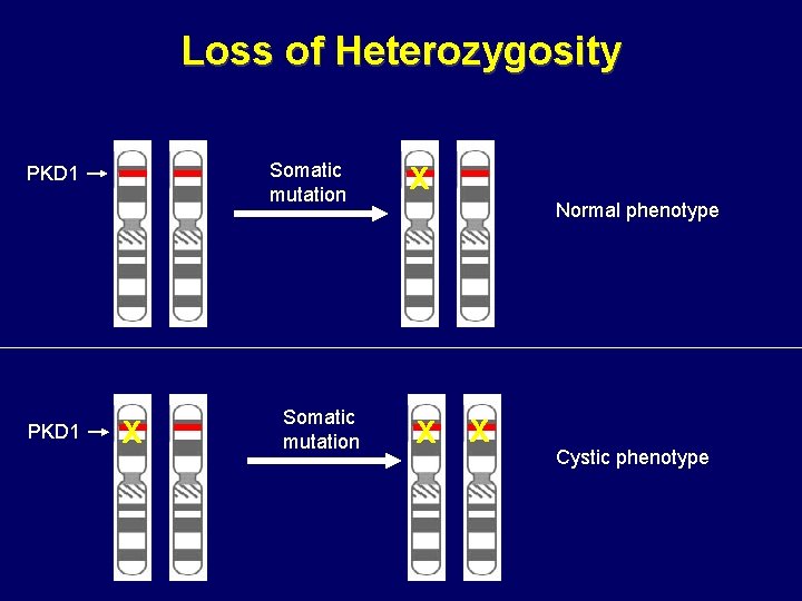
Loss of Heterozygosity Somatic mutation PKD 1 X Somatic mutation X Normal phenotype X X Cystic phenotype
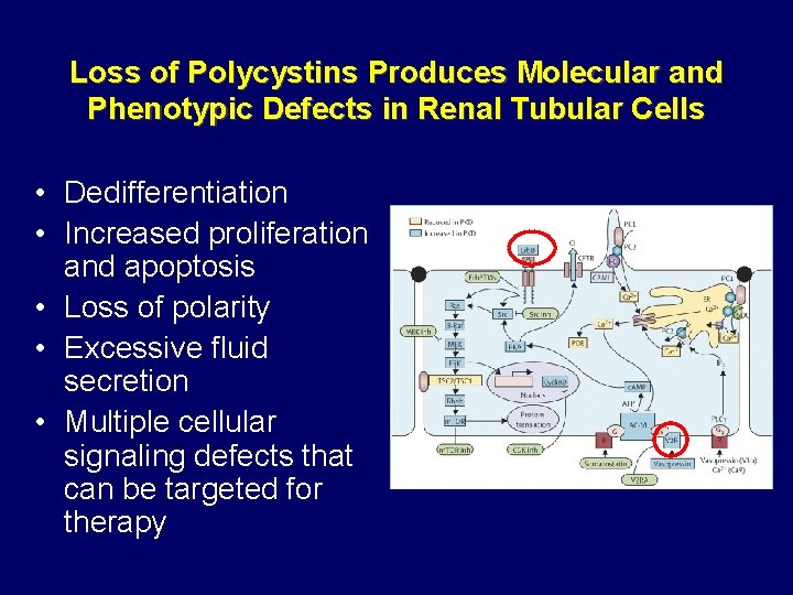
Loss of Polycystins Produces Molecular and Phenotypic Defects in Renal Tubular Cells • Dedifferentiation • Increased proliferation and apoptosis • Loss of polarity • Excessive fluid secretion • Multiple cellular signaling defects that can be targeted for therapy
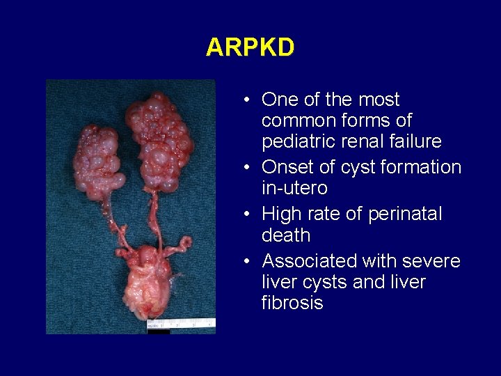
ARPKD • One of the most common forms of pediatric renal failure • Onset of cyst formation in-utero • High rate of perinatal death • Associated with severe liver cysts and liver fibrosis
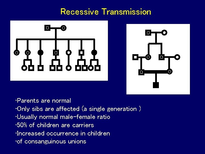
Recessive Transmission • Parents are normal • Only sibs are affected (a single generation ) • Usually normal male-female ratio • 50% of children are carriers • Increased occurrence in children • of consanguinous unions
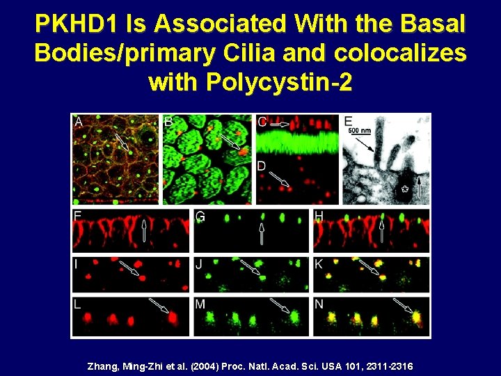
PKHD 1 Is Associated With the Basal Bodies/primary Cilia and colocalizes with Polycystin-2 Zhang, Ming-Zhi et al. (2004) Proc. Natl. Acad. Sci. USA 101, 2311 -2316
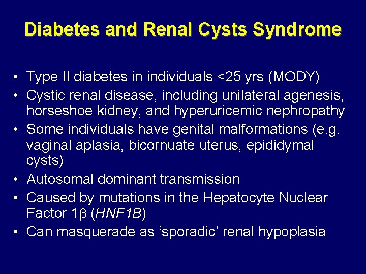
Diabetes and Renal Cysts Syndrome • Type II diabetes in individuals <25 yrs (MODY) • Cystic renal disease, including unilateral agenesis, horseshoe kidney, and hyperuricemic nephropathy • Some individuals have genital malformations (e. g. vaginal aplasia, bicornuate uterus, epididymal cysts) • Autosomal dominant transmission • Caused by mutations in the Hepatocyte Nuclear Factor 1 (HNF 1 B) • Can masquerade as ‘sporadic’ renal hypoplasia
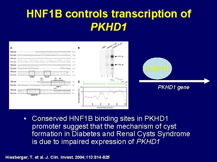
HNF 1 B controls transcription of PKHD 1 • HNF 1 B PKHD 1 gene • Conserved HNF 1 B binding sites in PKHD 1 promoter suggest that the mechanism of cyst formation in Diabetes and Renal Cysts Syndrome is due to impaired expression of PKHD 1 Hiesberger, T. et al. J. Clin. Invest. 2004; 113: 814 -825
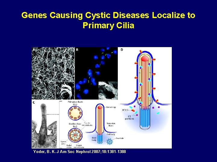
Genes Causing Cystic Diseases Localize to Primary Cilia Yoder, B. K. J Am Soc Nephrol 2007; 18: 1381 -1388
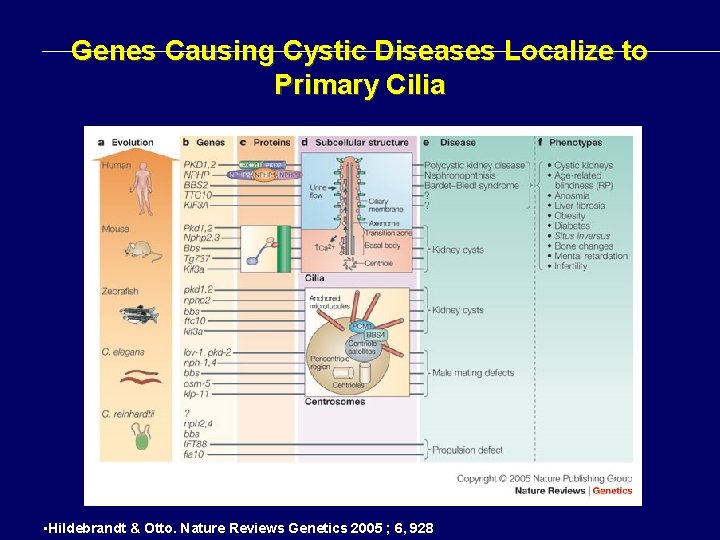
Genes Causing Cystic Diseases Localize to Primary Cilia • Hildebrandt & Otto. Nature Reviews Genetics 2005 ; 6, 928
Web References • Pathology Pictures – Columbia Pathology: http: //cpmcnet. columbia. edu/dept/curricpathology/pathology/pathoatlas/index. html – Pathology Education Instructional resources (PEIR) http: //peir. net/ • Human Genetics – OMIMTM - Online Mendelian Inheritance in Man. TM http: //www. ncbi. nlm. nih. gov/sites/entrez? db=OMIM
Further Reading • Woolf AS. et al. Evolving concepts in human renal dysplasia. J Am Soc Nephrol. 2004 : 998 • Genetic approaches to human renal agenesis/hypoplasia and dysplasia. Pediatr Nephrol. 2007 : 1675 • Torres et al. Autosomal Dominant Polycystic Kidney disease. Lancet 2007; 369: 1287 • Hildebrandt & Otto. Cilia and centrosomes: a unifying pathogenic concept for cystic kidney disease? Nature Reviews Genetics 2005 ; 6, 928