Shoulder Osteology Xray Muscles CTMRI Shoulder Osteology Xray


- Slides: 44
Shoulder • • Osteology X-ray Muscles CT/MRI
Shoulder • • Osteology X-ray Muscles CT/MRI
Shoulder X-ray, AP projection 1, Clavicle. 2, Acromion. 3, Greater tubercle. 4, Lesser tubercle. 5, Neck of Humerus. 6, Humerus. 7, Coracoid Process. 8, Axillary border of scapula. 9, Rib.
Shoulder X-ray: lateral view 1, Coracoid Process. 2, Clavicle. 3, Acromion. 4, Head of Humerus. 5, Humerus. 6, Axillary border of scapula.
Shoulder • • Osteology X-ray Muscles CT/MRI
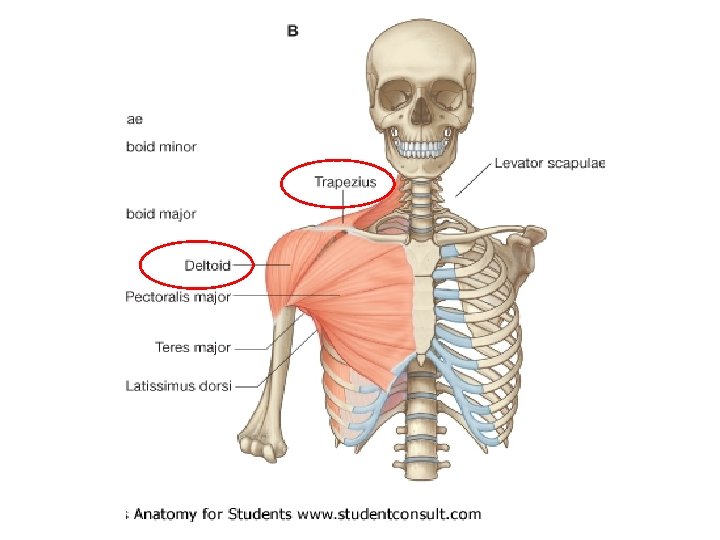
Serratus anterior
Short muscles
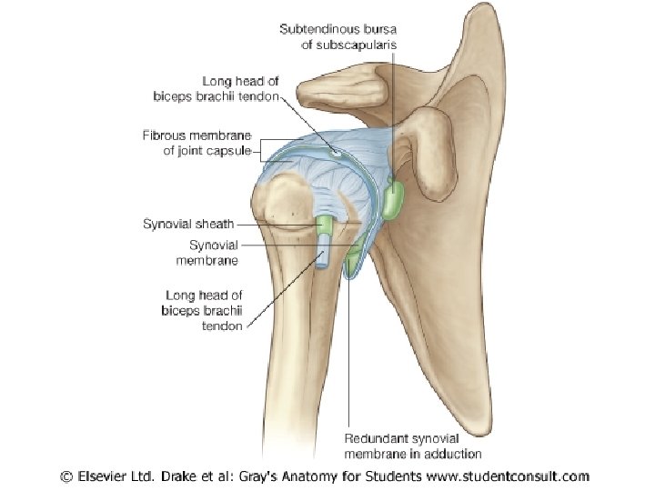
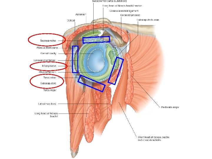
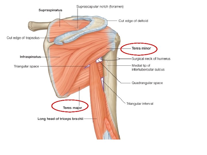
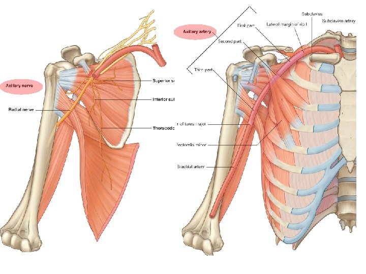
Rotator Cuff
Muscles of arm • Antererior group – Biceps brachii – Coracobrachialis
Posterior group – triceps brachii
Shoulder • • Osteology X-ray Muscles CT/ MRI
1. 2. 3. 4. 5. 6. 7. 8. 9. 10. coracoid subclavius pectoralis major clavicle Subscapularis Serratus anterior spine Deltoid muscle. Humeral head.
CT
1. 2. 3. 4. 5. 6. 7. 8. 9. humerus corachobrachialis Pectoralis minor Pectoralis major Serratus anterior trapezious infraspinatus Teres major triceps
CT
Anatomical planes
Anatomical Planes • Saggital – divides the body into right and left parts • Coronal or frontal– divides the body into anterior and posterior parts • Horizontal or transverse (cross section) – divides the body into superior and inferior parts
Saggital plane
Coronal Plane
Horizontal Plane
Body Planes 1 2 3
Slides: • http: //www. info-radiologie. ch/indexenglish. php • http: //www. meddean. luc. edu/lumen/meded/ grossanatomy/x_sec/mainx_sec. htm
Coronal/Frontal T 1
MRI of the shoulder. Coronal T 1 -weighted view. Image 2 1, Trapezius muscle. 2, Acromion. 3, Deltoid muscle. 4, Humeral head. 5, Infraspinatus muscle. 6, Teres minor muscle. 7, Teres major muscle. 8, Tricipital muscle. Arrow, Posterior humeral circumflex artery and axillary nerve.
Arrow: Posterior humeral circumflex artery and axillary nerve.
MRI of the shoulder. Coronal T 1 -weighted view. Image 6 1, Trapezius muscle. 2, Acromioclavicular joint. 3, Acromion. 4, Deltoid muscle. 5, Humeral head. 6, Supraspinatus muscle. 7, Spine of the scapula. 8, Infraspinatus muscle. 9, Scapula. 10, Subscapularis muscle. 11, Teres major muscle. .
MRI of the shoulder. Coronal T 1 -weighted view. Image 11 1, Trapezius muscle. 2, Clavicle. 3, Supraspinatus tendon. 4, Deltoid muscle. 5, Humeral head. 6, Glenoid. 7, Supraspinatus muscle. 8, Biceps tendon (long head). 9, Biceps and coracobrachialis muscle.
Axial / Transverse / Horizontal T 1
MRI of the shoulder. Axial T 1 -weighted view. Image 3 1, Pectoralis major muscle. 2, Axillary vein and artery. 3, Coracoid process. 4, Deltoid muscle. 5, Supraspinatus muscle. 6, Acromion. 7, Scapula. 8, Subscapularis muscle.
MRI of the shoulder. Axial T 1 -weighted view. Image 5 1, Pectoralis major muscle. 2, Deltoid muscle (anterior). 3, Coracoid process. 4, Humeral head. 5, Glenoid. 6, Supraspinatus muscle. 7, Scapula. 8, Deltoid muscle. 9, Infraspinatus muscle. 10, Subscapularis muscle. 11, Axillary vein and artery. 12, Pectoralis minor
MRI of the shoulder. Axial T 1 -weighted view. Image 7 1, Pectoralis major muscle. 2, Deltoid muscle. 3, Humeral head. 4, Deltoid muscle. 5, Glenoid. 6, Infraspinatus muscle. 7, Subscapularis muscle. 8, Axillary vein and artery. 9, Pectoralis minor muscle.
MRI of the shoulder. Axial T 1 -weighted view. Image 19 1, Pectoralis major muscle. 2, Biceps muscle (short head). 3, Humerus. 4, Deltoid muscle. 5, Tricipital muscle. 6, Scapula. 7, Subscapularis muscle. 8, Coracobrachialis muscle. 9, Pectoralis minor muscle.
Sagittal T 2
MRI of the shoulder. Sagittal T 2 FATSAT. Image 9 1, Coracobrachialis. 2, Subscapularis tendon. 3, Humeral head. 4, Coracoid process. 5, Deltoid muscle. 6, Biceps tendon (long head). 7, Clavicle. 8, Acromioclavicular joint. 9, Supraspinatus muscle. 10, Acromion. 11, Infraspinatus muscle. 12, Deltoid muscle. 13, Teres minor muscle
MRI of the shoulder. Sagittal T 2 FATSAT. Image 11 1, Teres minor muscle and tendon. 2, Infraspinatus tendon. 3, Acromion. 4, Supraspinatus tendon. 5, Biceps tendon (long head). 6, Subscapularis tendon. Rotator Cuff Muscles