XRay Xray Films The film used in dental


 between adjacent areas")


- Slides: 26
X-Ray ﺃﻤﻞ ﺭﺅﻮﻑ. ﺩ ============= X-ray Films: The film used in dental radiography is photographic film that has been adapted for dental use. A photographic image is produced on dental X- ray film when it is exposed to X- ray that have passed through teeth and adjacent structures.
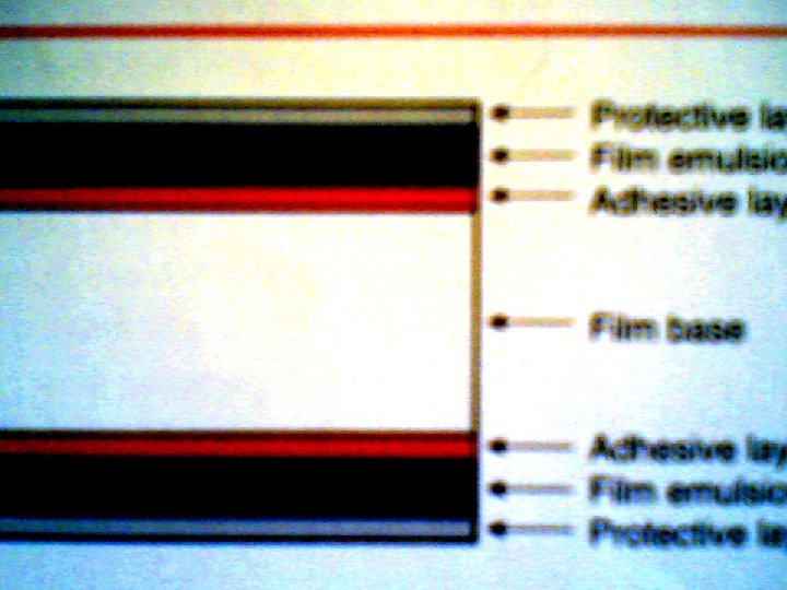
Types of X- ray films : 1. Intraoral X- ray film 2. Extraoral X- ray film 1. Intra oral x- ray film : in dentistry has four basic components: A-Film Base: is a flexible piece made of cellulose acetate measures 0. 2 mm thick and is constructed to withstand heat , moisture , and chemical exposure. The primary purpose of the film base is to provide a stable support for the delicate emulsion , it also provides strength
B-Adhesive layer : Added to the film base before the emulsion is applied and serve to attach the emulsion to the base. C-Film Emulsion : is a coating attached to both sides of the film base by the adhesive layer to give the film greater sensitivity to X- radiation. The emulsion is a homogeneous mixture of gelatin and silver halide crystals. During film processing , the gelatin serves to absorb the processing solution and allows the chemical to react with the silver halide crystals
A halide is a chemical compound that is sensitive to radiation or light. The halides used in dental X-ray film are made up of the element silver , either bromine or iodine , silver bromide ( Ag. Br) and silver iodide (Ag. I) are two types of silver halide crystals found in the film emulsion. The silver halide crystals absorb radiation during xray exposure and store energy from the radiation. D-Protective Layer : is a thin , transparent coating placed over the emulsion. It serves to protect the emulsion surface from manipulation as well as mechanical and processing damage.
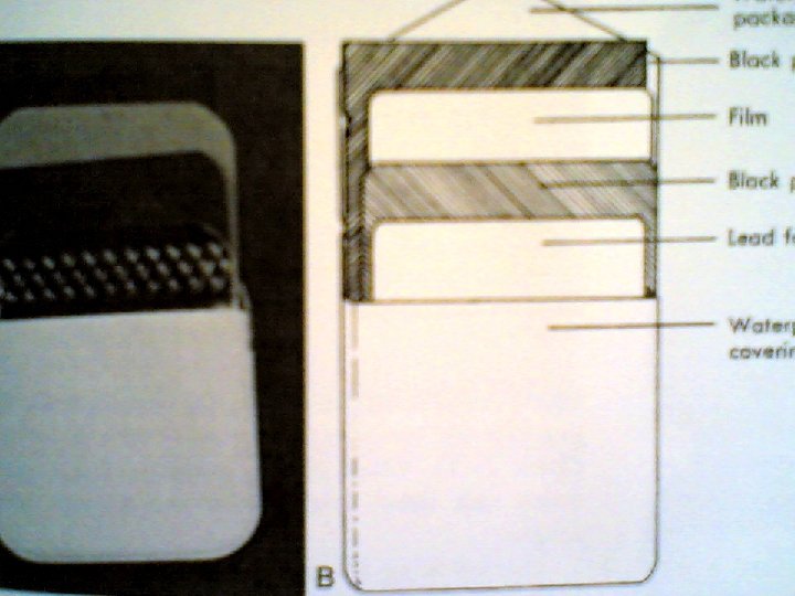
X- Ray film packet : The intraoral X- ray film is a double – emulsion (emulsion on both sides ) , double – emulsion film is used instead of single – emulsion (emulsion on one side ) film , because it requires less radiation exposure to produce an image. A film packet may contain one film (one – film packet ) or two films ( two – film packet ). A two – film packet produces two identical radiographs with the same amount of exposure necessary to produce a single radiograph.
Identification dot : In one corner of the intraoral x- ray film , a small raised bump known as the identification dot is found. The raised bump is used to determine film orientation. After the film is processed , the raised identification dot is used to distinguish between the left and right sides of the patient. Paper film wrapper: The paper film wrapper within the film packet is a black paper protective sheet that covers the film and shield the film from light.
Lead foil sheet: The lead foil sheet is a single piece of lead foil that is found within the film packet and is located behind the film wrapped in black protective paper. The thin lead foil sheet is positioned behind the film to shield the film from back – scattered (secondary )radiation that results in film fog.
Outer package wrapping: The outer package wrapping is a soft vinyl or paper wrapper that hermetically seals the film packet , protective black paper , and lead foil sheet. This outer wrapper serves to protect the film from exposure to light and saliva. The outer wrapper of the film packet has two sides : 1. Tube side : is solid white and has a raised bump in one corner that corresponding to the identification dot on the x- ray film. Whine placed in the mouth , the tube side is of the film packet must face the teeth and the tube head 2. Label side : this side has a flap that is used to open the film packet prior to processing
X –Ray Films Type and size : Type I : Intraoral X – ray film which called periapical film which used to examine the apical area of the tooth and the surrounding structure. This type present in different sizes from( 0 , 1 , 2)
Type II : Which called bitwing film and it used to examine the crowns of both the maxillary and mandibular teeth on one film. The bite – wing film is useful in examining the interproximal , or adjacent , tooth surfaces. This type is present in different sizes from (0 , 1 , 2 , 3 , ).
Type III : Called occlusal film that used to demonstrate an area of greater dimension than area appear in P. A film , and this occlusal film has just one size which is ( 3. 4 ). So that the intraoral film size and type have been standerized on a numerical basis
Intraoral Film Speed : It means the sensitivity of X- ray film to X- radiation , or the amount of radiation required to produce a radiograph of standard density. Film speed , or sensitivity , is determined by the followings : 1. the size of the silver halide crystals 2. the thickness of the emulsion 3. the presence of special radiosensitive dyes. Film speed determines how much radiation and how much exposure time are necessary to produce an image on a film. So a fast film requires less radiation exposure because the film responds more quickly , because the silver halide crystals in the emulsion are larger. the larger the crystals , the faster the film speed.
Intraoral film speeds have been standerized on alphabetical basis. So film speed will range from ( A) to ( F ), film speed ( A ) is slower while film speed ( F ) is faster one. In general the faster film speed the much less amount of X- radiation required to produce the photographic image, and this mean a reduction in patient exposure.
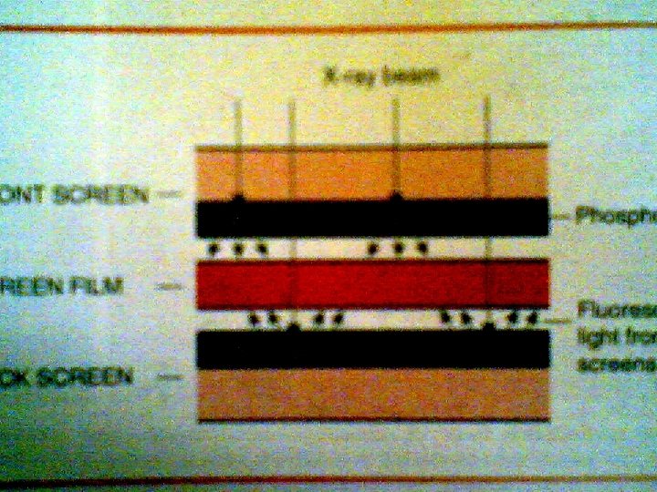
Extraoral Film: Is one that is placed outside of the mouth during Xray exposure. Extraoral film are used to examine large areas of the skull or jaws. Types of extaoral film : A-Screen Film : The majority of extra oral films are screen films. A screen film is a film that requires the use of screen for exposure. The emulsion of the film is sensitive to visible light and more specifically to blue light of visible light spectrum. The screen film placed between 2 florescent screen in a rigid holder or cassette. These screens are made of ( Ting calcium Tungstate )bound together in a uniform layer on a film base. When the cassette is exposed to X- ray , the screen convert the X- ray energy into light , which in turn exposes the screen film.
Films used in a screen film combination are sensitive to specific colors of fluorescent light. some screen films are sensitive to blue light , whereas others are sensitive to green light. Blue – sensitive film must be paired with screen that produce blue light , and green – sensitive film must be paired with screen that produce green light. The screen film must be in intimate contact with fluorescent screen and any space between them will result in un sharp image in the radiograph. Intimate film screen contact is obtained through the use of spring or clamps. The sizes of screen film include 5 x 7 , 8 x 10 and 10 x 12 inches.
B- Non –Screen film : Is an extraoral film that dose not require the use of screen for exposure. The non – screen film is exposed directly to X- rays than to the light , because the emulsion is sensitive to direct X- ray exposure rather than fluorescent light. Non screen film either have one emulsion layer or may have a double emulsion like intraoral film , but with greater thickness than intraoral film , so a non screen film requires more exposure time than screen film so is not recommended for use in dental radiography. The non screen film place in a cardboard holder or envelope and the size of the film used are 5 x 7 , 8 x 10 inches.
Film Properties : These include A-Density : Is the degree of blackness present in the processed film. It is measured in terms of light transmission on a percentage or logarithmic scale. for examples if a film permit only (110)of a beam light to pass through it , the film is said to have a density of (log 10 =1). If the film is blacker and allow only (100) of a beam of light to pass through it , the film said to have density of (log 100=2). Basically , diagnostic radiography uses a film density range of (0. 25 -2). Film density best shows by sensitometric or by H and D curve, this curve is obtained by plotting film density against exposure time.
Factors affect film density : 1 -Exposure time : increase exposure time will increase the film density 2 -Milliamper: increase m. A value cause increase film density. 3 -Kilovoltage : increase k. V cause increase density 4 - Developing time : increase developing time cause increase density 5 -Distance : increase distance between source of radiation and the film cause decrease film density.
B- Contrast : The difference in the degrees of blackness (densities) between adjacent areas on a dental radiograph is termed contrast. High contrast image : image of short scale contrast , it has black and white with few gray shade Low contrast image : image of long scale contrast , have a wide range of gray shade with stable contrast differences.
Factors affect of contrast : 1. k. V : increase k. V of x- ray machine will decrease the contrast 2. Processing solution temperature : increase the temp. cause increase the contrast. C. Resolution : Also called details , definition , sharpness. It is the ability of a film to produce sharp outline of an object.
Factors affect on Resolution 1. Focal spot size : it must be as small as possible in order to produce sharp image 2. Size of film grain : increase the size of the film grain produce less sharp radiographic image. 3. Movement of the patient or tube or film during exposure cause un sharp image 4. Source – film distance : which should be as great as possible , otherwise the image will be un sharp 5. Object – Film distance ; which should be as small as possible to produce sharp image 6. Screen-film contact: poor contact cause un sharp image
D- Latitude: refers to the range of useful densities that are recorded on the film.