Shoulder Shoulder Shoulder Dislocations Most common type flattened


 – Detects pathological conditions")


 – Tightness or")


- Slides: 41
Shoulder
Shoulder
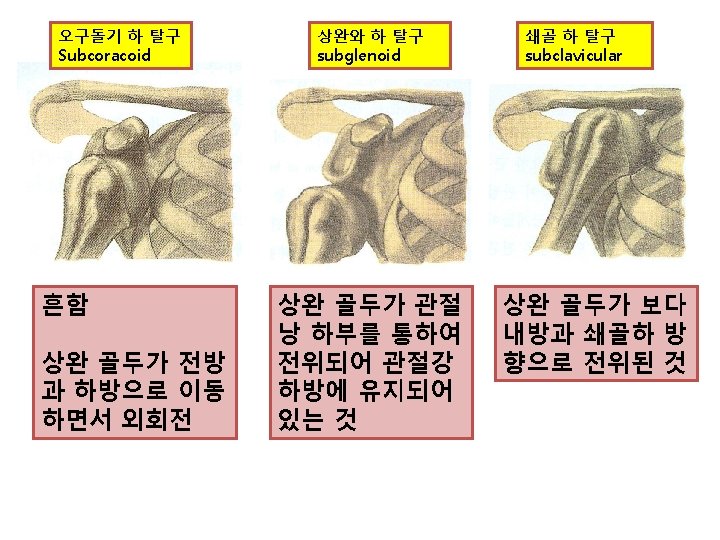
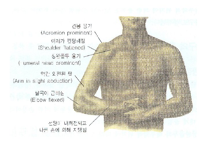
Shoulder Dislocations • • Most common type flattened deltoid, prominent humeral head in axilla arm carried in slight abduction and external rotation moderate pain and disability
Anatomy of the Shoulder Rotator Cuff
Rotator Cuff Origin: supraspinous fossa of scapula Insertion: greater tubercle of humerus Action: assists deltoid muscle in abducting arm at shoulder joint, Initiates the first 30 -60 degrees of abduction at which point the deltoid takes over Origin: subscapular fossa of scapula Insertion: lesser tubercle of humerus Action: medially rotates arm at shoulder joint Origin: Infraspinous fossa of scapula Insertion: greater tubercle of humerus Action: laterally rotates and adducts arm at shoulder joint Origin: Inferior lateral border of scapula Insertion: greater tubercle of humerus Action: laterally rotates, extends, and adducts arm at shoulder joint
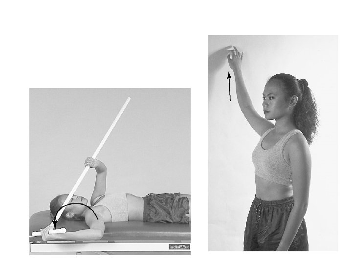
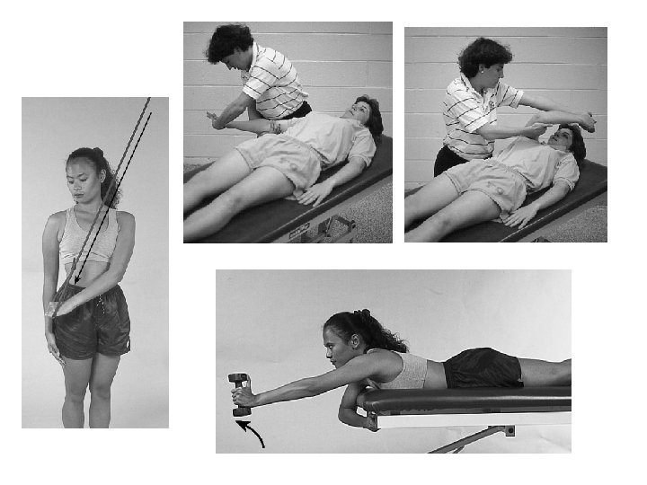
• Flexibility – Codman’s pendulum exercises – Progress to active assisted ROM in pain free – Should be performed in rotator cuff and scapula strengthening exercises
• Strengthening Exercises
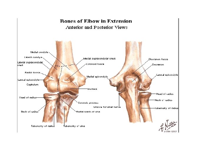
Elbow
Elbow Dislocation • Typically results from hyperextension, trochlea levered over coronoid process • Most common direction is posterolateral • Involve injury to most ligament structures, and potential for injury to neurovascular structures
• Signs and Symptoms – Swelling, severe pain, disability – Median and radial nerves may be compromised – Blood vessels may be compromised – Often a radial head fracture is involved
Kendall’s Test tests for rectus femoris tightness
Thomas Test for hip flexor contractures
Test for Hip and Sacroiliac Joint • Patrick Test (FABER) – Detects pathological conditions of the hip and SI joint – Pain may be felt in the hip or SI joint
• Gaenslen’s Test – Test works to push SI joint into extension – Test is positive if hyperextension on affected side increases pain
Testing the Tensor Fasciae Latae and Iliotibial Band • Renne’s test – Athlete stands w/ knee bent at 30 40 degrees – Positive response of TFL tightness occurs when pain is felt at lateral femoral condyle
• Ober’s Test – Used to determine presence of contracted TFL or ITband – Thigh will remain in abducted position, not falling into adduction
Trendelenburg’s Test - Iliac crest on unaffected side should be higher when standing on one leg - Test is positive when affected side is higher indicating weak abductors (glut medius) positive
• Piriformis Test – Hip is internally rotated(with knee flexion) – Tightness or pain is indicative of piriformis tightness
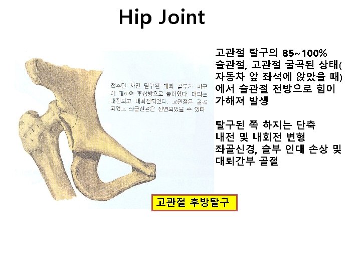
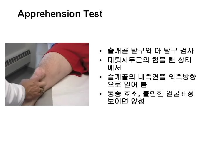
Dislocation of the Patella • Patella usually dislocates to lateral side • May reduce spontaneously during splinting 38
Patellar Dislocation/Subluxation
Patellar Dislocation/Subluxation • Pathology • Lateral deviation of patella • Tearing of the medial retinaculum • Trauma to joint surfaces • Signs and Symptoms • Pain • Deformity • When you see it, you will know it