
ELEMENTS OF OSTEOLOGY Definitions Osteology osteon bone logia

ELEMENTS OF OSTEOLOGY Definitions: Osteology osteon = bone; logia = study of bones and their components.

Skeleton: • A skeleton is collective term for continuously joined bones, cartilages, ligaments and tendons for the formation of the body framework. • The human skeleton is found inside (interior) the body and therefore endoskeleton as compared to the outside (exterior) skeleton of an insect which is known as exoskeleton. • Bones, cartilages, ligaments and tendons are commonly known as skeletal components.
Functions of the skeleton: • • • formation of the body framework and lever protection of organs (mostly internal organs) muscle attachment hence locomotion mineral reseivoir houses bone marrow for haemopoiesis provides a mechanism body balancing and weight reduction ( skull pneumatic bones)
Parts of the human skeleton • As it is for the body, a human skeleton is divided into two major parts. i. axial skeleton which include skull - head skeleton vertebral column - cervical, thoracic, lumbar, sacrum and coccyx. chest cage - ribs and stenum. ii. appendicular skeleton which include the upper limbs– shoulder girdle, arm, forearm and hand the lower limbs – pelvic girdle, thigh, leg and foot.
BONES: • These are major skeletal components and hardened structures (tissue) of the skeleton. • The hard consistence is due to mineralization taking place during development but maintained by metabolic balances throughout in life. • In the skeleton, bones are covered by other soft tissues like connective tissue fibrous membrane called periosteum, deep fasciae and attached tendons and ligaments. • Dry skeleton has only macerated and articulated bones.
Types of bones: • Three categories are used to classify bones: a). by morphology; this bases on the gross appearance of the bone that involves the size, shape and plateaus. In this category, six types of bones are recognized which are: i. long bones, these bones are characterized by having two ends (epiphysis) and a shaft (diaphysis). The two areas are separated by a cartilaginous material plate called metaphysis or metaphyseal plate. They are normally bones of the appendicular region. eg femur, humerus and fibular. ii. Short bones, these have approximately six surfaces at least four surfaces for muscle attachment and the rest for articulation. eg. carpal and tarsal bones
Cont……. . iii. Flat bones, these have a spongy layer (diploe) enclosed in between the two compact layers. eg cranial skull bones, scapula iv. Irregular bones, these have no particular shape to compare eg hip bones, vertebrae. v. Sesamoid bones, these are those bones developing in the tendon for the purpose of reducing friction between a particular tendon and its underlined hard structure like bones or a ground surface. eg patella, pisiform vi. Pneumatic bones, all bones having air cavities. Big cavities are known as sinuses while smaller cavities are called air cells. Sinuses in the facial skull bones help to reduce unnecessary weight of the head give resonance to voice production and equalize the atmospheric and intercranial pressures. eg facial skull bones (sinuses), mastoid process of temporal bone( air cells).
Cont……… b). by histology; this bases on the histological features of the bones. Each individual bone demonstrates all histological characters such that all histological types bones are found in each individual bone. There are two types of bones in this category: i. compact bone, bone matrix is fully mineralized (compact) such that no spaces which are available except for the canals and canaliculi. ii. spongy (cancellous) bone, this is characterized by having spongy appearance whereby the matrix has interconnected bars (trabeculae).
Cont…. . c). by development; this bases on the modes of bone formation (ossification). There are two ways by which bones are forming, these are intermembranous and endochondral ossification. In intermembranous, a layer of mesenchyme differentiates into bone tissue. At first, the mesenchymal cells differentiate into osteoprogenitor cells (bone forming primitive cell) which start to lay down the bone matrix. From this mode, the resultant is a i. membranous bone. In endochondral ossification, at first there is a cartilage model which is invaded by the bone forming cells then the model is replaced by a newly formed bone. From this mode, the resultant is a ii. Cartilage bone. Therefore two types of embryological bones are membranous and cartilage bones.
Bone development and growth: • Bone development (refer to embryological type of bones and bone histology) • Bone growth is supported by factors like good nutrition (vitamin, protein and minerals), hormones and environmental status. • It passes through two modes which are i. interstitial or auxetic growth, here is where the whole tissue expansion occurs such that after a certain period a tissue increased markedly on its size. This occurs in the long bones at the junction between the epiphyseal plate and the diaphysis or epiphysis. Epiphyseal plate cartilage has an ability to undergo auxetic growth.
Cont……. . ii. Appositional or accretional growth, here there is an additional of materials on the surface of the tissue. But the previous tissue remains unchanged in its size though there is a remarkable increase in size. • NB: multiplicative growth is not possible to a bone as it has a hard and mineralized matrix with scattered cell group(s) found in the lacunae. • Functions of the bones. Refer to the functions of the skeleton.
ELEMENTS OF ARTHROLOGY. • Definition: Arthrology : arthrons = joint logia = study of -study of joints and their associated movements. • Joints are junctions between two or more skeletal components which facilitate the levering action of the skeleton.
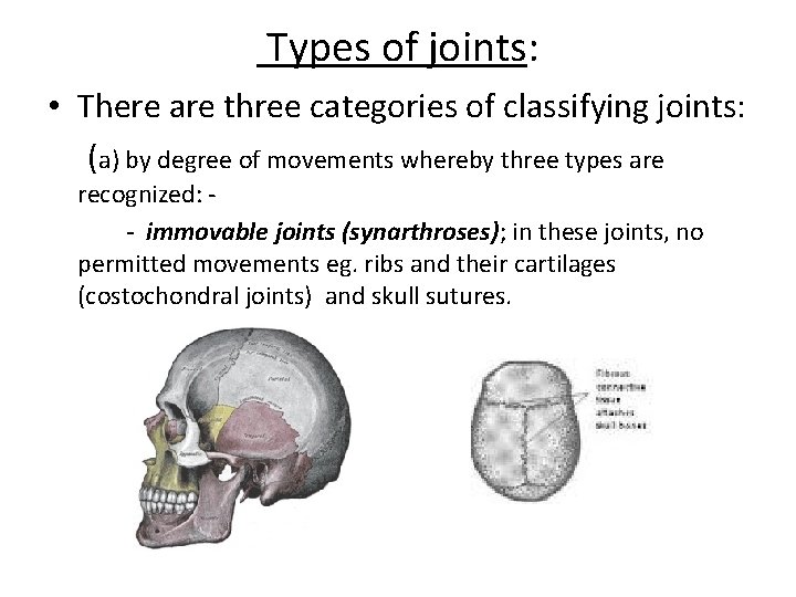
Types of joints: • There are three categories of classifying joints: (a) by degree of movements whereby three types are recognized: - - immovable joints (synarthroses); in these joints, no permitted movements eg. ribs and their cartilages (costochondral joints) and skull sutures.
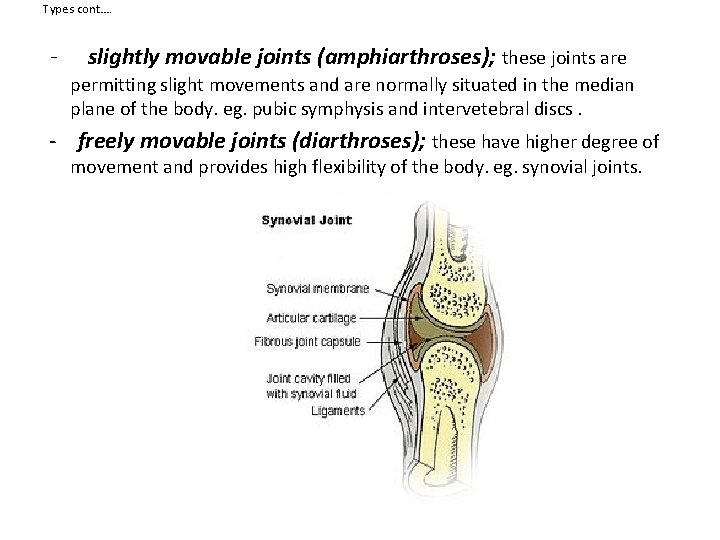
Types cont…. - slightly movable joints (amphiarthroses); these joints are permitting slight movements and are normally situated in the median plane of the body. eg. pubic symphysis and intervetebral discs. - freely movable joints (diarthroses); these have higher degree of movement and provides high flexibility of the body. eg. synovial joints.
Cont…… • (b) by plane(s) or axis of movement(s) whereby three types are recognized - uniaxial joints; only one plane of movements is possible. eg. flexion and extension. - biaxial joints; two planes of movements are allowed eg. flexion / extension and adduction / abduction - multiaxial joints; more than two planes of movements are permitted eg. rotation and others ie. Circumduction.
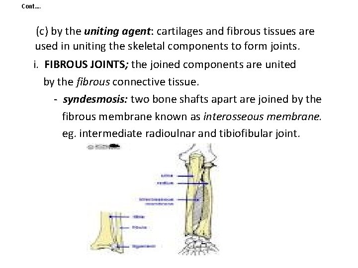
Cont…. (c) by the uniting agent: cartilages and fibrous tissues are used in uniting the skeletal components to form joints. i. FIBROUS JOINTS; the joined components are united by the fibrous connective tissue. - syndesmosis: two bone shafts apart are joined by the fibrous membrane known as interosseous membrane. eg. intermediate radioulnar and tibiofibular joint.
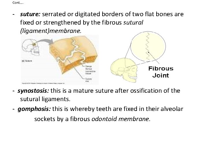
Cont…. . - suture: serrated or digitated borders of two flat bones are fixed or strengthened by the fibrous sutural (ligament)membrane. - synostosis: this is a mature suture after ossification of the sutural ligaments. - gomphosis: this is whereby teeth are fixed in their alveolar sockets by a fibrous odontoid membrane.
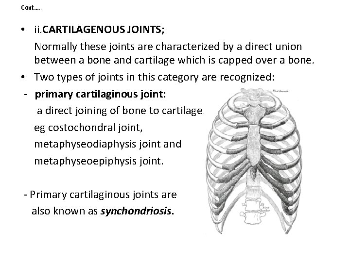
Cont…. . • ii. CARTILAGENOUS JOINTS; Normally these joints are characterized by a direct union between a bone and cartilage which is capped over a bone. • Two types of joints in this category are recognized: - primary cartilaginous joint: a direct joining of bone to cartilage. eg costochondral joint, metaphyseodiaphysis joint and metaphyseoepiphysis joint. - Primary cartilaginous joints are also known as synchondriosis.
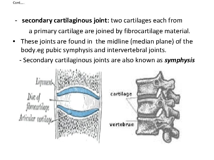
Cont…. . - secondary cartilaginous joint: two cartilages each from a primary cartilage are joined by fibrocartilage material. • These joints are found in the midline (median plane) of the body. eg pubic symphysis and intervertebral joints. - Secondary cartilaginous joints are also known as symphysis
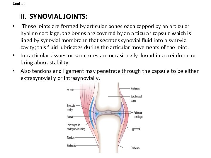
Cont…. . iii. SYNOVIAL JOINTS: • These joints are formed by articular bones each capped by an articular hyaline cartilage, the bones are covered by an articular capsule which is lined by synovial membrane that secretes synovial fluid into a synovial cavity; this fluid lubricates during the articular movements of the joint. • Intrarticular tissues or structures are occasionally found in to reinforce or bring about stability. • Also tendons and ligament may penetrate through the capsule to be either extrasynovially or intrasynovially.
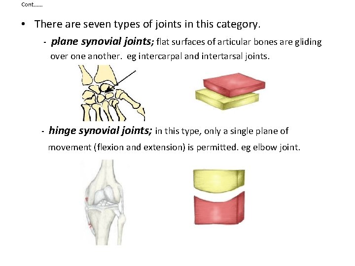
Cont…… • There are seven types of joints in this category. - plane synovial joints; flat surfaces of articular bones are gliding over one another. eg intercarpal and intertarsal joints. - hinge synovial joints; in this type, only a single plane of movement (flexion and extension) is permitted. eg elbow joint.
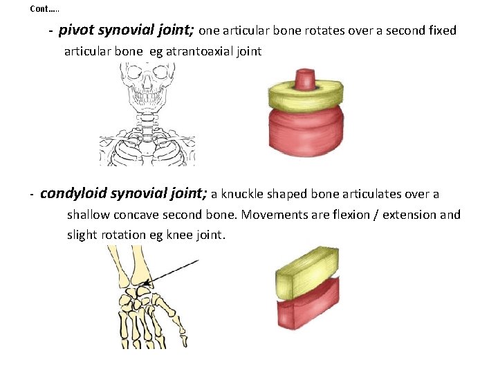
Cont…. . - pivot synovial joint; one articular bone rotates over a second fixed articular bone eg atrantoaxial joint - condyloid synovial joint; a knuckle shaped bone articulates over a shallow concave second bone. Movements are flexion / extension and slight rotation eg knee joint.
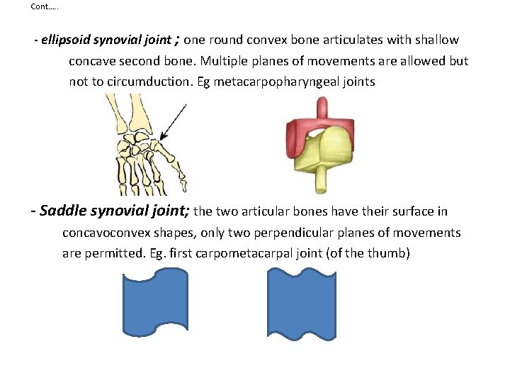
Cont…. . - ellipsoid synovial joint ; one round convex bone articulates with shallow concave second bone. Multiple planes of movements are allowed but not to circumduction. Eg metacarpopharyngeal joints - Saddle synovial joint; the two articular bones have their surface in concavoconvex shapes, only two perpendicular planes of movements are permitted. Eg. first carpometacarpal joint (of the thumb)
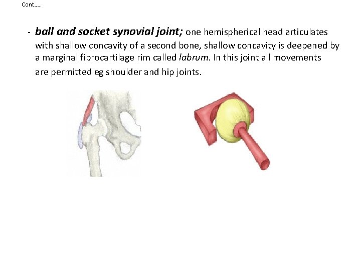
Cont…. . - ball and socket synovial joint; one hemispherical head articulates with shallow concavity of a second bone, shallow concavity is deepened by a marginal fibrocartilage rim called labrum. In this joint all movements are permitted eg shoulder and hip joints.
Joint stability • The stability of many freely movable joints depend much on three main factors: - Articular surfaces, Ligaments and Muscles i. Articular surfaces: the shape, size, and the arrangement of the articular surfaces contribute much in avoiding over movements, misalignments and dislocations eg. The ball and socket of the hip joint and the mortise arrangement of the ankle joint. ii. Ligaments: Fibrous and elastic ligaments prevent excessive movements (fibrous) as well as over stretchibility (elastic). But this is only when the period of the joint under tension does not prolong much. iii. Muscle tone: tonicity of muscles is also a major factor for controlling joint stability. Eg Rotator cuff muscles of the shoulder joint keep the hemispherical head of the humerus into the shallow glenoid cavity (fossa) of the scapula. Without these muscles , a little force would be required to dislocate the shoulder joint.
Nerve supply of joints • A nerve supply of the muscle moving the joint and the supply to the skin overlying these muscles and skin overlying the joint send twigs into the joint • This is HILTON’S LAW.
Joint disorders • • A joint disorder is termed an arthropathy and when involving inflammation of one or more joints the disorder is called an arthritis. Most joint disorders involve arthritis (mostly to people over 55 years of age) , but joint damage by external physical trauma is typically not termed arthritis. Arthropathies are called polyarticular when involving many joints and monoarticular when involving only one single joint. • Forms of arthritis i. Osteoarthritis (also known as degenerative joint disease) occurs following trauma to the joint, following an infection of the joint or simply as a result of aging. ii. Rheumatoid arthritis and psoriatic arthritis, which are autoimmune diseases in which the body is attacking itself. iii. Septic arthritis is caused by joint infection. iv. Gouty arthritis is caused by deposition of uric acid crystals in the joint that results in subsequent inflammation. v. pseudogout (calculus) is caused by the formation of rhomboidal shaped crystals of calcium pyrophosphate.
- Slides: 27