IMAGE QUALITY AND ARTIFACTS 1 WHY YOU SEE


 Prime Controlling Factor • m. As Influencing Factors •")


 Prime Controlling Factor • m. As Influencing Factors •")


 ON SCATTER 26")


 54")
 55")


- Slides: 104
IMAGE QUALITY AND ARTIFACTS 1
WHY YOU SEE WHAT YOU SEE… • The films or images have different levels of density – different shades of gray • X-rays show different features of the body in various shades of gray. • The gray is darkest in those areas that do not absorb Xrays well – and allow it to pass through • The images are lighter in dense areas (like bones) that absorb more of the X-rays. 2
IMAGE PRODUCTION 1. Primary Beam– The beam of photons, B 4 it interacts with the pt’s body. 2. Remnant (exit) Beam-The resulting beam that is able to exit from the patient. 3. Scatter Radiation– Radiation that interacts with matter & only continues in a different direction – not useful for image production. 4. Attenuated Beam– Primary radiation that is changed (partially absorbed) as it travels through the pt. 3
IMAGE QUALITY AND ARTIFACTS Density Contrast Detail Distortion Artifacts 4
IMAGE QUALITY AND ARTIFACTS Pho tog rap hic Density Contrast Qua lity Distortion Detail Artifacts 5
IMAGE QUALITY AND ARTIFACTS G eom etri Density c. Q uali Contrast ty Detail Distortion Artifacts 6
DENSITY THE OVERALL DARKENING OF THE IMAGE 7
DENSITY (OPTICAL DENSITY, IMAGE DENSITY) Prime Controlling Factor • m. As Influencing Factors • • • k. Vp SID Beam Filtration Beam restriction Body part thickness grids 8
10 m. A 1000 m. A 9
10
DENSITY (OPTICAL DENSITY, IMAGE DENSITY) Prime Controlling Factor • m. As Influencing Factors • • • k. Vp SID Beam Filtration Beam restriction Body part thickness grids 11
KVP MORE ENERGY = MORE PHOTONS PASSING THOUGH TISSUE & STRIKING THE IMAGE á 15% k. Vp = doubling of exposure to the film 15% k. Vp = halving of exposure to the film 15% rule will also change the contrast of the image because k. V is the primary method of changing image contrast. Remember : 15% change ( ) KVP has the same effect as doubling or ½ the MAS on density 12
This is an actual arm tatoo. Now that’s dedication ! I a n w v e r s e s q u a r e I a n w v v e e r r s s e e s q u a r e 13
COLLIMATORS Always collimate smaller than the image receptor 14
3 DIFFERENT BODY HABITUS HYPERSTHENIC HYPOSTHENIC 15
GOAL: PRODUCING OPTIMAL RADIOGRAPHS DENSITY Too dark Too light 16
CONTRAST THE RANGE OF DARK, LIGHTS, GRAYS IN THE IMAGE 17
RADIOGRAPHIC CONTRAST Primary controlling factor • k. Vp Influencing Factors • • • Subject contrast m. As SID Filtration Beam restriction grids 18
Patient Interactions CASCADE 19
Patient Interactions COMPTON SCATTERING 1. Outer shell electron in body 2. Interacts with x-ray photon from the tube 3. Moderate energy electron 20
SCALE OF CONTRAST? WHICH ONE IS “BETTER” HOW DOES THE KVP AFFECT THESE IMAGES? 21
RADIOGRAPHIC CONTRAST • Subject Contrast • • • Tissue thickness Tissue density Tissue type (atomic #) Contrast agents Scatter radiation 22
10 m. A 1000 m. A 23
This is an actual arm tatoo. Now that’s dedication ! I a n w v e r s e s q u a r e I a n w v v e e r r s s e e s q u a r e 24
RADIOGRAPHIC CONTRAST Primary controlling factor • k. Vp Influencing Factors • • • Subject contrast m. As SID Filtration Beam restriction grids 25
EFFECTS OF COLLIMATION (BEAM RESTRICTION) ON SCATTER 26
GRIDS • A device with lead strips that is placed between the patient and the cassette • Used on larger body parts to reduce the number of scattering photons from reaching the image 27
GRIDS 28
GRIDS ABSORB SCATTER – PREVENTS IT FROM REACHING THE IMAGE 29
GRID NO GRID CONTROLS CONTRAST 30
DETAIL RESOLUTION, ABILITY TO DISTINGUISH SHAPES 31
DETAIL Factors that affect detail • Image sharpness • Spatial resolution • Motion • Smallest separation of • SID two lines or edges • Focal spot size • Measured by lp/mm • OID • Image receptor type 32
RECORDED DETAIL • The degree of sharpness in an object’s borders and structural details. • How “clear” the object looks on the radiograph 33
RESOLUTION TEST TOOLS LINE PAIRS/ MM Depicts how well you can see the differences in structures More lines=more detail 34
35
MOTION • Can be voluntary or involuntary • Best controlled by short exposure times • Use of careful instructions to the pt. • Suspension of pt. respiration • Immobilization devices 36
Blurring of image due to patient movement during exposure. 37
38
SID • Shine a flashlight on a 3 -D object, shadow borders will appear “fuzzy” -On a radiograph called penumbra • Penumbra (fuzziness) obscures true border • Farther the flashlight from object = sharper borders. Same with radiography. 39
40
OID OBJECT TO IMAGE DISTANCE • The closer the object to the film, the sharper the detail. • OID , penumbra , sharpness • Structures located deep in the body, radiographer must know how to position to get the object closest to the film. 41
42
The position of the structure in the body will influence how magnified it will be seen on the image The farther away – the more magnified 43
DISTORTION THE IMAGING OF THE TRUE SHAPE AND SIZE OF THE OBJECT 44
DISTORTION FACTOR INFLUENCING DISTORTION • Misrepresentation of the size or shape of an object • Two types: • Size Distortion • magnification • Shape distortion • • SID OID Beam Angulation Body Part-Beam alignment • Foreshortening • elongation 45
46
40” SID VS 72” SID WHICH ONE IS WHICH? 47
Which one was taken at 72”? 48
Minimal magnification small OID 49
50
SIZE DISTORTION & OID • If source is kept constant, OID will affect magnification • As OID , magnification • The farther the object is from the film, the more magnification 51
DISTORTION FACTOR INFLUENCING DISTORTION • Misrepresentation of the size or shape of an object • Two types: • Size Distortion • magnification • Shape distortion • • SID OID Beam Angulation Body Part-Beam alignment • Foreshortening • elongation 52
DISTORTION FACTOR INFLUENCING DISTORTION • Misrepresentation of the size or shape of an object • Two types: • Size Distortion • magnification • Shape distortion • • SID OID Beam Angulation Body Part-Beam alignment • Foreshortening • elongation 53
A = TRUE SHAPE B & C = SHAPE DISTORTION (ELONGATION OF PART) 54
D & E = SHAPE DISTORTION (FORESHORTENING OF PART) 55
IMAGE DISTORTION • When the part to be imaged – does not lay FLAT with the IR (cassette) • If the Central Ray is not perpendicular to the IR • CR should be at right angle with the cassette 56
57
58
ELONGATION FORESHORTENED NORMAL 59
Which in normal? 60
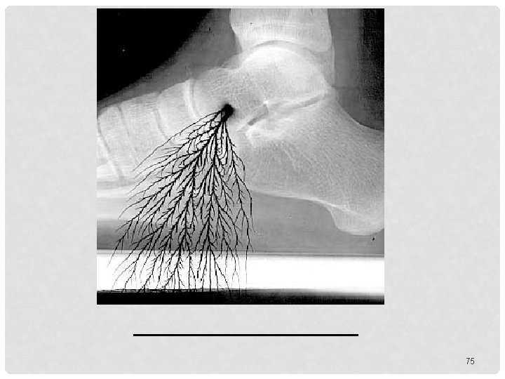
ARTIFACTS THINGS ON THE IMAGE THAT DISTRACT 61
ARTIFACTS: AN UNWANTED DENSITY ON THE FILM http: //www. xray 2000. co. uk/ 62
ARTIFACTS TYPES • Processing Artifacts • Exposure Artifacts • Handling and Storage Artifacts 63
PROCESSING ARTIFACTS • • • Emulsion pickoff Chemical fog Guide-shoe marks Water marks Chemical spots Guide-shoe & roller scratches 64
Developer spots 65
Water mark 66
Discolored film due to FIXER RETENTION Chemicals not washed off – over time will turn film brown 67
68
EXPOSURE ARTIFACTS • • Motion Improper patient position Wrong screen-film match Poor film/screen contact Double exposure Warped cassette Improper grid position Patient artifacts 69
ARTIFACT 70
71
HANDLING & STORAGE ARTIFACTS • • • Light fog Radiation fog Static Kink marks Scratches Dirty cassettes 72
73
74
76
77
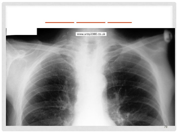
PATIENT ______ 80
81
DOUBLE EXPOSURE CHILD 82
POOR SCREEN CONTACT 83
DOUBLE EXPOSURE 84
EX TR A S HA E ND T O F S 85
86
87
2 chest tubes in the patient 88
Patient Swallowed Batteries 89
90
PATHOLOGY NOT ARTIFACT 91
NAME & CAUSE OF THIS? 92
Digital image Mis. Registration error 93
GRID ARTIFACT 94
The type of shield that is attached to the side of the collimator on a radiographic tube is : A. B. C. D. flat contact Detachable Shaped shield shadow REVIEW QUESTION REVIEW RADIATION BIOLOGY 95
On what date were x-rays discovered? A. January 4, 1886 B. November 8, 1895 C. October 9, 1898 D. November 18, 1808 REVIEW QUESTION REVIEW THE HISTORY OF X-RAYS 96
What agency regulates the quality of care provided to patients and the way the organization is supervised and operated for the purposes of providing continuous quality care? A. JRCERT B. ARRT C. JCAHO D. AHRA REVIEW QUESTION REVIEW THE REGULATORY BODIES 97
Which organization provides a C. R. T. in general Radiography and Fluoroscopy? A. Joint Commission on Accreditation of Health Care Organizations B. California Department of Public Health- Radiologic Health Branch C. American registry of Radiologic Technologists D. Joint Review Committee on Education in Diagnostic Medical Sonography REVIEW QUESTION REVIEW REGULATORY BODIES 98
The collimator controls the: A. Target material B. Amount of x-rays produced C. Type of x-rays produced D. Size of the x-ray field REVIEW QUESTIONS REVIEW X-RAY EQUIPMENT 99
During x-ray production when an electron is knocked out of its shell by a projectile electron. An electron from an outer shell fills the vacancy, what is produced? A. B. C. D. radio waves scatter radiation characteristic radiation brem’s radiation REVIEW X-RAY PRODUCTION 100
Thermionic emission at the filament releases: A. protons B. photons C. electrons D. neutrons REVIEW QUESTION REVIEW X-RAY PRODUCTION 101
This interaction an incoming photon enters with an energy, changes directions, and exits with the same amount of energy. A. B. C. D. Heat Brems Compton Coherent REVIEW QUESTION REVIEW PATIENT INTERACTIONS 102
All the following are radiation protection devices EXCEPT: A. B. C. D. lead apron contact shield film badge collimators REVIEW QUESTION REVIEW RADIATION BIOLOGY 103
What is the most radiosensitive component of the cell? A. B. C. D. protein cytoplasm nucleus mitochondria REVIEW QUESTION REVIEW RADIATION BIOLOGY 104