A CASE PRESENTATION MANAGEMENT DISCUSSION AND SHARING OF


- Slides: 70
A CASE PRESENTATION, MANAGEMENT, DISCUSSION AND SHARING OF INFORMATION ON PENETRATING CHEST INJURY BY: Jonathan R. Malabanan, M. D. Ospital ng Maynila Medical Center Department of Surgery
General Data: E. J. 29 –years- old Male Baseco, Port Area, Manila
Chief Complaint: Stab wound, 5 th ICS PAL, Left
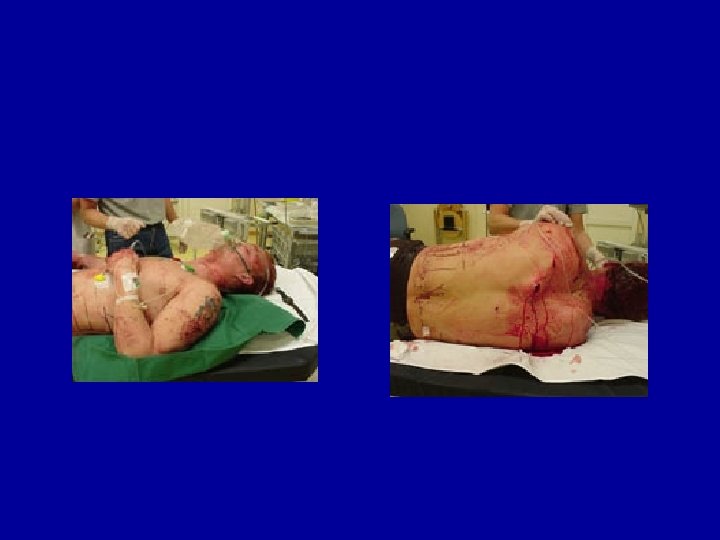
HISTORY OF PRESENT ILLNESS NOI: TOI: DOI: POI: stabbing 8: 00 pm October 4, 2006 Port Area, Manila Patient was walking alone after a drinking session when suddenly was approached by an unknown assailant and allegedly stabbed on the back by a “balisong”. Patient was then brought to our institution.
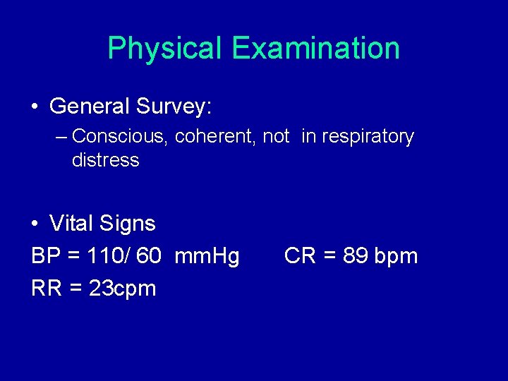
Physical Examination • General Survey: – Conscious, coherent, not in respiratory distress • Vital Signs BP = 110/ 60 mm. Hg RR = 23 cpm CR = 89 bpm
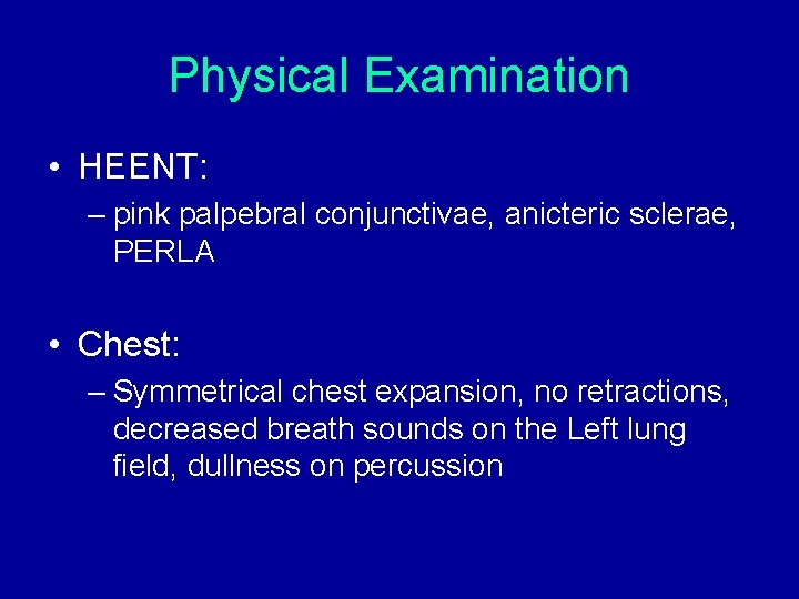
Physical Examination • HEENT: – pink palpebral conjunctivae, anicteric sclerae, PERLA • Chest: – Symmetrical chest expansion, no retractions, decreased breath sounds on the Left lung field, dullness on percussion
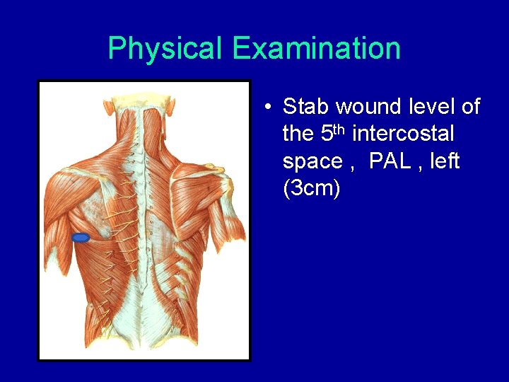
Physical Examination • Stab wound level of the 5 th intercostal space , PAL , left (3 cm)
Physical Examination • Cardiac: – Normal rate, regular rhythm, no murmur
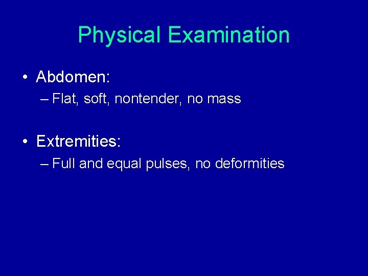
Physical Examination • Abdomen: – Flat, soft, nontender, no mass • Extremities: – Full and equal pulses, no deformities
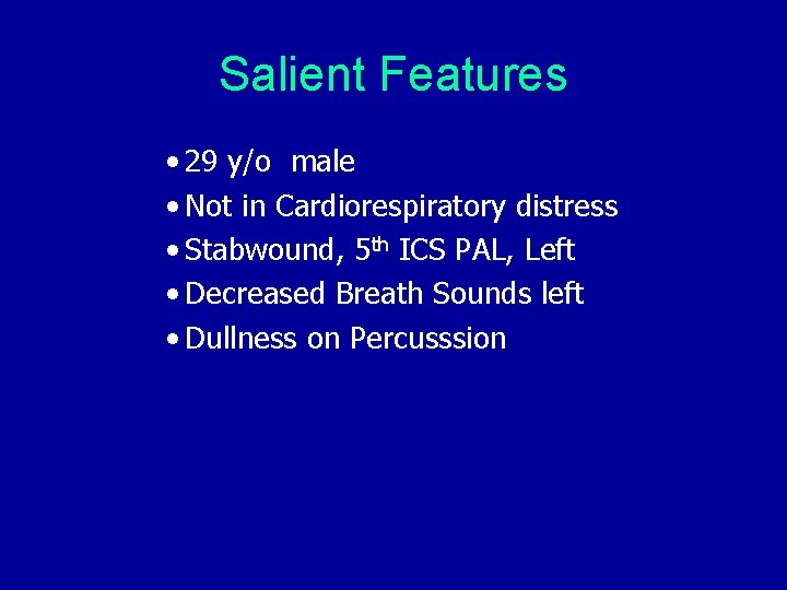
Salient Features • 29 y/o male • Not in Cardiorespiratory distress • Stabwound, 5 th ICS PAL, Left • Decreased Breath Sounds left • Dullness on Percusssion
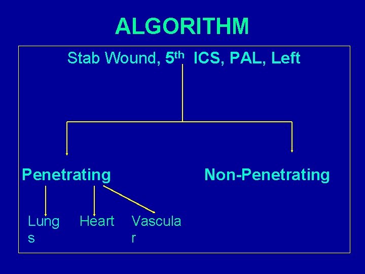
ALGORITHM Stab Wound, 5 th ICS, PAL, Left Penetrating Lung s Heart Non-Penetrating Vascula r
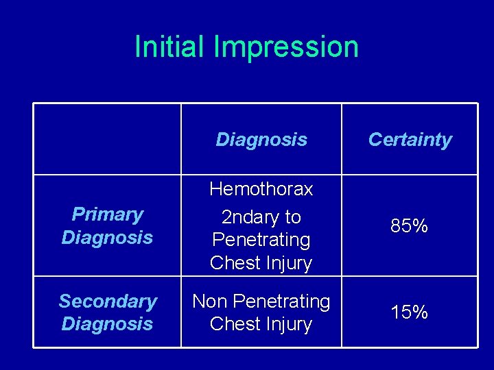
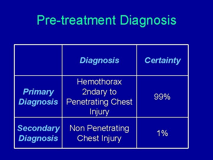
Initial Impression Diagnosis Certainty Primary Diagnosis Hemothorax 2 ndary to Penetrating Chest Injury 85% Secondary Diagnosis Non Penetrating Chest Injury 15%
Paraclinical Diagnostic Procedure • Do I need to perform a paraclinical diagnostic procedure? “Yes”
Goals of Paraclinical • To be more certain in diagnosis. • To determine my treatment plan.
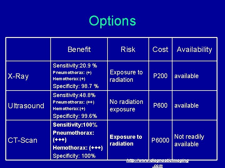
Options Benefit Sensitivity: 20. 9 % X-Ray Pneumothorax: (+) Hemothorax: (+) Specificity: 98. 7 % Sensitivity: 48. 8% Ultrasound Pneumothorax: (++) Hemothorax: (+) Specificity: 99. 6% CT-Scan Sensitivity: 100% Pneumothorax: (+++) Hemothorax: (+++) Specificity: 100% Risk Cost Availability Exposure to radiation P 200 available No radiation exposure P 600 available Exposure to radiation P 6000 Not readily available http: //www. diagnosticimaging. com
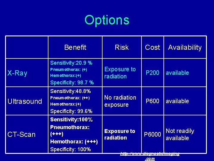
Options Benefit Sensitivity: 20. 9 % X-Ray Pneumothorax: (+) Hemothorax: (+) Specificity: 98. 7 % Sensitivity: 48. 8% Ultrasound Pneumothorax: (++) Hemothorax: (+) Specificity: 99. 6% CT-Scan Sensitivity: 100% Pneumothorax: (+++) Hemothorax: (+++) Specificity: 100% Risk Cost Availability Exposure to radiation P 200 available No radiation exposure P 600 available Exposure to radiation P 6000 Not readily available http: //www. diagnosticimaging. com
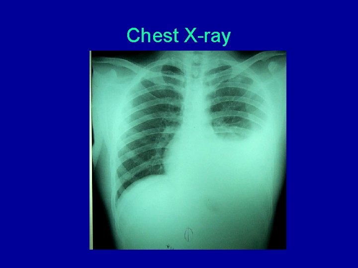
Chest X-ray
Pre-treatment Diagnosis Certainty Primary Diagnosis Hemothorax 2 ndary to Penetrating Chest Injury 99% Secondary Diagnosis Non Penetrating Chest Injury 1%
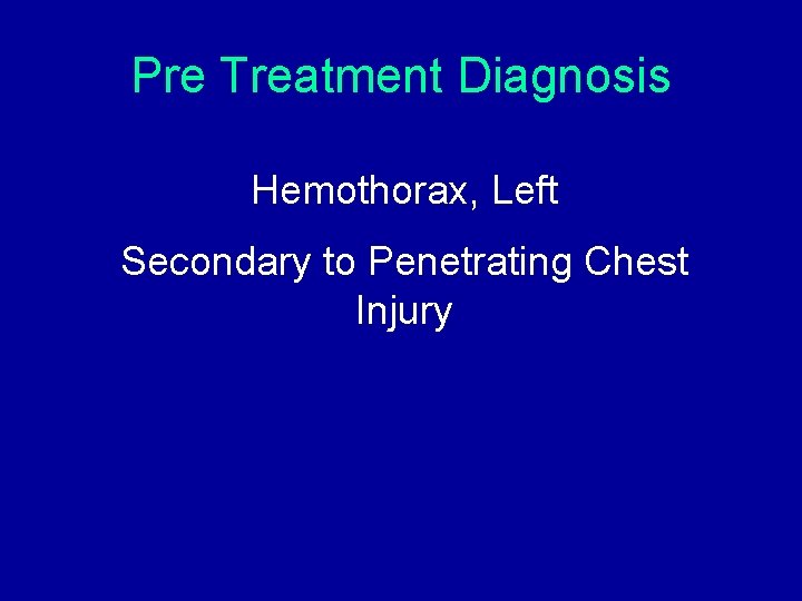
Pre Treatment Diagnosis Hemothorax, Left Secondary to Penetrating Chest Injury
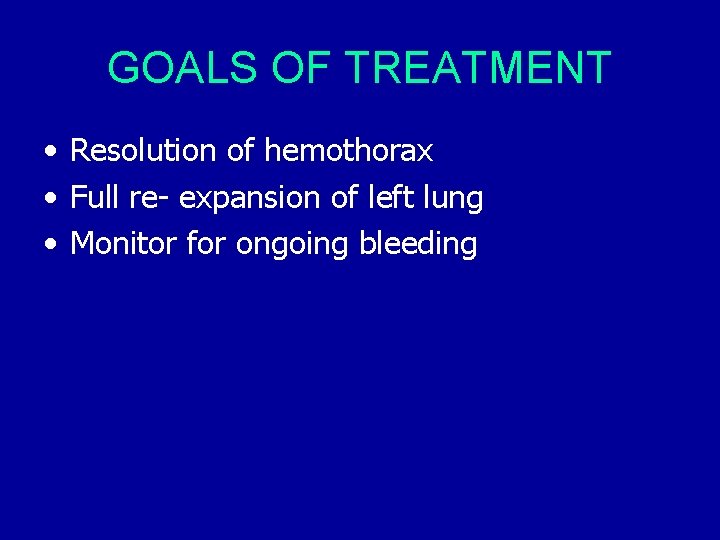
GOALS OF TREATMENT • Resolution of hemothorax • Full re- expansion of left lung • Monitor for ongoing bleeding
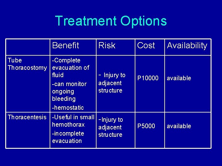
Treatment Options Benefit Tube -Complete Thoracostomy evacuation of fluid -can monitor ongoing bleeding -hemostatic Risk Cost Availability - Injury to P 10000 available P 5000 available adjacent structure Thoracentesis -Useful in small -Injury to hemothorax adjacent -incomplete structure evacuation
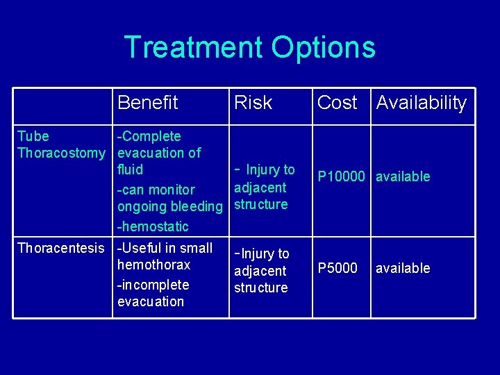
Treatment Options Benefit Risk Cost Availability Tube -Complete Thoracostomy evacuation of fluid - Injury to adjacent -can monitor ongoing bleeding structure -hemostatic P 10000 available Thoracentesis -Useful in small hemothorax -incomplete evacuation P 5000 -Injury to adjacent structure available
Management CHEST TUBE THORACOSTOMY, LEFT
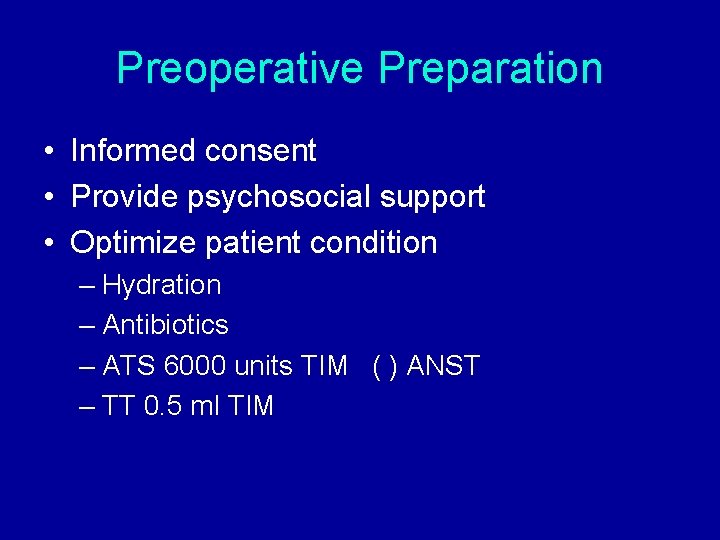
Preoperative Preparation • Informed consent • Provide psychosocial support • Optimize patient condition – Hydration – Antibiotics – ATS 6000 units TIM ( ) ANST – TT 0. 5 ml TIM
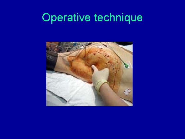
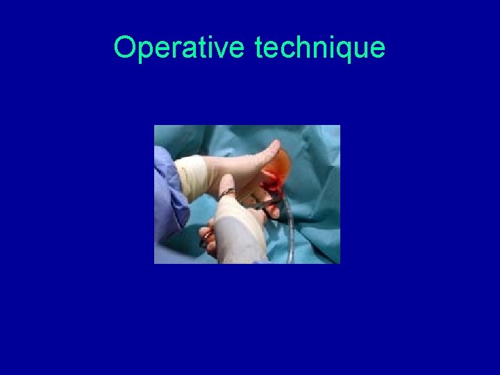
Operative technique • Patient semi-sitting with the ipsilateral arm placed above the head to expose the lateral aspect of the chest • chest prepared with antiseptic solution • draped to create a sterile field • large bore chest tube (F 36) placed to facilitate adequate drainage
Operative technique
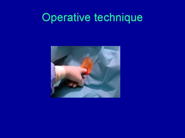
Operative technique • 5 th ICS midaxillary line identified and skin, periosteum, and pleura anesthesized with 1% lidocaine • transverse incision made over the underlying space • blunt dissection continued with Kelly clamp • clamp passed adjacent to the superior surface of the rib to prevent injury to the intercostals neurovascular bundle
Operative technique
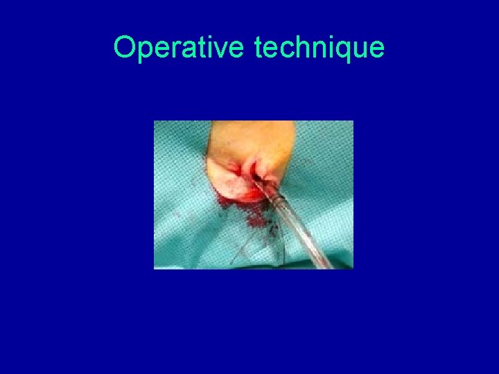
Operative technique • entry into the pleural space confirmed with rush of blood-filled fluid • finger inserted into the pleural space to identify any pleural adhesions • Fr 36 chest tube inserted into the pleural space on a Kelly clamp and directed posteriorly • tube secured with a silk 0 suture
Operative technique
Operative technique
Operative technique • attached to a water sealed thora-bottle • insertion site dressed gauze and covered with air-tight dressing • initial and subsequent drainage recorded • post-procedure chest film obtained
Operative Findings • 550 cc of fresh non clotted blood evacuated
Postoperative Management • Adequate analgesia • Monitoring of CT output
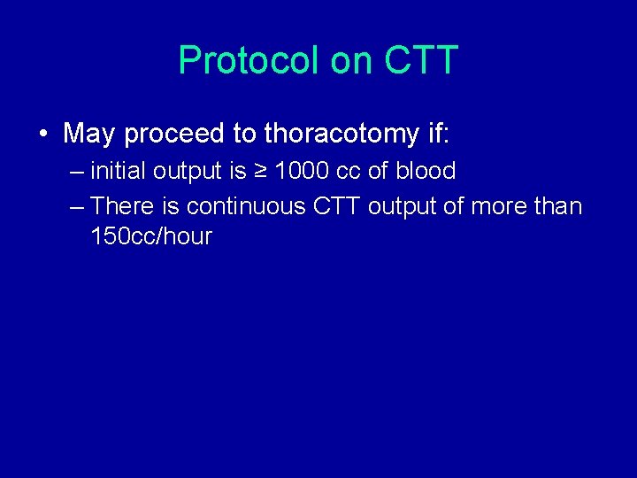
Protocol on CTT • May proceed to thoracotomy if: – initial output is ≥ 1000 cc of blood – There is continuous CTT output of more than 150 cc/hour
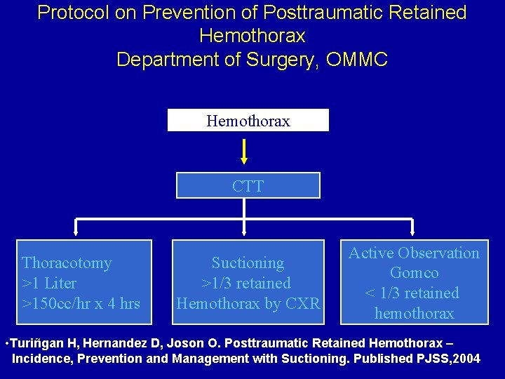
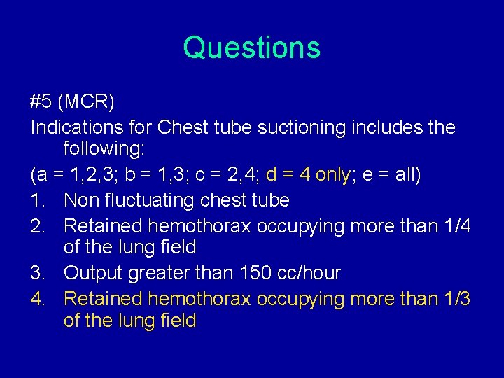
Protocol on Prevention of Posttraumatic Retained Hemothorax Department of Surgery, OMMC Hemothorax CTT Thoracotomy >1 Liter >150 cc/hr x 4 hrs Suctioning >1/3 retained Hemothorax by CXR Active Observation Gomco < 1/3 retained hemothorax • Turiñgan H, Hernandez D, Joson O. Posttraumatic Retained Hemothorax – Incidence, Prevention and Management with Suctioning. Published PJSS, 2004
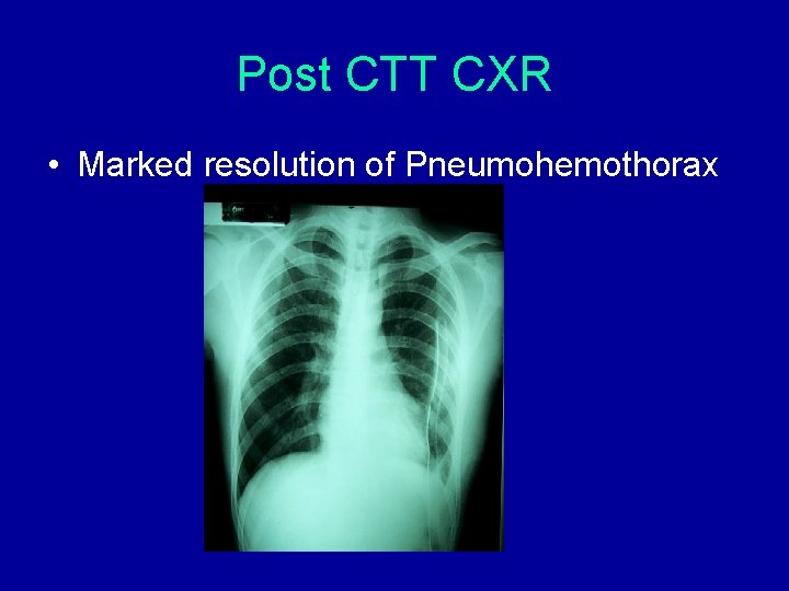
Post CTT CXR • Marked resolution of Pneumohemothorax
Final Diagnosis S/P Chest Tube Thoracostomy Left for Pneumohemothorax, Left Secondary to Penetrating Stab Wound 5 th ICS Posterior Axillary Line Left
COURSE IN THE WARD • 1 st Hospital Day – DAT – Moderate to high back rest – Adequate Antibiotic – Adequate Analgesia – Blow bottle exercises
COURSE IN THE WARD • 2 nd-3 rd Hospital Day – DAT – Moderate to high back rest – Adequate Antibiotic – Adequate Analgesia – Blow bottle exercises – Change of thora bottle with CT output monitoring
COURSE IN THE WARD • 4 th Hospital Day – Repeat CXR done – Chest tube removed
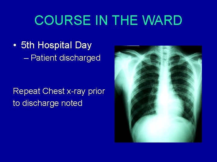
COURSE IN THE WARD • 5 th Hospital Day – Patient discharged Repeat Chest x-ray prior to discharge noted
PREVENTION AND HEALTH PROMOTION • Advise given to patient regarding – Possible complications – Proper wound care – OPD follow up after 7 days for removal of sutures
SHARING OF INFORMATI 0 N
Evaluation & Management 1. Assess airway & establish adequate ventilation. 2. Stabilize circulation. 3. Get good history, P. E.
THORACIC TRAUMA Potentially lethal injuries: – – – – Hemothorax Pneumothorax tension pneumothorax myocardial contusion sucking chest wound cardiac tamponade aortic rupture
Hemothorax • collection of blood in the pleural space • may be caused by blunt or penetrating trauma
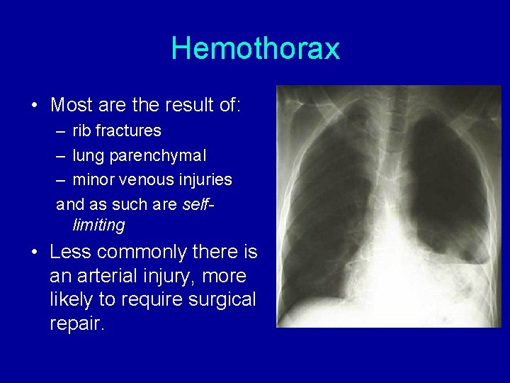
Hemothorax • Most are the result of: – rib fractures – lung parenchymal – minor venous injuries and as such are selflimiting • Less commonly there is an arterial injury, more likely to require surgical repair.
Pneumothorax • the collection of air in the pleural space • air may come from: – injury to the lung tissue – bronchial tear – chest wall injury allowing air to be sucked in from the outside.
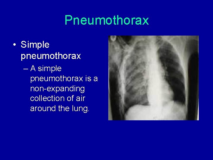
Pneumothorax • Simple pneumothorax – A simple pneumothorax is a non-expanding collection of air around the lung.
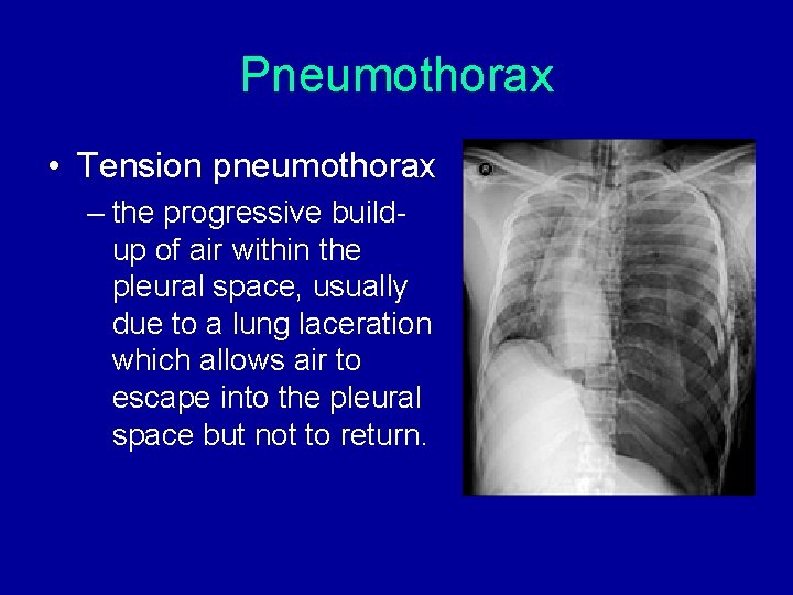
Pneumothorax • Tension pneumothorax – the progressive buildup of air within the pleural space, usually due to a lung laceration which allows air to escape into the pleural space but not to return.
Diagnosis of Hemothorax • Most small-moderate hemothoraces are not detectable by physical examination and will be identified only on Chest X-ray, FAST or CT scan. • Larger and more clinically significant hemothoraces may be identified clinically and should be treated promptly.
Physical examination • Chest examination may indicate the presence of significant thoracic trauma with external bruising or lacerations, or palpable crepitus indicating the presence of rib fractures. • There may be evidence of a penetrating injury over the affected hemithorax. • Don't forget to examine the back!
The classic signs of a hemothorax • decreased chest expansion • dullness to percussion • reduced breath sounds in the affected hemothorax.
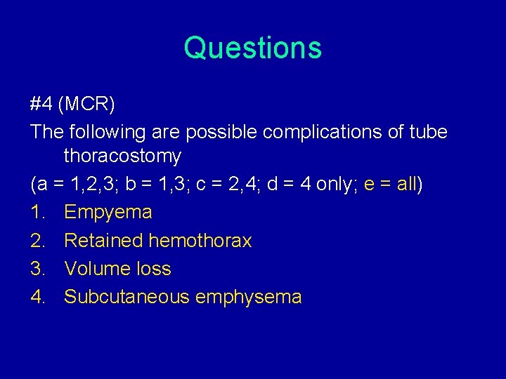
Complications • Retained Hemothorax • Empyema
Complications • Failure to adequately drain a haemothorax: – initially results in residual, clotted haemothorax which will not drain via a chest tube – left untreated, these retained haemothoraces may become infected and lead to empyema formation
Complications • If uninfected: – clot will organise and fibrose – resulting in a loss of lung volume impaired pulmonary function
References • Mattox KL, Allen MK. Systematic approach to pneumothorax, haemothorax, pneumomediastinum and subcutaneous emphysema. Injury. 1986; 17: 309312. • Fallon W, Barnosci A, Mancuso C, Injury to the Chest, Complications and Management: Experience at a Level I Trauma Center, Top Emerg Med 1990. • Turiñgan H, Hernandez D, Joson O. Posttraumatic Retained Hemothorax – Incidence, Prevention and Management with Suctioning Published PJSS, 2004. • Joson R. Management of a Surgical Patient, 2001
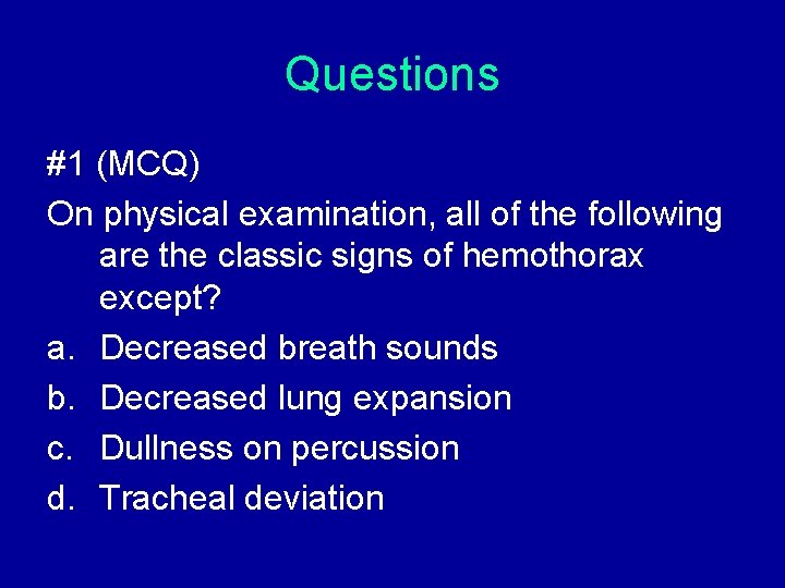
Questions #1 (MCQ) On physical examination, all of the following are the classic signs of hemothorax except? a. Decreased breath sounds b. Decreased lung expansion c. Dullness on percussion d. Tracheal deviation
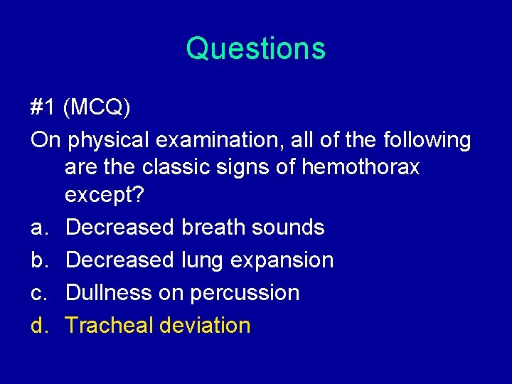
Questions #1 (MCQ) On physical examination, all of the following are the classic signs of hemothorax except? a. Decreased breath sounds b. Decreased lung expansion c. Dullness on percussion d. Tracheal deviation
Questions #2 (MCQ) The progressive build-up of air within the pleural space, usually due to a lung laceration which allows air to escape into the pleural space but not to return is also known as: a. Simple pneumothorax b. Open pneumothorax c. Tension pneumothorax d. All of the above
Questions #2 (MCQ) The progressive build-up of air within the pleural space, usually due to a lung laceration which allows air to escape into the pleural space but not to return is also known as: a. Simple pneumothorax b. Open pneumothorax c. Tension pneumothorax d. All of the above
Questions #3 (MCR) According to OMMC Department of Surgery Protocol, thoracotomy is indicated in the following conditions: (a = 1, 2, 3; b = 1, 3; c = 2, 4; d = 4 only; e = all) 1. Initial output of 1, 000 cc 2. Initial output of 1, 500 cc 3. Output of ≥ 150 cc/hour 4. Output of ≥ 200 cc/hour
Questions #3 (MCR) According to OMMC Department of Surgery Protocol, thoracotomy is indicated in the following conditions: (a = 1, 2, 3; b = 1, 3; c = 2, 4; d = 4 only; e = all) 1. Initial output of 1, 000 cc 2. Initial output of 1, 500 cc 3. Output of ≥ 150 cc/hour 4. Output of ≥ 200 cc/hour
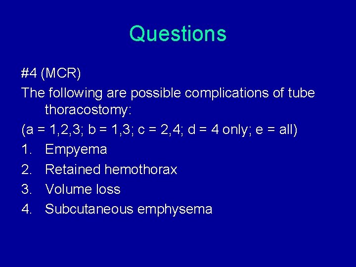
Questions #4 (MCR) The following are possible complications of tube thoracostomy: (a = 1, 2, 3; b = 1, 3; c = 2, 4; d = 4 only; e = all) 1. Empyema 2. Retained hemothorax 3. Volume loss 4. Subcutaneous emphysema
Questions #4 (MCR) The following are possible complications of tube thoracostomy (a = 1, 2, 3; b = 1, 3; c = 2, 4; d = 4 only; e = all) 1. Empyema 2. Retained hemothorax 3. Volume loss 4. Subcutaneous emphysema
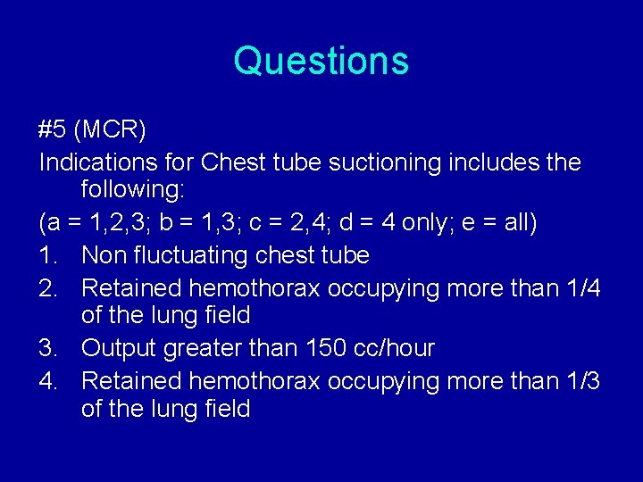
Questions #5 (MCR) Indications for Chest tube suctioning includes the following: (a = 1, 2, 3; b = 1, 3; c = 2, 4; d = 4 only; e = all) 1. Non fluctuating chest tube 2. Retained hemothorax occupying more than 1/4 of the lung field 3. Output greater than 150 cc/hour 4. Retained hemothorax occupying more than 1/3 of the lung field
Questions #5 (MCR) Indications for Chest tube suctioning includes the following: (a = 1, 2, 3; b = 1, 3; c = 2, 4; d = 4 only; e = all) 1. Non fluctuating chest tube 2. Retained hemothorax occupying more than 1/4 of the lung field 3. Output greater than 150 cc/hour 4. Retained hemothorax occupying more than 1/3 of the lung field
Thank you!