Preoperative Case Presentation Sharing of Information on Vomiting





- Slides: 81
Preoperative Case Presentation & Sharing of Information on Vomiting Jeffy G. Guerra, MD Level III Surgery Resident OMMC-Surgery 053006
General Data: C. P. , 68 F SAB, Mla
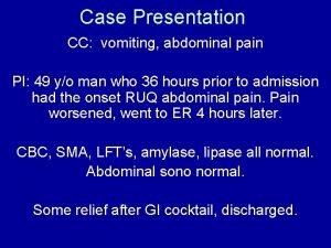
Chief Complaint: Vomiting
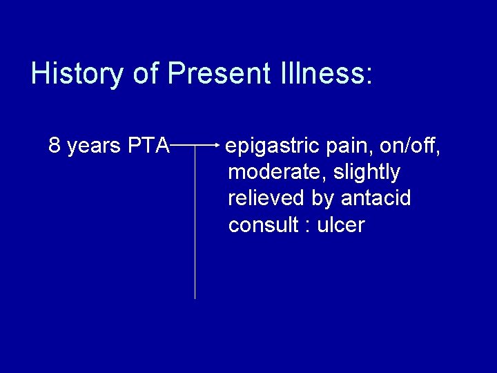
History of Present Illness: 8 years PTA epigastric pain, on/off, moderate, slightly relieved by antacid consult : ulcer
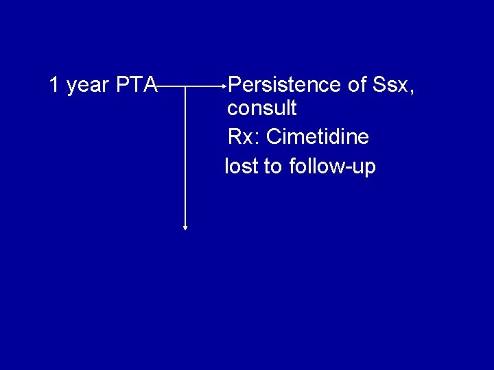
1 year PTA Persistence of Ssx, consult Rx: Cimetidine lost to follow-up
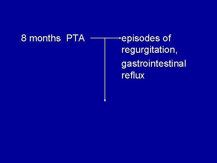
8 months PTA episodes of regurgitation, gastrointestinal reflux
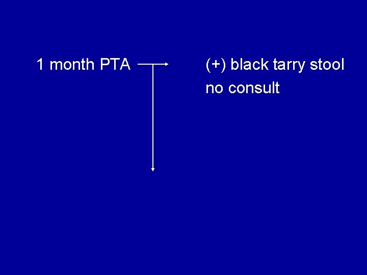
1 month PTA (+) black tarry stool no consult
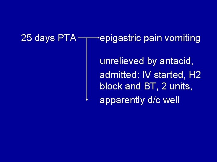
25 days PTA epigastric pain vomiting unrelieved by antacid, admitted: IV started, H 2 block and BT, 2 units, apparently d/c well
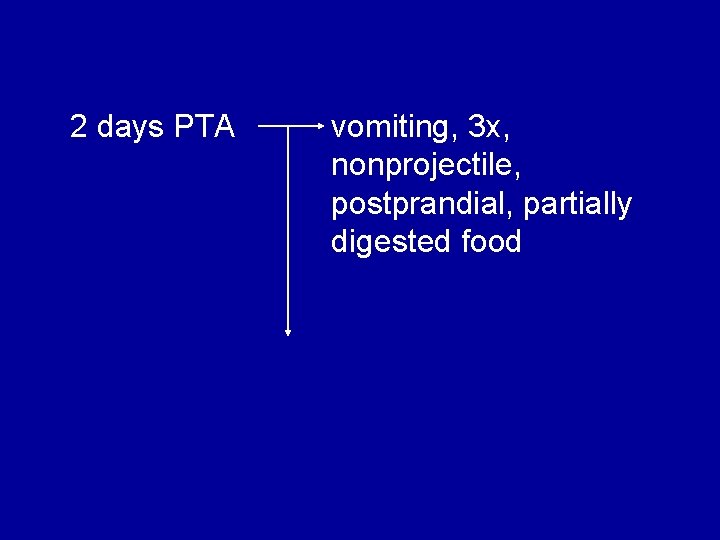
2 days PTA vomiting, 3 x, nonprojectile, postprandial, partially digested food
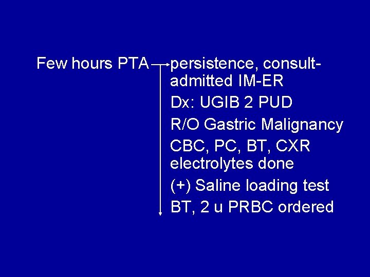
Few hours PTA persistence, consultadmitted IM-ER Dx: UGIB 2 PUD R/O Gastric Malignancy CBC, PC, BT, CXR electrolytes done (+) Saline loading test BT, 2 u PRBC ordered
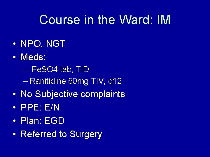
Course in the Ward: IM • NPO, NGT • Meds: – Fe. SO 4 tab, TID – Ranitidine 50 mg TIV, q 12 • • No Subjective complaints PPE: E/N Plan: EGD Referred to Surgery
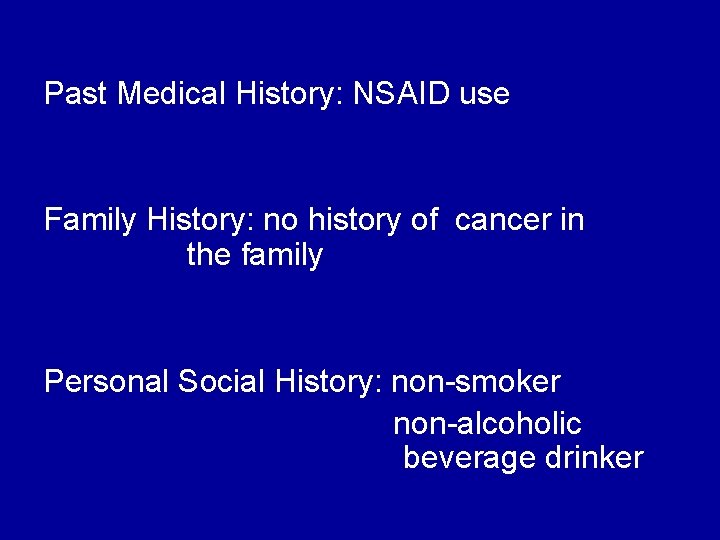
Past Medical History: NSAID use Family History: no history of cancer in the family Personal Social History: non-smoker non-alcoholic beverage drinker
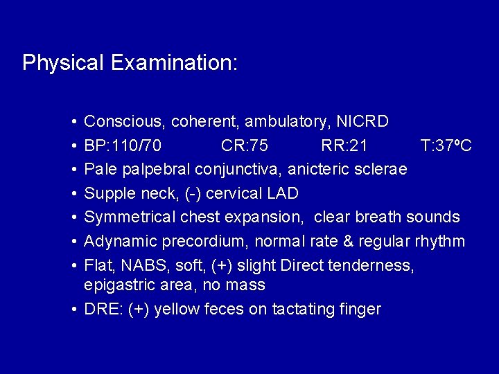
Physical Examination: • • Conscious, coherent, ambulatory, NICRD BP: 110/70 CR: 75 RR: 21 T: 37ºC Pale palpebral conjunctiva, anicteric sclerae Supple neck, (-) cervical LAD Symmetrical chest expansion, clear breath sounds Adynamic precordium, normal rate & regular rhythm Flat, NABS, soft, (+) slight Direct tenderness, epigastric area, no mass • DRE: (+) yellow feces on tactating finger
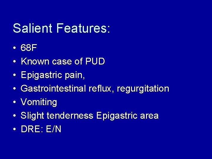
Salient Features: • • 68 F Known case of PUD Epigastric pain, Gastrointestinal reflux, regurgitation Vomiting Slight tenderness Epigastric area DRE: E/N
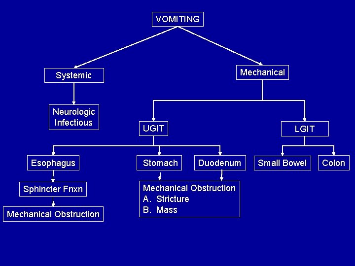
VOMITING Mechanical Systemic Neurologic Infectious Esophagus Sphincter Fnxn Mechanical Obstruction UGIT Stomach LGIT Duodenum Mechanical Obstruction A. Stricture B. Mass Small Bowel Colon
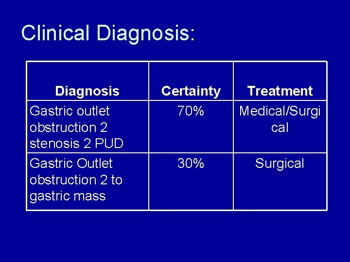
Clinical Diagnosis: Diagnosis Gastric outlet obstruction 2 stenosis 2 PUD Gastric Outlet obstruction 2 to gastric mass Certainty 70% Treatment Medical/Surgi cal 30% Surgical
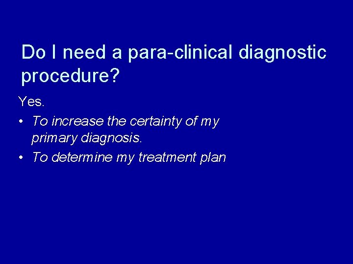
Do I need a para-clinical diagnostic procedure? Yes. • To increase the certainty of my primary diagnosis. • To determine my treatment plan
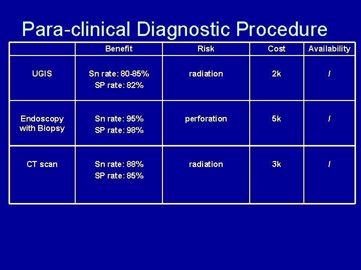
Para-clinical Diagnostic Procedure Benefit Risk Cost Availability UGIS Sn rate: 80 -85% SP rate: 82% radiation 2 k / Endoscopy with Biopsy Sn rate: 95% SP rate: 98% perforation 5 k / CT scan Sn rate: 88% SP rate: 85% radiation 3 k /
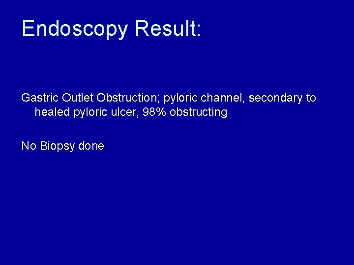
Endoscopy Result: Gastric Outlet Obstruction; pyloric channel, secondary to healed pyloric ulcer, 98% obstructing No Biopsy done
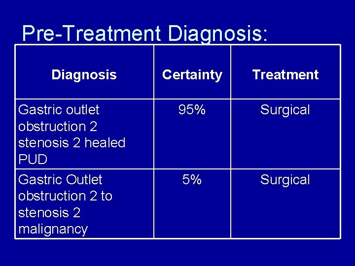
Pre-Treatment Diagnosis: Diagnosis Gastric outlet obstruction 2 stenosis 2 healed PUD Gastric Outlet obstruction 2 to stenosis 2 malignancy Certainty Treatment 95% Surgical
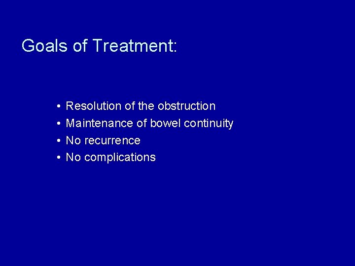
Goals of Treatment: • • Resolution of the obstruction Maintenance of bowel continuity No recurrence No complications
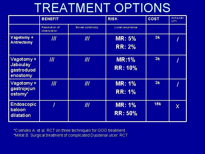
TREATMENT OPTIONS BENEFIT Resolution of obstruction Vagotomy + Antrectomy Vagotomy + Jaboulay gastroduod enostomy /// RISK COST AVAILABI LITY Bowel continuity Local recurrence /// MR: 5% RR: 2% 3 k / /// MR: 1% RR: 10% 3 k / Vagotomy + gastrojejun ostomy* /// MR: 1% RR: 1% 3 k / Endoscopic baloon dilatation / /// MR: 1% RR: 50% 15 k x *Csendes A. et al. RCT on three techniques for GOO treatment. *Millat B. Surgical treatment of complicated Duodenal ulcer: RCT
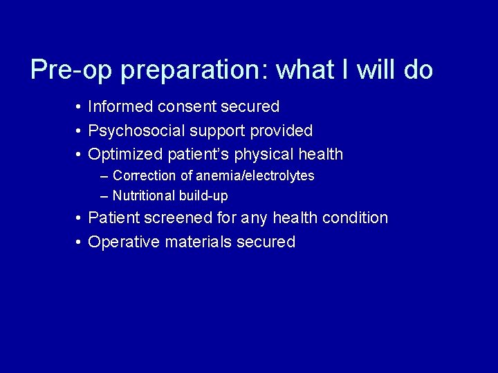
Pre-op preparation: what I will do • Informed consent secured • Psychosocial support provided • Optimized patient’s physical health – Correction of anemia/electrolytes – Nutritional build-up • Patient screened for any health condition • Operative materials secured
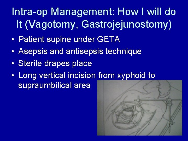
Intra-op Management: How I will do It (Vagotomy, Gastrojejunostomy) • • Patient supine under GETA Asepsis and antisepsis technique Sterile drapes place Long vertical incision from xyphoid to supraumbilical area
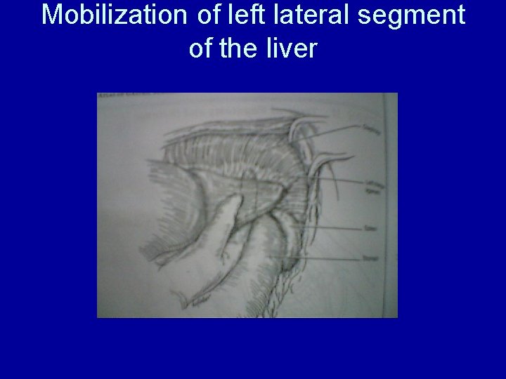
Mobilization of left lateral segment of the liver
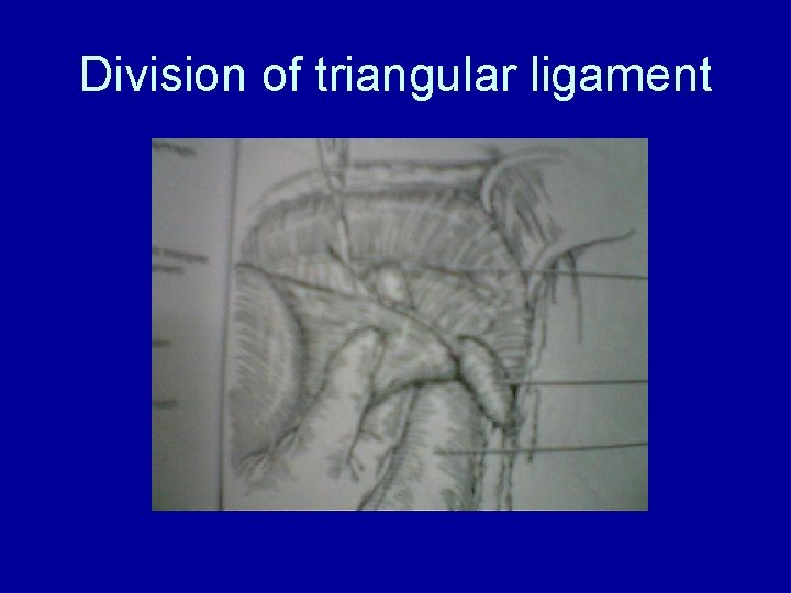
Division of triangular ligament
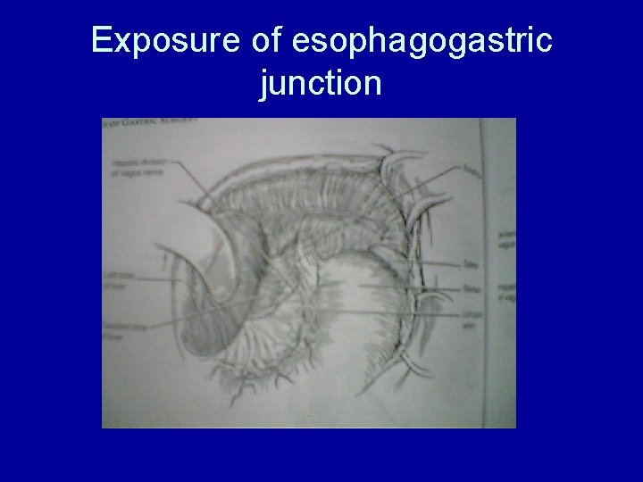
Exposure of esophagogastric junction
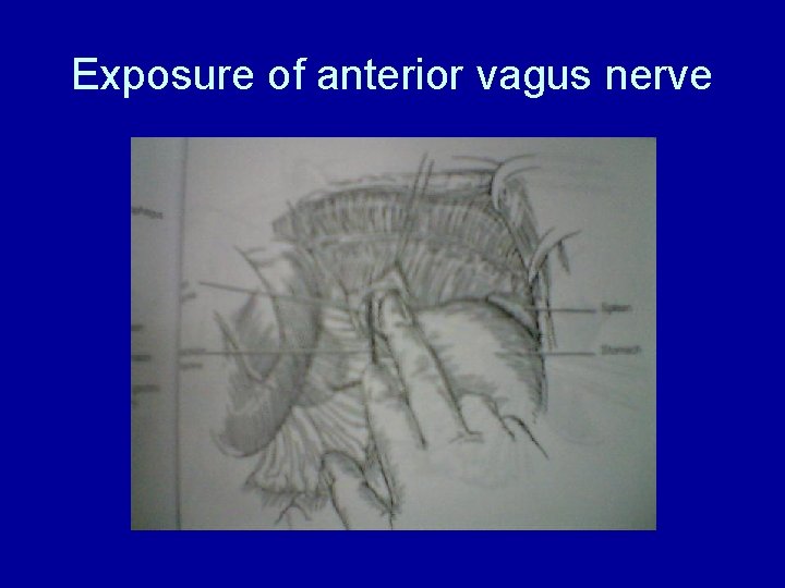
Exposure of anterior vagus nerve
Isolation/ligation of nerve trunk, anterior, posterior and esophageal branches • Anterior vagal trunk is encircled with hook and dissected sharply from esophageal musculature • Nerve trunk is ligated proximally and distally
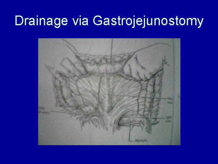
Drainage via Gastrojejunostomy
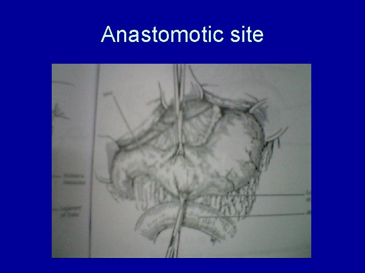
Anastomotic site
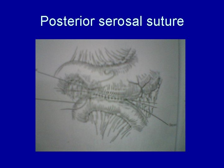
Posterior serosal suture
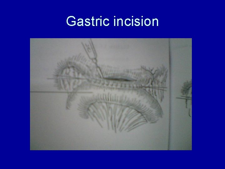
Gastric incision
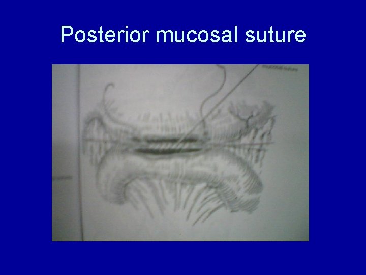
Posterior mucosal suture
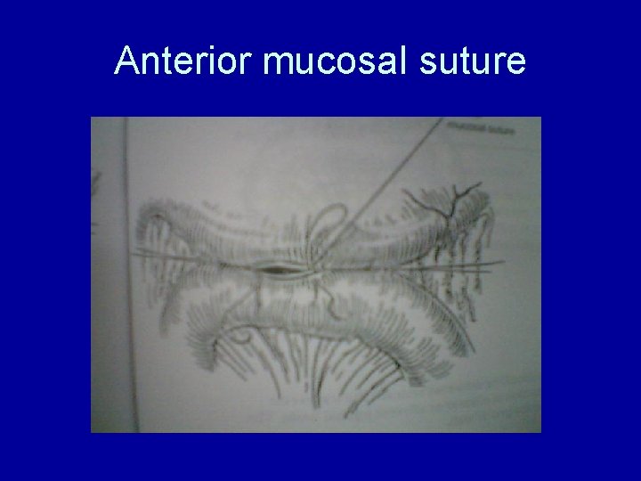
Anterior mucosal suture
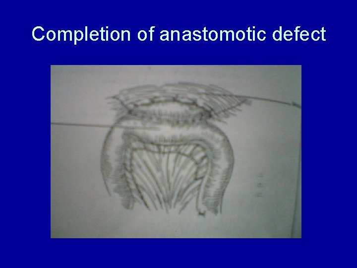
Completion of anastomotic defect
Post-op Care
• Postoperative care: – Intravenous fluids – nasogastric decompression – Analgesics – hemodynamics • The nasogastric tube is removed upon return of gastrointestinal transit, and feeding is slowly begun.
Outcome: • • • Resolution of obstruction Live patient No complications Satisfied patient No medico-legal suit
Sharing of information
• • SURGERY FOR PEPTIC ULCER DISEASE(PUD) Ulcer in the GIT is characterized by an interruption in the mucosa stretching through the muscularis mucosa into the submucosa or deeper Location - in order of decreasing frequency – Duodenum – Stomach – Esophagus
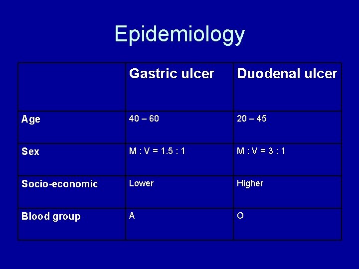
Epidemiology Gastric ulcer Duodenal ulcer Age 40 – 60 20 – 45 Sex M : V = 1. 5 : 1 M : V = 3 : 1 Socio-economic Lower Higher Blood group A O
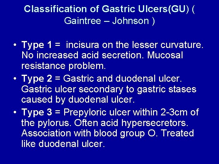
Classification of Gastric Ulcers(GU) ( Gaintree – Johnson ) • Type 1 = incisura on the lesser curvature. No increased acid secretion. Mucosal resistance problem. • Type 2 = Gastric and duodenal ulcer. Gastric ulcer secondary to gastric stases caused by duodenal ulcer. • Type 3 = Prepyloric ulcer within 2 -3 cm of the pylorus. Often acid hypersecretors. Association with blood group O. Treated like duodenal ulcer.
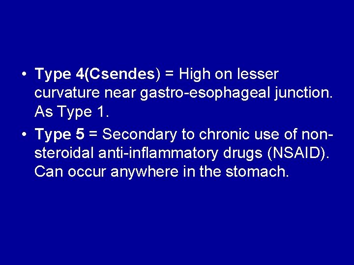
• Type 4(Csendes) = High on lesser curvature near gastro-esophageal junction. As Type 1. • Type 5 = Secondary to chronic use of nonsteroidal anti-inflammatory drugs (NSAID). Can occur anywhere in the stomach.
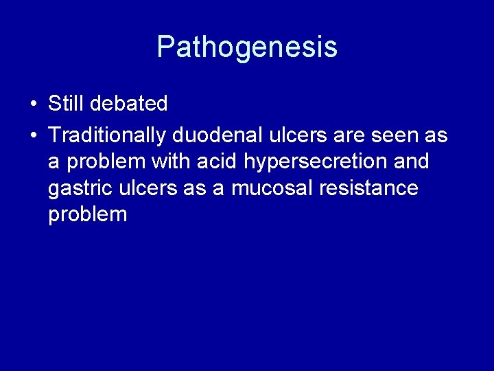
Pathogenesis • Still debated • Traditionally duodenal ulcers are seen as a problem with acid hypersecretion and gastric ulcers as a mucosal resistance problem
Gastric acid. Central in pathogenesis – no benign ulceration occurs without gastric acid Gastric stases. Delayed emptying of normal amounts of acid with increased exposure
Enviromental factors are very important. a) Helicobacter pylori infection. 90% of patients with DU and 50% of patients with GU b) NSAID use. The mucus gel layer contains bicarbonate. This layer adheres to the gastric mucosa. It protects the mucosa against back diffusion of hydrogen ions. NSAID’s suppress mucus cell function. c) Smoking
4) Mucosal resistance 5) Genetic predisposition
Clinical Picture
DUODENAL ULCER 1) Epigastric pain – Central or slightly to the right Burning or gnawing Can spread to the back Relieved by ingestion of food or anti-acid Pain occurs when patient is hungry
2) Different degrees of nausea and vomiting 3) Weight gain ( Pain relieved by ingestion of food) 4) Epigastric tenderness just to the right of the midline, may be absent.
GASTRIC ULCER 1) Epigastric pain – Brought on by meals often within 30 minutes 2) Nausea and vomiting 3) Weight loss 4) Epigastric tenderness
Complications 1) Bleeding 2) Perforation 3) Gastric outlet obstruction 4) Penetration
Management • Surgery is indicated and for the following: 1) Non-healing ulcer ( 8 – 12 weeks for GU, DU can be managed conservatively for longer since the risk for malignancy is low) 2) Complications • a) Perforation b) Bleeding if massive, c) Gastric outlet obstruction that does not clear up on conservative management.
Surgical principle for definitive ulcer surgery
Definitive ulcer operations for GU • Type 1 GU partial gastrectomy. Vagotomy not done. • Type 2 and 3 GU treated as DU. HSV contra-indicated due to high ulcer recurrence with prepyloric ulcers. • Type 4 GU treated with partial gastrectomy and excision of a long tongue of lesser curvature including the ulcer(Pauchet procedure).
Gastric outlet obstruction • Cycles of inflammation and repair may cause obstruction at the gastroduodenal junction as a result of edema, muscular spasm and fibroses.
• Edema and spasm can resolve with medical treatment. • Obstruction is mainly caused by DU and prepiloric GU. • Malignant tumors is the other important cause of gastric outlet obstruction.
• normal pylorus is about 20 mm in diameter and can distend to 25 mm • gastric outlet obstruction occur when the diameter of the antroduodenal segment is below 10 mm • A saline load test can be utilized in the objective measurement of outlet obstruction or gastric atony and the assessment of response to therapy
• The major benign causes of GOO are PUD, gastric polyps, ingestion of caustics, pyloric stenosis, congenital duodenal webs, gallstone obstruction (Bouveret syndrome), pancreatic pseudocysts, and bezoars
Clinical picture • Longstanding history of PUD • Progressive worsening of ulcer pain and early satiety. • Vomiting after meals of partially digested food without bile ( food eaten earlier the day or the previous day). • Dehydration and severe weight loss.
• Visible peristalses of the dilated stomach (rarely). • Succussion splash audible with to and fro movement of abdomen. • Tetany in cases of advanced alkaloses. • Develop hyponatremic, hypokalemic, hypochloremic metabolic alkaloses
Management 1) Resussitation initially with 0. 9% sodium chloride. Potassium supplementation only after good urine output is established. 2) Gastric lavage with thick stomach tube ( 32 F) to remove food residue. 3) Diagnostic tests after gastric lavage : Gastroscopy with biopsies with or without barium meal to rule out malignancy.
4) IV H 2 -blockers or proton pump inhibitors. 5) A nasogastric tube is passed. The patient may drink water. The amount of oral intake and drainage is charted. This gives an impression whether the obstruction is resolving. 6) Balloon dilatation of pyloric channel is possible but seldom produces a final solution.
7) Surgery is indicated if the obstruction does not resolve after one week of conservative treatment. Mostly a truncal vagotomy and antrectomy is done although truncal vagotomy with a drainage procedure is sometimes performed.
Complications of PUD surgery
Complications due to vagotomy • Intraoperative complications can occur with injury to adjacent structures. • Early post-operative complication – delayed gastric emptying – dysphagia and lesser curve necroses( lesser curve necroses specific to HSV). • Late complications include postvagotomy diarrhea, reflux esophagitis and gallstones
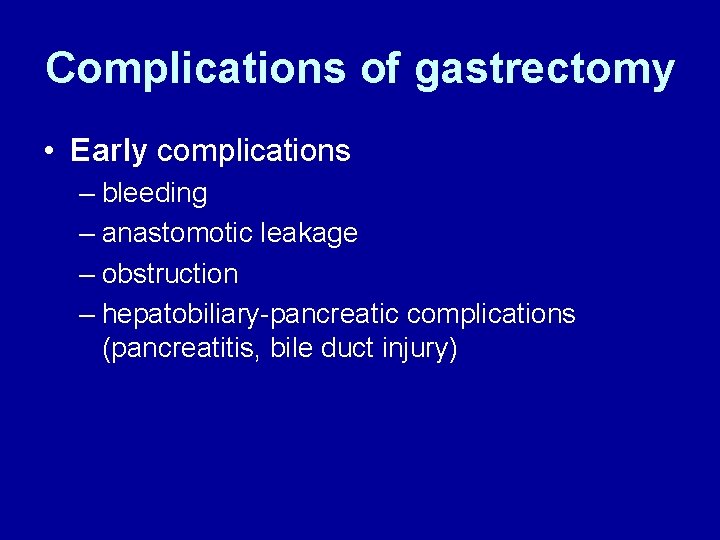
Complications of gastrectomy • Early complications – bleeding – anastomotic leakage – obstruction – hepatobiliary-pancreatic complications (pancreatitis, bile duct injury)
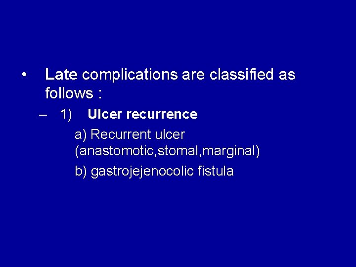
• Late complications are classified as follows : – 1) Ulcer recurrence a) Recurrent ulcer (anastomotic, stomal, marginal) b) gastrojejenocolic fistula
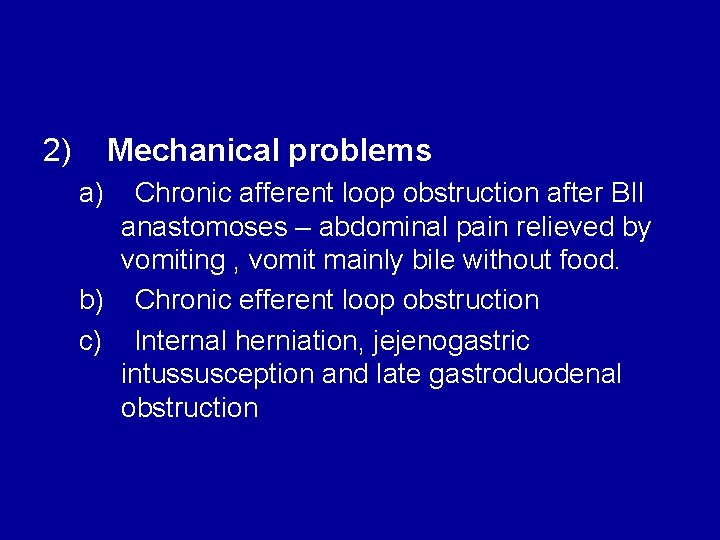
2) Mechanical problems a) Chronic afferent loop obstruction after BII anastomoses – abdominal pain relieved by vomiting , vomit mainly bile without food. b) Chronic efferent loop obstruction c) Internal herniation, jejenogastric intussusception and late gastroduodenal obstruction
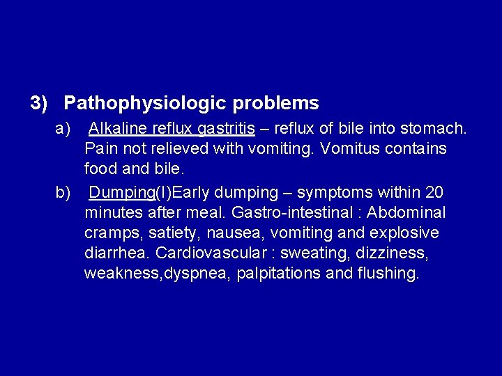
3) Pathophysiologic problems a) Alkaline reflux gastritis – reflux of bile into stomach. Pain not relieved with vomiting. Vomitus contains food and bile. b) Dumping(I)Early dumping – symptoms within 20 minutes after meal. Gastro-intestinal : Abdominal cramps, satiety, nausea, vomiting and explosive diarrhea. Cardiovascular : sweating, dizziness, weakness, dyspnea, palpitations and flushing.
– Due to sudden release of high osmolality chyme into duodenum with fluid shifts and release of gastro-intestinal hormones. • (II) Late dumping – only vasomotor symptoms. Caused by enteroglucagon secretion which leads to increased and prolonged insulin secretion with resultant hypoglycaemia.
4) Malabsorption and Nutritional problems a) Malabsorption of protein, carbohydrates and fat b) Early satiety c) Anemia : Fe, folate and B 12 deficiency. B 12 problems mostly after total or near total gastrectomy. d) Osteopmalacia
References: 1. Csendes A. Maluenda F. et al. Prospective randomized controlled trial comparing three surgical techniques for the treatment of gastric outlet obstruction secondary to duodenal ulcer. Am J Surg. 1993 Jul 166: 45 -49 2. Edwards LW, Herrington JL Jr. Vagotomy and gastroenterostomy— vagotomy and conservative gastrectomy. Ann Surg, 1953; 137: 873– 83. 3. Emas S, Fernstrom M. Prospective, randomized trial of selective vagotomy with pyloroplasty and selective proximal vagotomy with and without pyloroplasty in the treatment of duodenal, pyloric and prepyloric ulcers. Am J Surg, 1985; 149: 236– 43. 4. Fischer AB. Twenty-five years after Billroth II gastrectomy for duodenal ulcer. World J Surg, 1984; 8: 293– 302. 5. Kuwada, S et al. Long-term outcome of endoscopic dilation of nonmalignant pyloric stenosis. Gastrointestinal Endoscopy 1995; 41(1) 15 -17. 6. Gibson JB, Behrman SW, Fabian TC: Gastric outlet obstruction resulting from peptic ulcer disease requiring surgical intervention is infrequently associated with Helicobacter pylori infection. J Am Coll Surg 2000 Jul; 191(1): 32 -7[Medline].
7. Millat B, Fingerhut A et al. surgical treatment of complicated duodenal ulcer. Controlled trial. World J Surg. 2000 Mar. 24(3) 299 -306. 8. Siu WT, Tang CN, Law BK, et al: Vagotomy and gastrojejunostomy for benign gastric outlet obstruction. J Laparoendosc Adv Surg Tech A 2004 Oct; 14(5): 2669[Medline]. 9. Haglund UH, Jansson RL, Lindhagen JG, Lundell LR, Svartholm EG, Olbe LC. Primary Roux-Y gastrojejunostomy versus gastroduodenostomy after antrectomy and selective vagotomy. Am J Surg. 1992 Apr; 163(4): 457 -8.
Questions 1. Gastric Outlet Obstruction secondary to healed pyloric ulcer may present with which of the following? a. b. c. d. e. vomiting hyponatremia hypochloremia epigastric pain All of the above
2. What is the most common complication of peptic ulcer disease? a. b. c. d. bleeding perforation intractability obstruction
3. The following statements is/are true regarding gastric outlet obstruction. 1. Cycles of inflammation and repair may cause obstruction at the gastroduodenal junction as a result of edema, muscular spasm and fibroses. 2. Edema and spasm can resolve with medical treatment. 3. Obstruction is mainly caused by DU and prepiloric GU. 4. Malignant tumors is the other important cause of gastric outlet obstruction.
4. Which of the following choices is/are late complication/s of vagotomy? 1. postvagotomy diarrhea, 2. reflux esophagitis and 3. Gallstones 4. Delayed gastric emptying
5. Which of the following is/are not early complication of gastric surgery ? 1. 2. 3. Bleeding anastomotic leakage hepatobiliary-pancreatic complications (pancreatitis, bile duct injury) 4. gastrojejenocolic fistula
Thank you!