Movements Of Gastrointestinal Tract Dr G S Deepa

 Translatory •")


")
 DEGLUTION OCCURS IN THREE STAGES ORAL STAGE – Food from mouth enter")


 and vagus nerve(esophageal stage) Reach soft palate")

 Mechanical obstruction Tumour Stricture Diverticular hernia 2) Neurological disorders causing")


")
 Regurgitation of acidic contents into the esophagus CAUSE: weakness")


 Stimulus: Fat Action:")


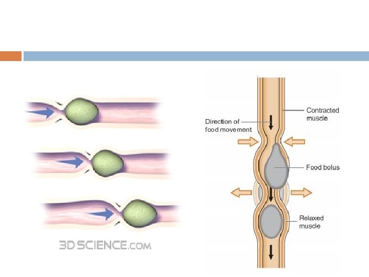
 2. Propulsive contractions (peristalsis)")


 Contraction of ileocecal sphincter Inhibition of ileal peristalsis")
")


 Internal anal")


- Slides: 114
Movements Of Gastrointestinal Tract Dr. G S Deepa Assistant Professor Dept. of Physiology and Biochemistry
CLASSIFICATION • Movements that propel onwards and propels the food mass A) Translatory • eg: - peristalsis, anti-peristalsis, and mass peristalsis. movements • Present in every part of GIT but not the stationary movements. : B) Stationary movements : • Movements remain localized and do not move onwards. • eg: - segmentation, tonic contraction
Causes Neurogenic : eg: peristalsis and antiperistalsis Myogenic : eg: segmentation or pendulum
NATUR E FREQUEN CY RATE CAUSE FUNCTIO NS
Mastication
Mastication DEFINITI ON Cutting the food substances into small particles and grinding them into a soft bolus
SIGNIFICANCE Breakdown of food stuffs into small particles especially raw veg. and fruits as they have undigestible cellulose memb. around them. Digestive enzymes can act only on the surfaces of food particles, so rate of digestion depends on surface area Mixing of food with saliva. Lubricating and moistening the food with saliva. Appreciation of taste.
Teeth are designed for chewing Anterior teeth - Incisors for cutting Posterior teeth – Molars for grinding
Teeth
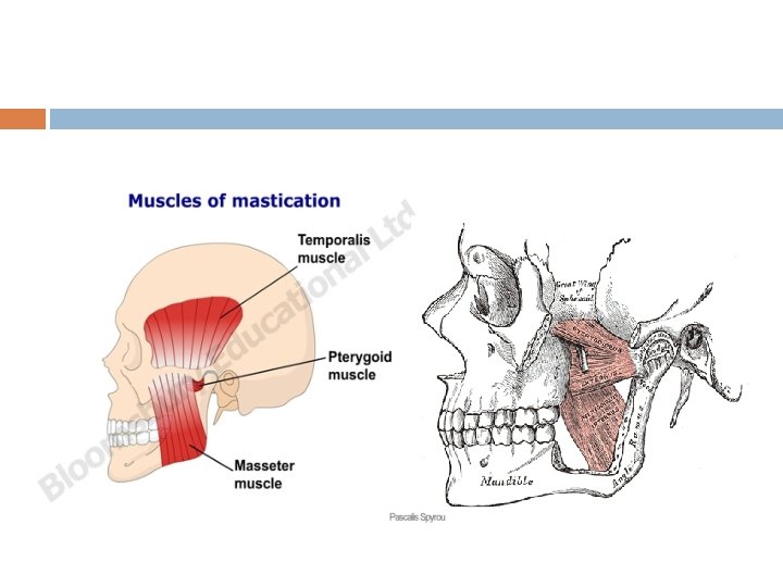
MUSCLES OF MASTICATION Massetor Temporalis Pterygoid Buccinator
• Muscles are supplied by Nervous mandibular division of V cranial nerve Regulation [trigeminal]. Center for mastication Chewing reflex • Medulla and cerebral cortex • Bolus of food in mouth initiates reflex inhibition of muscles of mastication
Chewing reflex Compress es the bolus against mouth Food in mouth Inhibition of muscles of masticatio n Raises the jaw, closure of mouth Rebound contractio n Dropping of lower jaw Initiates stretch reflex in jaw muscles
DEGLUTITION (SWALLOWING)
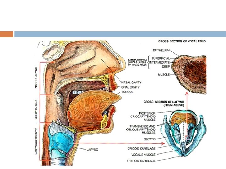
DEGLUTITION (SWALLOWING) DEGLUTION OCCURS IN THREE STAGES ORAL STAGE – Food from mouth enter pharynx PHARYNGEAL STAGE – Pharynx to esophagus OESOPHAGEAL STAGE — Esophagus to stomach
The initial stages of eating and swallowing are under voluntary control. This means that it is governed by the brain.
Once food enters the mouth the teeth break it down into smaller and smaller pieces. This has the dual function of making the food easier to swallow and increasing the surface area of food on which the saliva can act.
The tongue, lips and cheeks assist the teeth in the process by allowing the food to be "rolled" around the oral cavity. The mechanical action described above produces a softened bolus of food which is now ready to be swallowed. The correct biological term for swallowing is deglutition.
The picture on the left shows the voluntary stage of deglutition. Here the bolus is pushed into the upper part of the pharynx (known as the oropharynx) by the action of the tongue. The pharyngeal stage of deglutiton is stimulated when the bolus enters the oropharynx. This stage of swallowing is mainly due to a reflex response. Various nerve receptors send messages to the deglutition centre of the
This sets off muscular contractions in the pharynx. The soft palate closes off the nasopharynx. The vocal cords in the larynx are moved up and towards the front of the throat thus closing it off to the passage of food. This is extremely important in preventing food from
Another effect of the process is to widen the opening of the oesophagus thus making the passage of the bolus along the alimentary canal easier. As the bolus pushes it's way into the oesophagus it automatically pushes the epiglottis downwards further closing off the airway.
The bolus then enters the oesophagus and the final stage of swallowing begins. This final stage is known as the oesophageal stage of deglutiton.
Deglutition Reflex Though the beginning of swallowing is a voluntary action later it becomes involuntary and carried out by a reflex mechanism known as Deglutition reflex Bolus in oropharyngeal region Stimulates the receptors Afferent impulses from oropharengeal receptors to deglutition center(at the floor of IVth ventricle in medulla)via glossopharyngeal nerve
Deglutition Reflex Efferent impulses glossopharyngeal (pharyngeal stage) and vagus nerve(esophageal stage) Reach soft palate pharynx esophagus Cause upward movement of soft palate, upward movement of larynx, occurance of peristalsis.
Difficulty in swallowing CAUSES First stage: Inflammation of any oral structure Paralysis of tongue congenital defects of any oral structure Second stage: acute pharyngitis tonsillitis diphtheria
Third stage: 1) Mechanical obstruction Tumour Stricture Diverticular hernia 2) Neurological disorders causing decreased movements of esophagus eg: - Parkinsonism 3) Muscular disorders
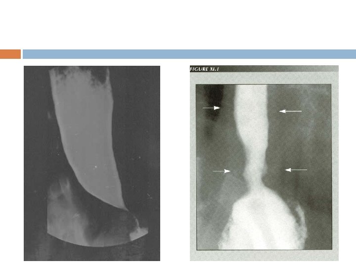
Hiatus Hernia
Condition in which lower esophageal sphincter fails to relax during swallowing. Food passage from esophagus to stomach is prevented
CAUSES Damage to myentric plexus in lower two thirds of esophagus. Musculature of esophagus remains spastically contracted.
Infection Ulceration of esophageal mucosa In severe cases, accumulation of food causes enlargement of esophagus until can hold 1 liter of food which become putridly infected Severe substernal pain Rupture
Management Stretching of lower end of esophagus by means of balloon inflated on the end of a swallowed esophageal tube Anti-spasmotic drugs
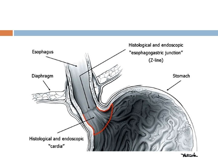
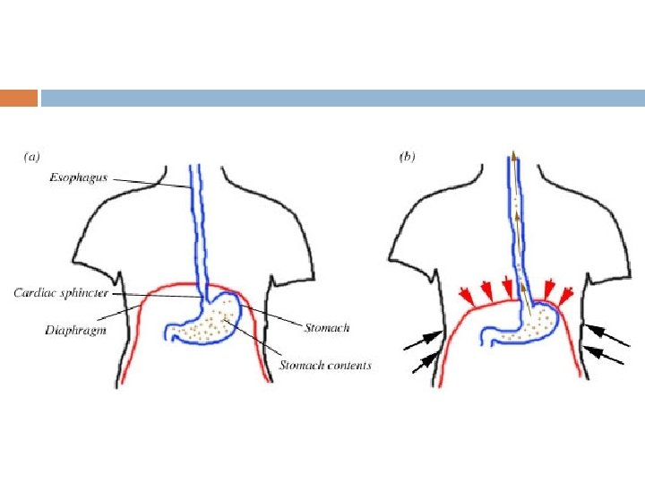
Gastro Esophageal Reflux Disease (GERD)
Gastro Esophageal Reflux Disease (GERD) Regurgitation of acidic contents into the esophagus CAUSE: weakness or incompetence (Failure to constrict) of lower esophageal sphincter
Gastro Esophageal Reflux Disease FEATUR ES: Heart burn or Pyrosis Dysphagi a Esophageal ulcers or cancer (chronic cases) Cough and change of voice
Movemen ts Of Stomach
Movements Of Stomach Hunger Contractions In fasting conditions three types of movements occur
TYPE 1 CONTRACTION OR TONUS RHYTHM First contractions appear in the empty stomach when the stomach is empty Rhythmic contraction occur at the rate of 3 per minute Lasts for about 20 seconds Interval between the contractions-3 -4 seconds Pressure produced-5 cm of water
TYPE 2 CONTRACTIO NS OR HUNGER CONTRACTIO N • Occurs when the tonus of stomach is stronger • Lasts for 20 seconds • No interval between contractions • Pressure produced is 10 -15 cm of water
TYPE 3 HUNGER CONTRACTION INCOMPLETE TETANUS • Successive contraction fuse together to cause a continuing tetanic contraction Occurs when the hunger becomes severe and the tonus increased to great extent • Lasts for 1 to 5 min • Pressure rises to 10 to 20 cm of water • When stomach is empty type 1 contraction occurs followed by type 2 and food intake still postponed type 3 occurs
Hunger contraction more intense in young healthy people who have high degrees of GIT tonus Also increased by low level of blood sugar Hunger Pangs: Pain occurs in the pit of stomach due to hunger contractions Usually will not occur till 12 to 24 hours after the last ingestion of food, attains greatest intensity in 3 or 4 days and then gradually decreases
Movemen ts after a meal
Movements after a meal Two different kinds of movements seen in two halves of stomach after the entry of food Pyloric part has different type of movement than the fundus and body of the stomach
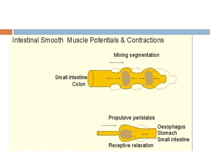
RECEPTIVE RELAXTION OF STOMACH Relaxation of upper portion of stomach when the bolus enters the stomach from esophagus It involves fundus and upper part of of body of stomach Significance: To accommodate food without much increase in pressure inside the stomach
Peristaltic wave Starts from longitudinal muscle in the greater curvature as a pace maker begin in the mid portion of the stomach wall(mixing waves) and passes over the body, antrum and pylorus at a rate of 3 per minute Rate of progress 1 cm per sec in the muscle of the body and accelerated in the antrum to 3 -4 cm per sec The wave spreads from longitudinal to circular muscle , more marked distally(antral systole)
As each wave passes down the antral wall towards pylorus it digs deeply into the food contents in the antrum. Opening of pylorus is still small enough that only a few millimeters or less antral contents are expelled into the duodenum with each wave As the wave approaches the pylorus the pyloric muscle itself contracts which further impedes emptying through pylorus
Most of the contents are squeezed upstream through peristaltic ring toward body and not toward pylorus Thus moving peristaltic movement along with this upstream squeezing action called retropulsion is an important mixing movement
Movements of Pylorus Terminal antrum and the pylorus contract with arrival of wave in their region known as systolic contraction Due to viscous property of gastric contents and pressing of gastric contents into the terminal part of antrum by the peristaltic wave antral pressure rises which overcome the pressure barriers in pylorus and thus antral contents are passed into duodenum
Factors Influencing the Emptying of Stomach Volume of gastric content Gastric emptying is directly propotional to the volume Consistency of gastric content Directly proportional to the consistency liquids pass more easily whereas solid will not pass easily unless it is not converted to semi fluid or fluid
Chemical composition Carbohydrates leave easier than proteins and proteins move easier than fats PH strong acid content empty slowly and accelerated by neutralizing it , thus protecting duodenal mucosa from acidity
Regulation of gastric Emptying is mainly regulated by signals from both the stomach and the duodenum.
Gastric factors that promote emptying Increased food volume increases emptying Increased food volume Causes stretching of wall of the stomach Elicit local myentric reflexes in the wall Increases the activity of the pyloric pump and slightly inhibit the pylorus
Duodenal factors inhibiting stomach emptying Entero gastric nervous reflexes from the Duodenum: Food entering duodenum Multiple nervous reflexes from duodenal wall Passes back to stomach Slows or stop stomach emptying when the volume of chyme in duodenum is too much
• Duodenum to stomach Reflexes through enteric mediate nervous system d • Through extrinsic through nerves three • Slight extent through routes: vagus nerve
1. 2. 3. 4. 5. Factors that can excite enterogastric reflexes: Degree of distension of duodenum presence of any degree of irritation of the duodenal mucosa Degree of acidity of duodenal chyme(>3. 5 _4) Degree of osmolality of the chyme Presence of certain break down products esp breakdown products of protiens and lesser extent to fat
HORMONAL FACTORS
STIMULUS: FATS Fat in duodenum Extract different hormones from duodenal and jejunal epithelium Blood Stomach Inhibit pyloric pump and increase pyloric contraction
MOST POTENT HORMONES CAUSE HORMONAL FEEDBACK INHIBITION Cholecystokinin (most important) Stimulus: Fat Action: Inhibit stomach motility caused by gastrin Other Hormones: Secretin GIP(Gastric inhibitory peptide) VIP PEPTIDE YY SOMATOSTATIN
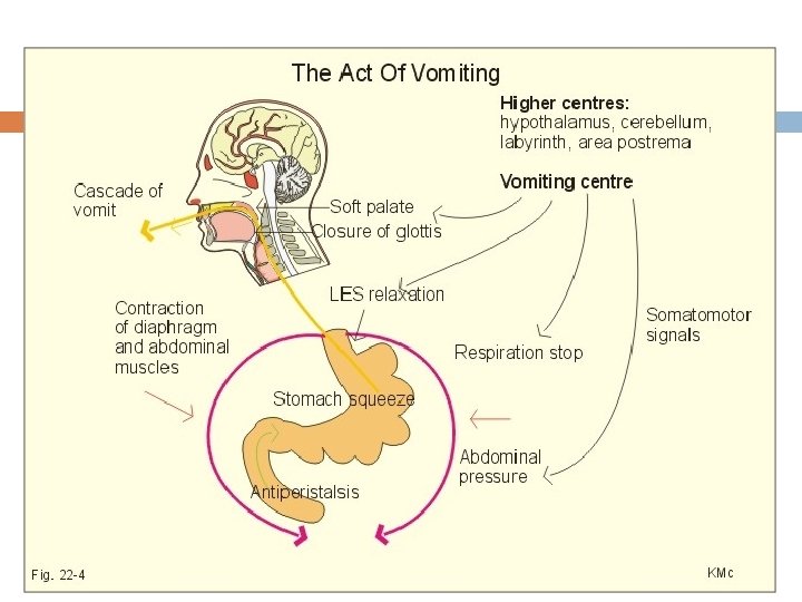
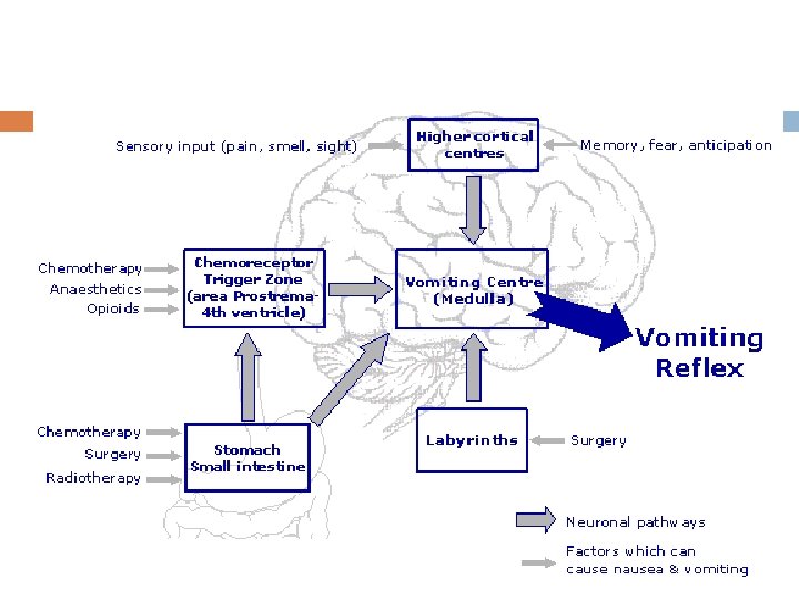
VOMITING DEFINITION: Vomiting is a reflex which serves to relieve the upper GIT by forcible expulsion of gastric contents through mouth
Causes: 1. 2. 3. 4. 5. 6. 7. 8. 9. 10. Presence of irritating contents of GIT Mechanical irritation of pharynx Pregnancy Excess alcohol Nauseating sight, odour or taste Unusual stimulation of Labyrinthine apparatus as in the case of sea sickness, air sickness, car sickness or swinging Drugs like antibiotics opiates Any GI disorder Acute infections like urinary tract infection Metabolic disturbances like carbohydrate starvation and ketosis
Mechanism of vomiting Nausea : Vomiting is always preceded by nausea. Nausea is the unpleasant sensation which induces the desire for vomiting. Characterized by secretion of large amount of saliva containing more amount of mucus. Retching : Strong involuntary movements in GIT before actual vomiting and intensify the feeling of vomiting is called retching. vomiting occurs few minutes after this.
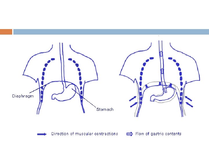
Anti peristalsis , the prelude to vomiting In early stages of gastrointestinal irritation Antiperistalsis begins to occur often many minutes before vomiting appears. Antiperistalsis starts in the ileum and travel backwards up the intestine at the rate of 2 - 3 cm/sec pushing large amount of intestinal contents back to duodenum and stomach within 3 -5 minutes Over distension of upper portion of GIT especially duodenum becomes the exciting factor that initiates actual vomiting act
At the onset of vomiting strong intrinsic contraction occurs in both the duodenum and stomach along with partial relaxation of lower esophageal sphincter allowing vomitus to move into the esophagus From here a specific vomiting act involving abdominal muscles takes over and expels the vomitus to exterior
Vomiting act: Once the vomiting center sufficiently stimulated and the vomiting act instituted the first effects are 1. Deep breath 2. Raising of hyoid bone and larynx to pull the upper esophageal sphincter open 3. Closing of the glottis 4. Lifting of soft palate to close the posterior nares
Next comes the strong downward contraction of diaphragm along with contraction of all the abdominal wall muscles Squeezes the stomach between diaphragm and abdominal muscles High intragastric pressure Complete relaxation of lower esophageal sphincter Expulsion of gastric contents upward through esophagus
Vomiting reflex: Irritation or distension of upper part of GIT or other organs Sensory impulses are carried by vagus and sympathetic afferent nerve fibers Vomiting center located bilaterally in medulla oblongata near nucleus solitaris Motor impulses through V, VII, IX, X, and XII cranial nerves Upper part of GIT Through Spinal nerves to diaphragm and abdominal muscles
Chemoreceptor trigger zone Center for vomiting during motion sickness or vomiting induced by drugs like morphine apomorphine etc is on the floor of IVth ventricle Center for psychic stimuli induced vomiting: Nauseating odour sight or noise is cerebral cortex
Movements of small intestine 1. Mixing contractions (segmentation contractions) 2. Propulsive contractions (peristalsis)
Movements of small intestine 1. Mixing contractions: Portion of SI distended with food , stretching of intestinal wall Elicit localized concentric contractions spaced at intervals Divides the intestine into many segments(chain of sausages lasting for a fraction of minute) Relaxation of contracted sites New set of contraction at new points between the previous contraction sites causing chopping of the food
CS of Small Intestine
PERISTALTIC MOVEMENT: A wave of contraction followed by a wave of relaxation Stimulation of smooth muscles of intestine initiates peristalsis It travels from point of stimulation to both the directions But normally that travels in aboral direction persists and to oral direction is inhibited
Starlings law of intestine : Depending upon the direction of peristalsis Starlings law of intestine is put forward by Starling The response of the intestine for a local stimulus consists of contraction of smooth muscle above and relaxation below the stimulated area
Peristaltic wave starts at any point of intestine and move anal ward at a velocity of 0. 5 to 2. 0 cm/sec Much faster in proximal intestine and much slower in terminal intestine Normally very weak and usually die after traveling 3 -5 cms , very rarely upto 10 cms So net movement along small intestine normally averages 1 cm/sec Thus it takes 3 -5 hours for passage of chyme from pylorus to ileocecal valve
Control of peristalsis by nervous and hormonal signals: Peristalsis greatly increased after a meal Partly by the entry of chyme and partly by gastroenteric reflex Gastroenteric reflex: Distension of stomach Stimulates Myentric plexus Impulses carried through the wall of the intestine
Hormonal factors: Increasing intestinal motility: Gastrin CCK Insulin Serotonin Inhibiting intestinal motility: Secretin Glucagon
Peristaltic rush: -Powerful peristaltic contraction caused by excessive irritation of intestinal mucosa or excessive distension -Begins In duodenum passes the entire length of SI and reaches ileocecal valve within a few minutes -Sweeps the contents of intestine into the colon relieving the intestine off either irritants or excessive distension
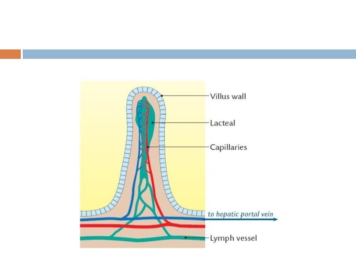
Movements of villi : These movements are due to contraction of of strips of muscle fibers which proceed from muscularis mucosae and attached to the sides of the villus Generally two types of movements: 1. Side to side movement 2. Pumping movements
Microscopic villi
Side to side movement: In these movements villi bent to one side due to contraction of muscle strip on that side Function: Admixture Absorption
Pumping movements: Muscle strips around the villus contracts simultaneously and the villus shorten in length as a whole At the height of digestion and abporption the movement takes place at the rate of 1 -6 /min Function: To increase the flow of bood and lymph
Factors controlling movements of villi: 1. Mechanical irritation of food acting through local nervous plexus 2. Neural stimuli: Stimulation of sympathetic nerves augments the movements of villi Stimulationof sympathetic nerves casuses contraction of muscularis mucosae
3. Chemical stimuli: Products of food digestion esp amino acid Yeast extracts 4. Villikinin: It is a hormone that can be extracted from the intestinal mucosa Structure not definitely known It is believed that during active digestion it is absorbed through blood and stimulates the movements of villi
Function of ileoceacal valve: � Principle function is to prevent back flow of fecal contents from the colon into small intestine. � The lips of ileocecal valve protrude into the lumen of cecum therefore forcefully closes when excess pressure builds up in the cecum and tries to push cecal contents backward against the lips � The valve usually can resist reverse pressure 5060 cms of water.
Ileocecal valve has thickened muscular coat called Ileocecal sphincter. The sphincter normally remains constricted and slows emptying But after a meal it relaxes as gastroileal reflex intensifies the peristalsis in the ileum The resistance to emptying prolongs the stay of chyme in the ileum and thereby facilitates absorption Normally about 1500 ml of chyme empty into cecum every day.
Feed back control of ileo cecal sphincter: The degree of contraction of ileo cecal sphincter and peristalsis in terminal ileum are controlled by reflexes from cecum
Distension of cecum or any irritation(appendicitis) Contraction of ileocecal sphincter Inhibition of ileal peristalsis Emptying of additional chyme from ileum delayed
Reflexes are mediated by: Myentric nervous plexus Extrinsic autonomic nerves( Prevertebral sympathetic ganglia)
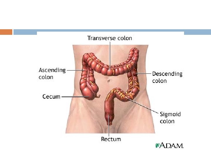
MOVEMENTS OF LARGE INTESTINE
Movements of Large Intestine Principle function: 1. Absorption of water and electrolytes from chyme to form solid chyme 2. Storage of fecal matter until it can be expelled Proximal half- concerned with absorption Distal half- concerned with storage As its function does not needs intense movements normally it will be sluggish and can be divided into mixing and propulsive
Mixing movement: Haustral contraction: bulging of wall of colon in between teniae, reaches its peak in 30 sec and disappears within 60 secs Propulsive movements: 1. Peristalsis : Same as in small intestine except in frequency Power of contraction greater in descending colon due to dry and hard character of the material to be moved
2. Mass peristalsis: - It is not true peristalsis and it involes large segmants of colon - First a constrictive ring occurs in response to distended or irritated point usually in transverse colon - Rapidly there after 20 or more cms of colon distal to constriction lose their haustrations and contract as a unit forcing down the fecal material.
Lasts for 30 secs and then relax for 2 -3 min The whole series persists upto 10 -30 min When they have forced a mass of feces into rectum desire for defecaton occurs It occurs one to three times a day
Initiation of mass movements: Facilitated by Gastro colic reflex Irritation of stomach or Duodenocolic reflex duodenum Distention of
DEFAECATION Defaeaction is an act of emptying of entire distal colon from the splenic flexure through the anal orifice into the exterior, which is a reflex process. Fecal matter stored in sigmod and pelvic colon Rectum is devoid of feces mostly 1. Partially from the fact a weak functional sphincter exists about 20 cms at the junction between sigmoid colon and rectum 2. Sharp angulation
When a mass movement forces feces into rectum Desire for defecation occurs Reflex contraction of rectum and relaxation of anal sphincter Usually the desire occurs when the pressure rises to 20 -25 cm H 2 O-
Anatomy of Anorectum
Ordinarily defecation initiated by defecation reflexes Intrinsic reflex: Mediated by Local nervous plexus comparatively very weak Parasympathetic defecation reflex: Involves sacral segments of spinal cord is more effective
Continual dribbling of fecal matter through anus is prevented by 1) Internal anal sphincter : Made up of smooth muscle and innervated by parasympathetic nerve fibers via Pelvic N 2) External anal sphincter: Composed of skeletal muscle and controlled by somatic N fibers pass through Pudendal N. It keeps external sphincter constricted always and it relaxes only when it is inhibited
Para sympathetic defecation reflex Due to mass movement , feces enters to rectum Distention of rectum Sensory nerve endings are stimulated Impulses transmitted via afferent fibers of pelvic nerve to defecation center (sacral segmants of spinal cord) Impulse via efferent fibers of pelvic N to descending colon, sigmoid colon and rectum Cause cantraction and relaxation of internal anal sphincter Voluntary relaxation of external sphincter due to inhibition of pudendal N arising from cerebral cortex
INTESTINAL GAS A considerable amount of gas is present in the gastrointestinal contents of all animals, and much of this is eliminated through the anus as flatus. Five gases constitute greater than 99% of the gases passed as flatus: N 2, O 2, CO 2, H 2 and methane. None of these gases has an odor, and the characteristic odor of feces is due to very small quantities of a few other gases, including hydrogen sulfide, scatols and indoles.
In normal adult humans, the rate of excretion of gas per rectum ranges between 200 and 2000 ml per day. Ingestion of certain foods, beans being the classical example, is widely recognized to increase the rate of gas production
Constipation: Failure of voidingof feces Causes: dietery Irregular bowel habit Spasm of sigmoid colon Diseases Dysfunction of myentric plexus- mega colon Drugs
eas Than k. U All