Gastrointestinal Physiology General Principles of Gastrointestinal Chapter 62
 Dec 26 th 2014 Mohammed")


 Mixing movements (segmentation) - Blend")
")
, they produce mucus. 2.")


 Ø Enzymes i. -amylase (from parotid glands) • cleaves -1")
 2. Mucus: Lubricating and Protective Properties of Mucus: • Mucus")
 v. The basic unit “salivon” consists of: Acinus -initial secretory process")


 Dr. Mohammed Alzoghaibi zzoghaibi@gmail. com")
 • Swallowing is initiated voluntarily in the mouth, but thereafter")
 Bolus of food Tongue Uvula Pharynx Bolus Epiglottis Glottis Esophagus Trachea (a)")

 Pancreatic Secretion Mohammed Alzoghaibi, Ph. D")


 • Pancreatic secretions contain many enzymes for digesting proteins, carbohydrates, and")
 When first synthesized in the pancreatic cells, the proteolytic digestive enzymes")

 • The pancreatic enzyme for digesting carbohydrates is pancreatic amylase, which")


 Secretin Stimulates Secretion of Copious Quantities of Bicarbonate Ions—Neutralization")
 Cholecystokinin—Its Contribution to Control of Digestive Enzyme Secretion by")


 • At a neutral")


- Slides: 54
Gastrointestinal Physiology General Principles of Gastrointestinal (Chapter 62 -65) Dec 26 th 2014 Mohammed Alzoghaibi, Ph. D. zzoghaibi@gmail. com Whats. App, SMS: 0506338400
Required Textbook of Medical Physiology Eleventh Edition Guyton & Hall Published by Elsevier Saunders 2011
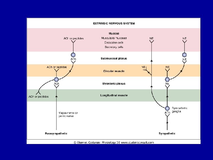
General Principles of Gastrointestinal Motility • Physiologic Anatomy of the Gastrointestinal Wall The following layers structure the GI wall from outer surface inward: (1) the serosa, (2) a longitudinal muscle layer, (3) a circular muscle layer, (4) the submucosa, and (5) the mucosa. In addition, sparse bundles of smooth muscle fibers, the mucosal muscle, lie in the deeper layers of the mucosa.
Hormone Site of Secretion Stimuli for Secretion Actions Gastrin G cells of the stomach Small peptides and ↑ Gastric H+ secretion amino acids Stimulates growth of gastric Distention of the mucosa stomach Vagal stimulation (GRP) Cholecystokinin (CCK) I cells of the duodenum and jejunum Small peptides and ↑ Pancreatic enzyme secretion amino acids ↑ Pancreatic HCO 3 - secretion Fatty acids Stimulates contraction of the gallbladder and relaxation of the sphincter of Oddi Stimulates growth of the exocrine pancreas and gallbladder Inhibits gastric emptying Secretin S cells of the duodenum H+ in the duodenum ↑ Pancreatic HCO 3 - secretion Fatty acids in the ↑ Biliary HCO 3 - secretion duodenum ↓ Gastric H+ secretion Inhibits trophic effect of gastrin on gastric mucosa Glucose-Dependent Insulinotropic Peptide (GIP) K cells of the Duodenum and jejunum Fatty acids Amino acids Oral glucose ↑ Insulin secretion from pancreatic β cells ↓ Gastric H+ secretion Motilin M cells of the duodenum and jejunum Fat Acid Nerve Stimulates: Gastric motility Intestinal motility
• Functional Types of Movements in the Gastrointestinal Tract Two types of movements occur in the gastrointestinal tract: (1) propulsive movements • Organizes propulsion of material over variable distances within the GI lumen • Usual stimulus is distention. Other stimuli that can initiate peristalsis include chemical or physical irritation of the epithelial lining in the gut. • Myenteric plexus is important • Atropine (cholinergic blocker) depresses propulsis v Receiving segment---contraction (longitudinal M. ) ---relaxation (circular M. ) Peristaltic Reflex and the---contraction "Law of the Gut. " When a segment of the intestinal tract is excited v Propulsive segment (circular M. ) by distention and thereby initiates peristalsis, the contractile ring causing the peristalsis normally ----relaxation begins on the orad side of the distended(longitudinal segment and moves toward the distended segment, M. ) the intestinal contents in the anal direction for 5 to 10 centimeters before dying out. pushing
Functional Types of Movements in the Gastrointestinal Tract 2) Mixing movements (segmentation) - Blend different juices with the chyme - Bring products of digestion in contact with absorptive surfaces
Chapter 64; page 773 -777 Secretory Functions of the Alimentary Tract (Secretion of Saliva) Mohammed Alzoghaibi, Ph. D zzoghaibi@gmail. com
Anatomical Types of Glands 1. Single-cell mucous glands (goblet cells), they produce mucus. 2. Crypts of Lieberkühn at the mucosal pits. 3. Tubular glands (in the stomach and duodenum) 4. Salivary glands, pancreas, and liver
SALIVARY GLANDS • The principal glands of salivation are: 1. Parotid glands 2. Submandibular (Submaxillary) glands 3. Sublingual glands 4. Smaller glands in mucosa of tongue, palate, etc. • Daily secretion of saliva = 800 -1500 m. L with p. H = 6 -7
Secretion of Saliva and its Characteristics § Saliva contains two major types of secretion: 1. Aqueous fluids (a serous secretion) • Water, ions and enzymes such as ptyalin (an αamylase) Ø Parotid, Submandibular and Sublingual glands 2. Mucus secretion (mucin) Ø Submandibular and Sublingual glands
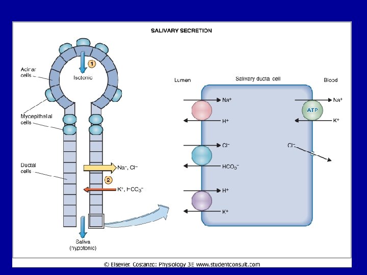
Composition of Saliva 1. Aqueous Fluids o H 2 O, K, HCO 3, Na, Cl, -amylase, lingual lipase, Ig. A, kallikrein, muramidase (lyses muramic acid of Staphylococcus), lactoferrin and epithelial growth factor (EGF) o Hypotonic Solution Ø Ions Na, K, CI, HCO 3: (the concentrations of these ions are altered with altered flow rates), e. g. , at low flow rate, the salivary secretions have: i. High K and HCO 3 ii. Low Na and CI−
Composition of Saliva (con’t) Ø Enzymes i. -amylase (from parotid glands) • cleaves -1 , 4 -glycosidic bonds • The optimal p. H for this enzyme to work properly is 7 • Inactivated at p. H 4 but continues to work for sometime in unmixed food in Orad portion of stomach ii. Lingual lipase • hydrolyzes lipids • continues working in the duodenum iii. Kallikrein (protease, from acinar cells) • Catalyzes production of bradykinin (good vasodilator) from globulin • Increases local blood flow Ø Water (0. 5 L saliva/day)
Composition of Saliva (con’t) 2. Mucus: Lubricating and Protective Properties of Mucus: • Mucus is a thick secretion composed mainly of water, electrolytes, and glycoproteins • The mucus is an excellent lubricant and a protectant for the wall of the gut because of the following: 1. It has adherent qualities that make it adhere tightly to the food. 2. It has sufficient body that it coats the wall of the gut and prevents actual contact of most food particles with the mucosa. 3. It has a low resistance for slippage. 4. It causes fecal particles to adhere to one another. 5. It is strongly resistant to digestion by the GI enzymes. 6. The glycoproteins of mucus have amphoteric
Secretory Unit (salivon) v. The basic unit “salivon” consists of: Acinus -initial secretory process 2. Intercalated duct -initial portion of duct 3. Striated duct -modification of secretory product 4. Myoepithelial cells 1. Ø surround acinus and intercalated duct Ø contraction moves saliva, prevents development of back pressure
Characteristics of Saliva and Flow Rate
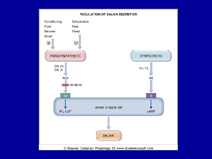
Control of Secretion • Unique aspect of control of salivary secretion Ø secretion rate depends entirely on neural control –autonomic nervous system (ANS) Ø both Parasympathetic and Sympathetic lead to increase secretion Ø Composition modified by Aldosterone: increases Na, Cl reabsoption ii. increases K secretion i.
Chapter 63: 763 -765 Swallowing (Deglutition) Dr. Mohammed Alzoghaibi zzoghaibi@gmail. com
Stages of Swallowing (Deglutition) • Swallowing is initiated voluntarily in the mouth, but thereafter is under involuntary or reflex control. The reflex portion is controlled by the swallowing center in the medulla. • Stages of Swallowing: 1. Oral Stage (voluntary) 2. Pharyngeal stage (involuntary) 3. Esophageal stage (involuntary)
Deglutition (Swallowing) Bolus of food Tongue Uvula Pharynx Bolus Epiglottis Glottis Esophagus Trachea (a) Upper esophageal sphincter contracted Bolus (c) Upper esophageal sphincter contracted (b) Upper esophageal sphincter relaxed Relaxed muscles Circular muscles contract, constricting passageway and pushing bolus down Bolus of food Gastroesophageal sphincter open Longitudinal muscles contract, shortening passageway ahead of bolus Gastroesophageal sphincter closed (d) Stomach (e)
Function of Gastroesophageal Sphincter ● Receptive Relaxation of the Stomach. When the esophageal peristaltic waves reaches the stomach, the stomach relaxes through inhibition of myenteric neurons which prepares the stomach to receive the food that is propelled into the esophagus during swallowing. ● Function of the Lower Esophageal Sphincter (Gastroesophageal Sphincter). The esophageal sphincter is formed by the esophageal circular muscle located in an area of ~ 3 cm upward of the junction with the stomach. This sphincter remains tonically constricted (protects the esophagus from the stomach acidic juices) until the peristaltic swallowing wave passes down the esophagus and causes a “receptive relaxation” of the sphincter and the emptying of the propelled food into the stomach. Failure of the sphincter to relax will result in achalasia. ● Additional Prevention of Esophageal Reflux by Valvelike Closure of the Distal End of the Esophagus. Receptive Relaxation of the Stomach. This is another protective mechanism (safety factor) that prevents reflux of gastric secretions into the lower portion of the esophagus. This mechanism involves a short portion of the esophagus that extends slightly into the stomach and that caves the esophagus inward in response to increased intra-abdominal pressure.
(Chapter 64; Pages 780 -783; Guyton & Hall) Pancreatic Secretion Mohammed Alzoghaibi, Ph. D zzoghaibi@gmail. com
Islet of Langerhans The pancreas, in addition to its digestive functions, secretes two important hormones: in the pancreas. 1. Insulin (Beta cells; 60%). 2. Glucagon (Alpha cells; ~25%). that are crucial for normal regulation of glucose, lipid, and protein metabolism. Also Somatostatin is secreted by delta cells ( form ~10% of islets’s Figure 78 -1. Copyright 2011, WB Saunders Elsevier, All Rights Reserved
Pancreatic Secretion • The major functions of pancreatic secretion: 1. 2. 3. To neutralize the acids in the duodenal chyme to optimum range (p. H= 7. 0 -8. 0) for activity of pancreatic enzymes. To prevent damage to duodenal mucosa by acid & pepsin To produce enzymes involved in the digestion of dietary carbohydrate, fat, and protein
Pancreatic secretion (continued) • Pancreatic secretions contain many enzymes for digesting proteins, carbohydrates, and fats, and large quantities of HCO 3 ions. • The most important pancreatic enzymes for digesting proteins are: 1. Trypsin. 2. Chymotrypsin. 3. Carboxypolypeptidase. • Trypsin and chymotrypsin split whole and partially digested proteins into peptides of various sizes but do not cause release of individual amino acids. • Carboxypolypeptidase splits some peptides into individual amino acids, thus completing digestion of some proteins to amino acids.
Pancreatic Enzymes (continued) When first synthesized in the pancreatic cells, the proteolytic digestive enzymes are in the inactive forms: 1. Trypsinogen 2. Chymotrypsinogen 3. Procarboxypolypeptidase These enzymes become activated only after they are secreted into the intestinal tract. 1. Trypsinogen is activated by: ● Enteropeptidase (enterokinase), an enzyme secreted by the intestinal mucosa when chyme comes in contact with the mucosa. ● Trypsinogen can be autocatalytically activated by trypsin formed from previously secreted trypsinogen. 2 & 3. Chymotrypsinogen and procarboxypolypeptidase are activated by trypsin to form chymotrypsin and carboxypolypeptidase.
Trypsin Inhibitor Secretion of Trypsin Inhibitor Prevents Digestion of the Pancreas Itself. ● Proteolytic enzymes of the pancreatic juice do not become activated until after they have been secreted into the intestine because the trypsin and the other enzymes would digest the pancreas itself. ● The same cells that secrete proteolytic enzymes into the acini of the pancreas secrete another substance called trypsin inhibitor, which is formed in the cytoplasm of the glandular cells, and it prevents activation of trypsin both inside the secretory cells and in the acini and ducts of the pancreas. ● Because trypsin activates the other pancreatic proteolytic enzymes, therefore trypsin inhibitor
Pancreatic secretion (continued) • The pancreatic enzyme for digesting carbohydrates is pancreatic amylase, which hydrolyzes starches, glycogen, and most other carbohydrates (except cellulose) to form mostly disaccharides and a few trisaccharides. • The main enzymes for fat digestion are: (1) Pancreatic lipase (2) Cholesterol esterase (3) Phospholipase
Pancreatic secretions are rich in bicarbonate ions • The pancreas secrets about 1 L/day of HCO 3 –rich fluid from the epithelial cells of the ductules and ducts. • The osmolarity of pancreatic fluid is equal to that of plasma • HCO 3 concentration increases with increasing secretion rate
Secretion of Isosmotic Sodium Bicarbonate Solution.
Flow Rate and pancreatic secretion
Pancreatic Secretion is Under Neural and Hormonal Control • Parasympathetic stimulation (through Ach on acinar cells) results in an increase in enzyme secretion-fluid and HCO 3 • Secretin tends to stimulate a HCO 3 rich secretion by activating ductal cells. • CCK stimulates a marked increase in enzyme secretion by stimulating the acinar cells. • Pancreatic secretion normally results from the combined effects of the multiple basic stimuli, not from one alone (potentiate each other).
Stimuli for Pancreatic Secretion 1. Acetylcholine: released from the parasympathetic vagus nerve endings and from other cholinergic nerves in the enteric nervous system. 2. Cholecystokinin: secreted by the duodenal and upper jejunal mucosa (I cells) when food enters the small intestine. 3. Secretin: also secreted by the duodenal and jejunal mucosa (S cells) when highly acidic chyme enters the small intestine. Acetylcholine and cholecystokinin stimulate the acinar cells of the pancreas, causing production of large quantities of pancreatic digestive enzymes but relatively small quantities of water and electrolytes to go with the enzymes.
Stimuli for Pancreatic Secretion (continued) Secretin Stimulates Secretion of Copious Quantities of Bicarbonate Ions—Neutralization of Acidic Stomach Chyme. ● Secretin is present in an inactive form, prosecretin (in S cells in the mucosa of the duodenum and jejunum). ● When acid chyme with p. H less than 4. 5 -5. 0 enters the duodenum from the stomach, it causes duodenal mucosal release and activation of secretin, which is then absorbed into the blood. ● Secretin causes the pancreas to secrete large quantities of fluid containing a high concentration of HCO 3 (up to 145 m. Eq/L = ~5 X normal) but a low concentration of Cl-. HCl + Na. HCO 3 → Na. Cl + H 2 CO 3
Stimuli for Pancreatic Secretion (continued) Cholecystokinin—Its Contribution to Control of Digestive Enzyme Secretion by the Pancreas. ● The presence of food in the upper small intestine causes cholecystokinin to be released from the I cells in the mucosa of the duodenum and upper jejunum. ● Release of cholecystokinin results especially from the presence of proteoses and peptones (products of partial protein digestion) and long-chain fatty acids in the chyme. ● Cholecystokinin, like secretin, passes by way of the blood to the pancreas and causes secretion of pancreatic digestive enzymes by the acinar cells. ● This effect is similar to that caused by vagal stimulation but even more pronounced, accounting for
Regulation of Pancreatic Secretion
Multiplicative or Potentiation Effects of Different Pancreatic Secretion Stimuli ● When all different stimuli of pancreatic secretion (acetylcholine, cholecystokinin, and secretin) occur at once, then the total secretion is far greater than the sum of the secretions caused by each stimulus separately. The stimuli are said to “multiply” or “potentiate” one another. ● Usually, pancreatic secretions are the result of multiple stimuli rather than one stimulus.
Pancreatic secretion is phasic Phase stimulus Cephalic phase Smell, taste, chewing and swallowing Gastric phase Mediators Release of Ach Protein, gastric Vago-vagal distention reflex Intestinal phase Acid in chyme, fatty acids Secretin, CCK and vago-vagal reflex
Chapter : 64; pages: 783 -786 Secretion of Bile by the Liver Functions of the Biliary Tree Mohammed Alzoghaibi, Ph. D zzoghaibi@gmail. com
What Are The Components of Bile? • 1. 2. 3. 4. 5. The components of bile are: Bile acids (bile salts) Cholesterol Phospholipids (Lecithin) Bile pigments Ions and water
Bile Secretion • Bile secretion is primarily regulated by a feedback mechanism, with secondary hormonal and neural controls Ø The major determinant of bile acid synthesis is its concentration in hepatic portal blood (feedback control) Ø CCK, Secretin and estrogen (hormonal control) Ø Parasympathetic and sympathetic nerves supply the biliary system. Parasympathetic (vagal) stimulation results in contraction of the gallbladder and relaxation of the sphincter of Oddi, as well as increased bile formation. Bilateral vagotomy results in reduced bile secretion after a meal, suggesting that the parasympathetic nervous system plays a role in mediating bile secretion. By contrast,
Secretion and enterohepatic circulation of bile salts
What is the bile acid ? What are the types of the bile acid?
Bile Acids Are Formed in the Liver From Cholesterol • Bile acids are formed in the liver from cholesterol. During the conversion, hydroxyl groups and a carboxyl group are added to the steroid nucleus. • Bile acids are classified as primary or secondary. The hepatocytes synthesize the primary bile acids, which include cholic acid and chenodeoxycholic acid. Bile acids are secreted as conjugates of taurine or glycine. When bile enters the GI tract, bacteria present in the lumen act on the primary bile acids and convert them to secondary bile acids by dehydroxylation. Cholic acid is converted to deoxycholic acid and
Bile Acids Are Formed in the Liver From Cholesterol (continued) • At a neutral p. H, the bile acids are mostly ionized and are referred to as bile salts. Conjugated bile acids ionize more readily than the unconjugated bile acids and, thus, usually exist as salts of various cations (e. g. , sodium glycocholate). • Bile salts are much more polar than bile acids and have greater difficulty penetrating cell membranes. Consequently, the small intestine absorbs bile salts much more poorly than bile acids. This property of bile salts is important because they play an integral role in the intestinal absorption of lipid. Therefore, it is important that the small intestine absorb bile
Function of Bile Salts in Fat Digestion and Absorption Bile acids are formed from cholesterol in the liver. Bile acids are conjugated with the amino acids glycine and taurine in the liver. At neutral p. H, the bile acids are mostly ionized and referred to as bile
Function of Bile Salts in Fat Digestion and Absorption
Function of Bile Salts in Fat Digestion and Absorption • They have a detergent action (emulsifying) on the fat particles in the food which decreases the surface tension of the particles. • They help in the absorption of fatty acids, monoglycerides, cholesterol, and other lipids from the intestinal tract.
Enterohepatic Circulation of Bile Salts Are Recycled Between the Small Intestine and the Liver • The enterohepatic circulation of bile salts is the recycling of bile salts between the small intestine and the liver. The total amount of bile acids in the body, primary or secondary, conjugated or free, at any time is defined as the total bile acid pool. In healthy people, the bile acid pool ranges from 2 to 4 g. The enterohepatic circulation of bile acids in this pool is physiologically extremely important. By cycling several times during a meal, a relatively small bile acid pool can provide the body with sufficient amounts of bile salts to promote lipid absorption. In a light eater, the bile acid pool may circulate three to five times a day; in a heavy eater, it may circulate 14 to 16 times a day. The intestine is normally extremely efficient in absorbing the bile salts by carriers located in the distal ileum. Inflammation of the ileum can lead to their malabsorption and result in the loss of large quantities of bile salts in the feces.
• 1. 2. 3. 4. The Enterohepatic circulation recycles bile salts between the small intestine and the liver Bile salts in the intestine lumen are absorbed via four pathways: Passive diffusion An active carriermediated process De-conjugation or transforming of bile salts to bile acids (by bacteria) Transforming the primary bile acids to
• Absorption of bile acids or bile salt back into hepatocytes Bile salts or bile acids in the portal circulation are absorbed via four pathways into hepatocytes: 1. An active carriermediated process: conjugated bile acids-Na co-transport. 2. Facilitated diffusion: Na -independent pathway. 3. Bile acid-HCO 3 or Bile acid-OH exchange.
The End