Anterior shoulder dislocation Findings Inferomedial subcoracoid displacement of
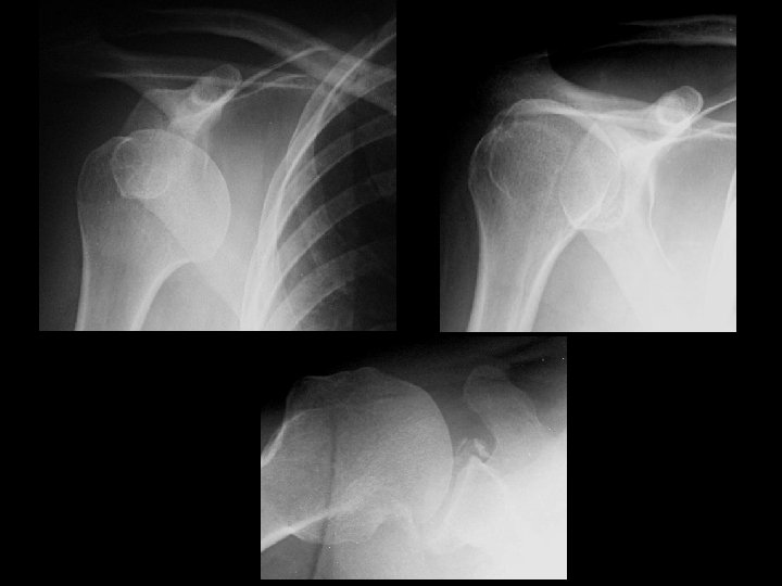

Anterior shoulder dislocation • Findings: – Inferomedial subcoracoid displacement of the humeral head – Too much overlap of humeral head and bony glenoid – Axillary view is diagnositic and shows Hill-Sachs and Bankhart lesions • ddx: – NONE! – This is an Aunt Minnie!
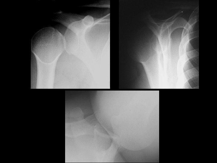

Posterior shoulder dislocation • Findings: – Lateral displacement of the humeral head – “Empty glenoid sign” – Axiallary and transscapular Y views are diagnostic – “Trough sign” and posterior bony glenoid impaction • ddx: – NONE! – This is an Aunt Minnie!
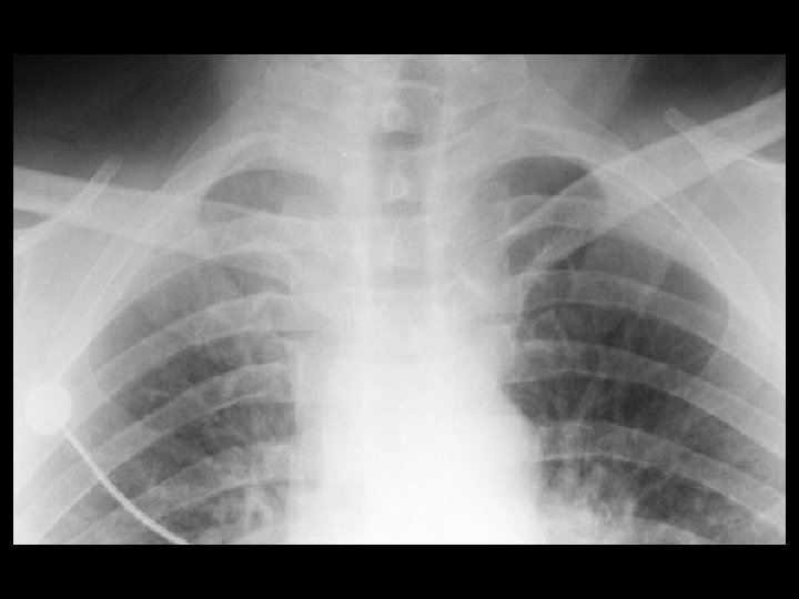

Sternoclavicular dislocation • Findings: – Asymmetric appearance of sternoclavicular joints – medial portion of left clavicle looks abnormal – Need to do CT to evaluate great vessels and mediastinum • ddx: – Occult fracture of sternum or medial clavicle
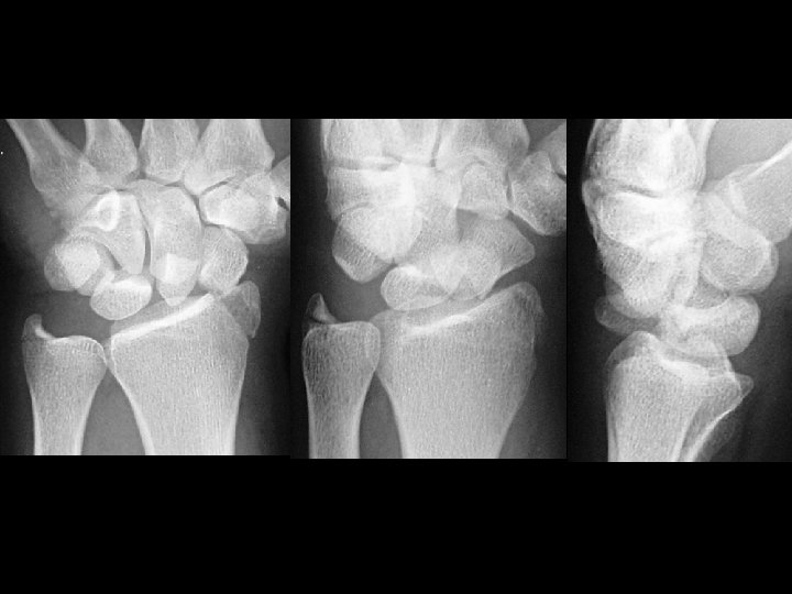

Trans-scaphoid perilunate dislocation • Findings: – Fracture of the radial styloid, scaphoid, and ulnar styloid – Posterior dislocation of the capitellum • ddx: – NONE! – This is an Aunt Minnie!
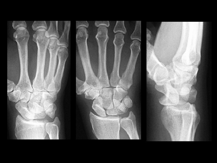

Lunate dislocation • Findings: – Disruption of proximal carpal row – “Pie-shaped lunate” – Lateral view is diagnostic • ddx: – NONE! – This is an Aunt Minnie!
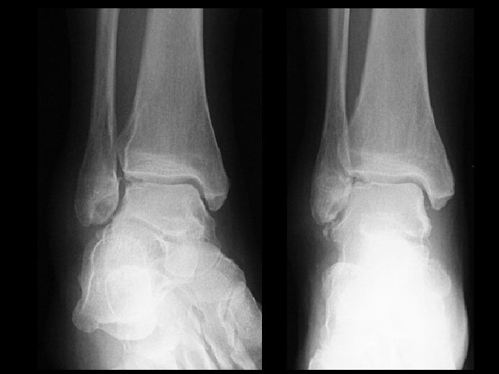

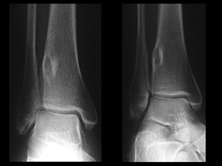
Talar osteochondral fracture • Findings: – Cortical irregularity of the lateral talar dome with bony fragments in the lateral mortise and widening of the distal tibiofibular syndesmosis – Check for proximal fibula fracture • ddx: – NONE! – This is an Aunt Minnie!
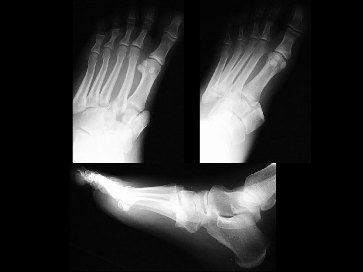

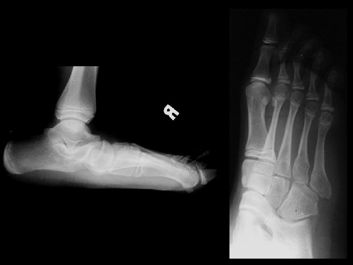
Homolateral Lisfranc fracture-dislocation • Findings: – Lateral dislocation of 1 st thru 5 th metatarsals at the Lisfranc joints – types • divergent • homolateral • partial incongruity • ddx: – NONE! – This is an Aunt Minnie!
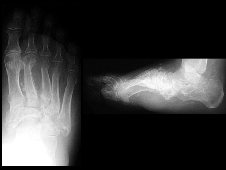

Diabetic Charcot foot & Lisfranc fracture dislocations • Findings: – Destruction, debris, dislocation, and increased density of Lisfranc joints – Pes planus – Vascular calcifications indicative of DM • ddx: – osteomyelitis
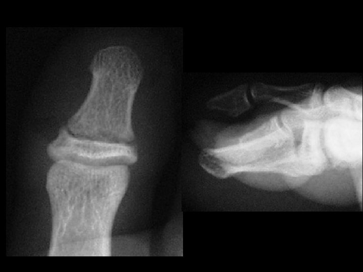

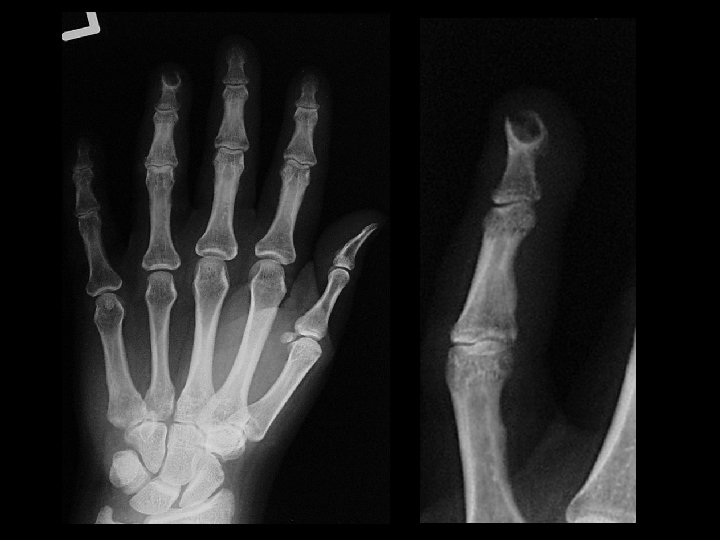
Nail bed injury • Findings: – Horizontal fracture at the base of the great toe distal phalanx – Consider this as an open fracture with a high incidence of osteomyelitis • ddx: – NONE! – This is an Aunt Minnie!
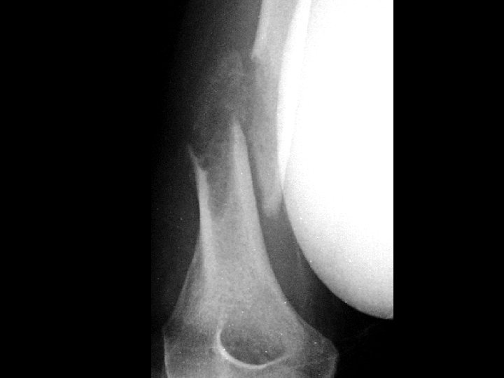

Pathologic fracture • Findings: – Lucent lesion in the medullary space of the distal humeral diaphysis with cortical scalloping and periosteal reaction • ddx: – NONE! – This is an Aunt Minnie!
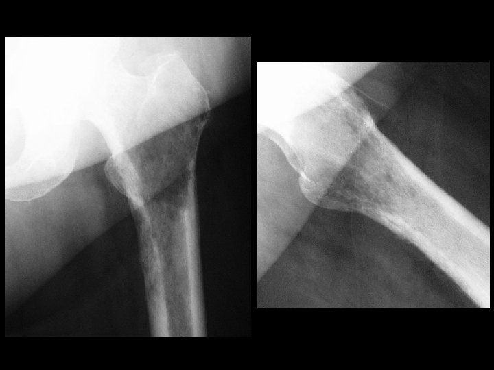

Lymphoma • Findings: – Diffuse permeative lesion of the proximal femur • ddx: – Metastases – Multiple myeloma – Osteomyelitis – Langerhan’s cell histiocytosis (peds)
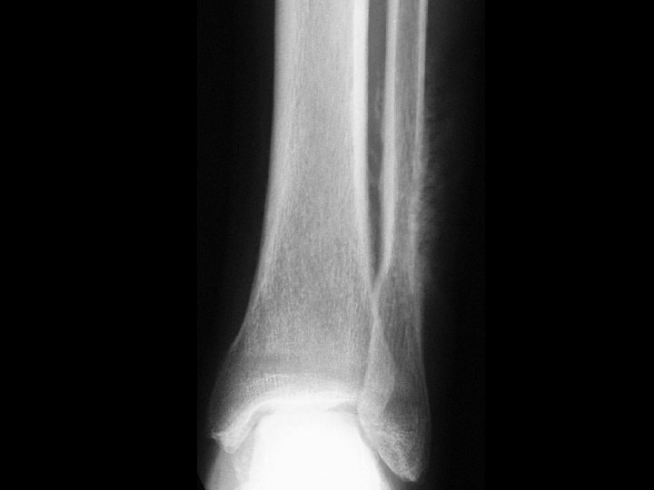

Ewings’s sarcoma • Findings: – “Sunburst” periosteal reaction of the distal fibular diaphysis • ddx: – Osteosarcoma – Osteomyelitis – Langerhan’s cell histiocytosis
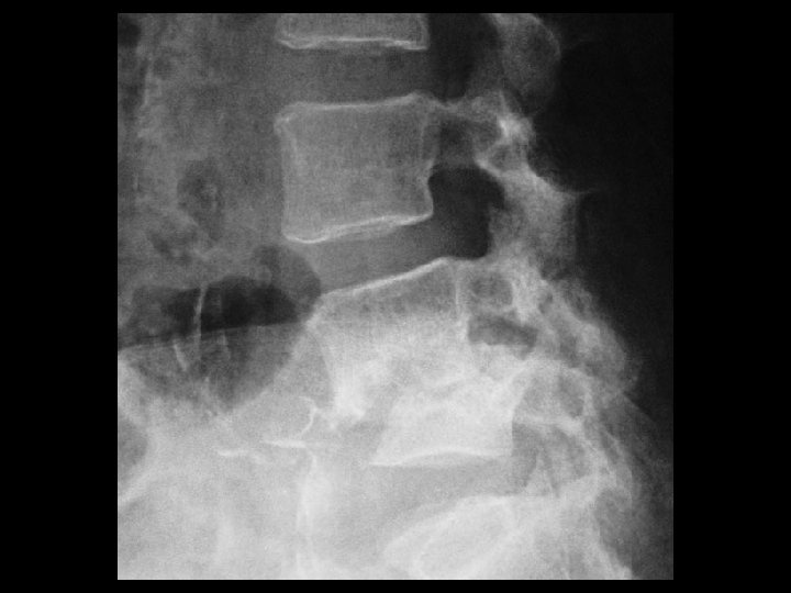

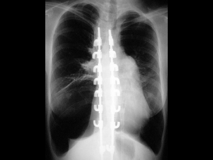
Discitis/Osteomyelitis • Findings: – L 4 -5 disc space and adjacent endplate sclerosis and destruction – ~50% anterolisthesis and gibbus deformity • ddx: – NONE! – This is an Aunt Minnie!

Rheumatoid arthritis • Findings: – Joint space narrowing – “rat bite” marginal erosions – No new bone formation – Soft tissue swelling • ddx: – Gout – Septic arthritis – psoriasis

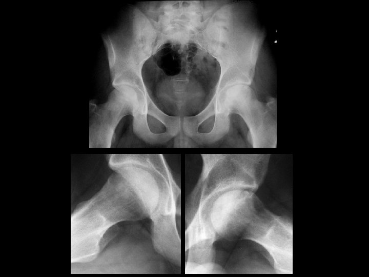
Right hip septic arthritis • Findings: – Increased distance form medial from medial femoral head to teardrop = teardrop distance • ddx: – Aseptic joint effusion – Synovitis – Hematoma
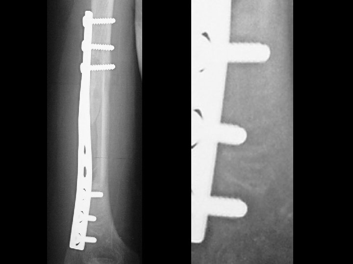

Hardware infection • Findings: – 2 mm lucency surrounding fixation hardware and screws • ddx: – Hardware loosening
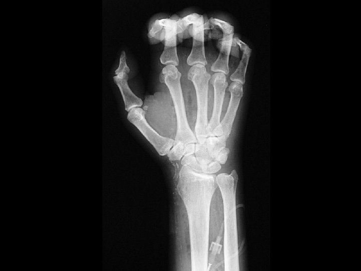

Necrotizing fasciitis • Findings: – Soft tissue swelling and gas overlying the wrist and proximal hand – Vascular Ca 2+ = DM • ddx: – Post-traumatic soft tissue gas

Asymmetric SI fusion • Findings: – Right SI joint fusion – Symmetric hip joint narrowing • ddx: (asymmetric) – Reiter syndrome – Psoriatic arthritis – Infection (symmetric) – Inflammatory bowel dz – Ankylosing spondylitis

Symmertic sacroiliitis & Ulcerative colitis • Findings: – Symmetric SI joint erosion and subchondral sclerosis = sacroiliitis – Mildly dilated ahaustral ascending colon • ddx: – NONE! – This is an Aunt Minnie!
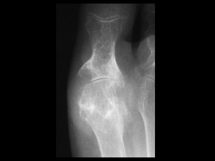

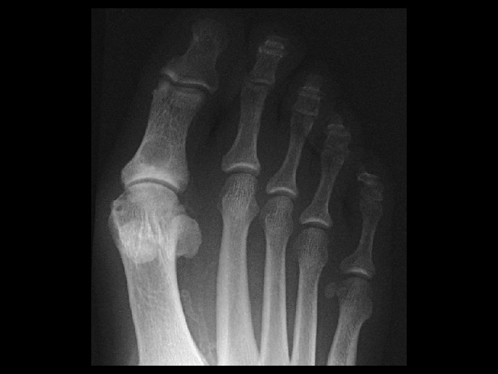
Gout • Findings: – Large erosions with overhanging margins at the great toe MTP joint – Dense periarticular swelling – Sparing of the joint space • ddx: – NONE! – This is an Aunt Minnie!
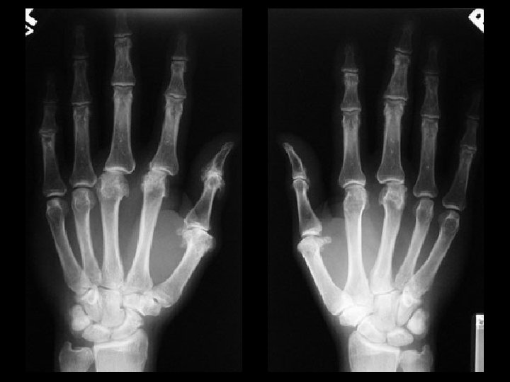

CPPD • Findings: – Bilateral wrist and 1 st – 3 rd MCP joint space narrowing – Small hook-like osteophytes of the MC heads – Chondrocalcinosis of the wrist • ddx: – hemochromotosis
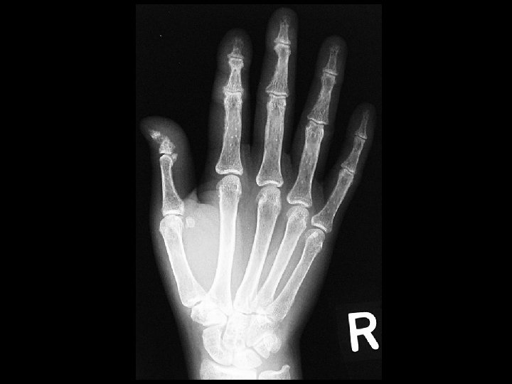

Hyperparathyroidism & Acro-osteolysis • Findings: – Erosion and resorption of digital tufts – Osteopenia with coarse trabecula – Subperiosteal bone resorption along the radial side • ddx: (acro-osteolysis) – Frostbite / Electrical burn – Polyviny chloride exposure – Pyknodysostosis – Neuropathy – Psoriatic arthritis – Hajdu-Cheney syndrome
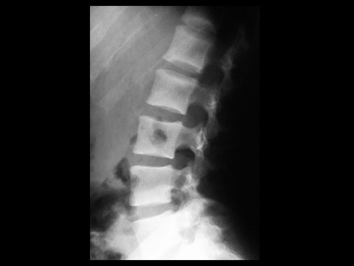

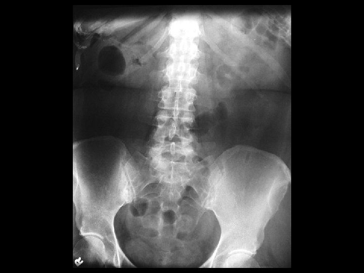
Renal osteodystrophy & Rugger jersey spine • Findings: – Trabecular rarefication within the central vertebral bodies and increased scleroisis of the endplates = “rugger jersey spine” • ddx: – NONE! – This is an Aunt Minnie!

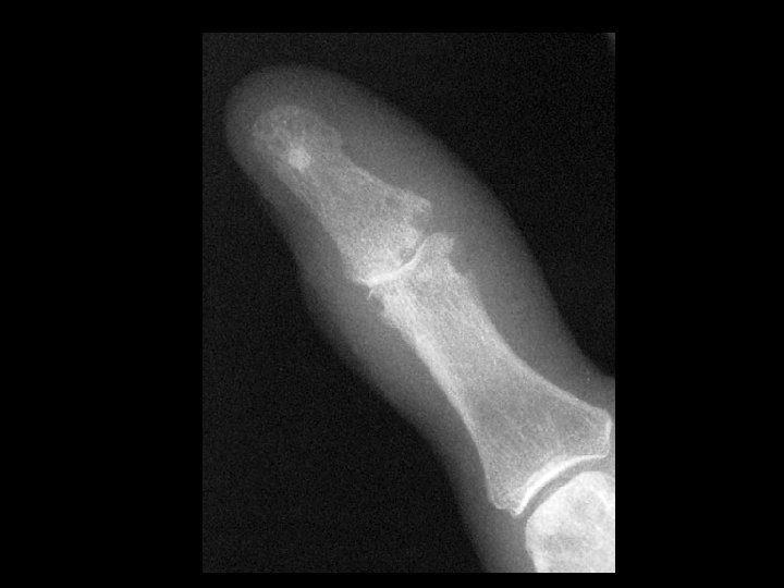
Hyperparathyroidism & Brown tumor • Findings: – Round lucent lesion in the proximal great toe phalanyx – Osteopenia – Marginal erosions – Vascular Ca 2+ = ESD • ddx: – Enchondroma – Fibrous dysplasia – Brodie’s abscess
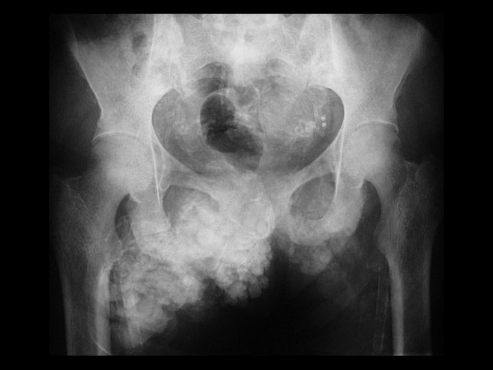

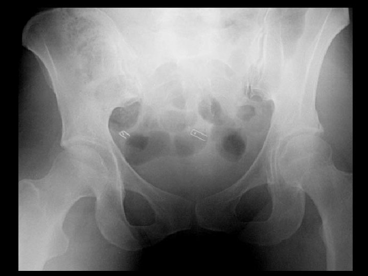
Hyperparathyroidism & Dystrophic soft tissue Ca+ • Findings: – Abnormal soft tissue Ca 2+ in the anterior pelvis – Vascular Ca 2+ • ddx: – Tumoral calcification – Myositis ossificans progressiva

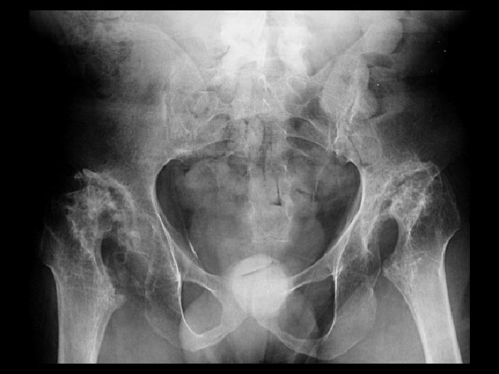
ESRD & amyloid arthropathy • Findings: – Osteopenia and fuzzy trabecula = osteomalacia – erosions on both sides of the hips joints – multiple joint involvement • ddx: (single joint) – synovial chondromatosis – PVNS
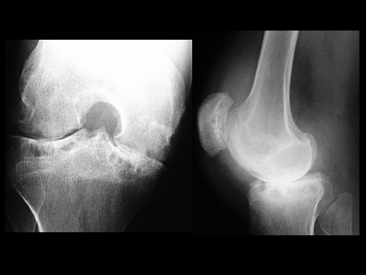

Hemophilia & secondary osteoarthritis • Findings: – severe secondary OA – flared metaphyses – widening of intercondylar tunnel – dense joint effusion • ddx: – JRA

Neouropathic hip joints & Spina bifida • Findings: – bilateral hip joint destruction with acetabular remodeling – subtle spinal dysraphism • ddx: – congenital hip dysplasia – bilateral femoral head AVN – bilateral septic arthritis (unlikely)

Ossifying non-ossifying fibroma • Findings: – cortical lucent lesion with a sharp margin and a thick sclerotic margin – no periosteal reaction or soft tissue mass • ddx: – fibrous dysplasia – EG – infection
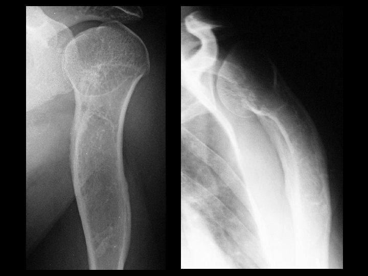

Fibrous dysplasia • Findings: – expansile, long lesion of the proximal humerus with bowing deformity – “ground glass matrix” • ddx: – NONE! – This is an Aunt Minnie!


Olier’s syndrome • Findings: – Multiple expansile lucent lesions of the wrist and digits • ddx: – polyostotic fibrous dysplasia

Tuft epidermoid • Findings: – very lucent round lesion in the tuft with a smooth sclerotic margin • ddx: – glomus tumor – giant cell of tendon sheath – simple cyst
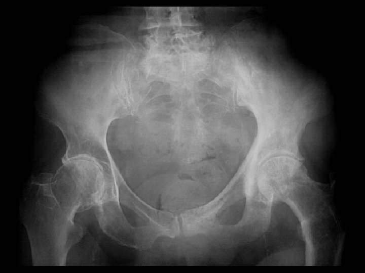

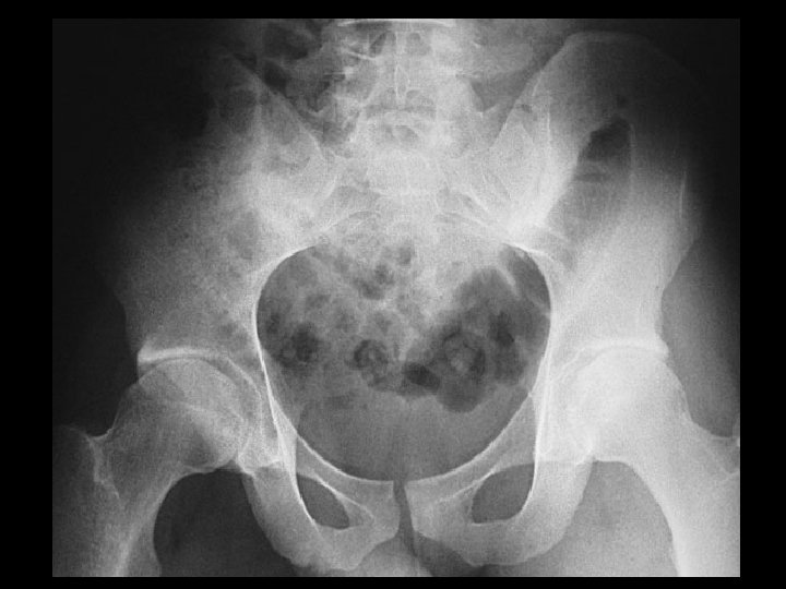
Paget’s disease • Findings: – trabecular and cortical thickening involving the left hemipelvis and proximal femur • ddx: – slcerotic mets (unlikely)

Osseous Calcaneonavicular coalition • Findings: – pes planus in a skeletally immature patient – enlongated anterior process of calcaneous = “anteater sign” – oblique view is diagnostic • ddx: – NONE! – This is an Aunt Minnie!
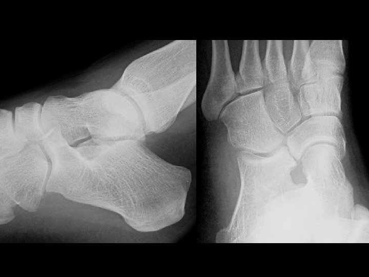

Fibrous Calcaneonavicular coalition • Findings: – pes planus in a skeletally immature patient – enlongated anterior process of calcaneous = “anteater sign” – oblique view is diagnostic • ddx: – NONE! – This is an Aunt Minnie!
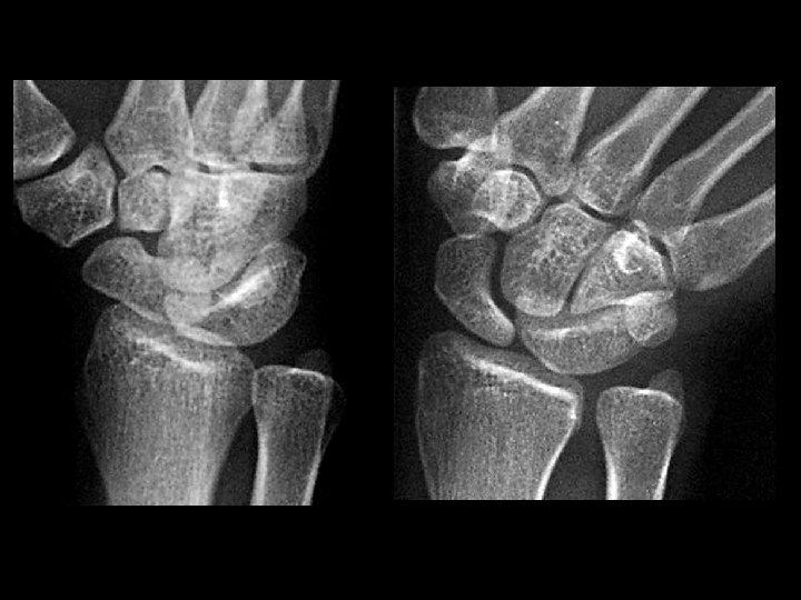
 • Findings: – fusion of the lunate and triquetrum • ddx:")
Carpal coalition (lunate-triquetrum) • Findings: – fusion of the lunate and triquetrum • ddx: – NONE! – This is an Aunt Minnie!

Arterial placement of central venous line • Findings: – central line from the right subclavian approach terminated to left of the spine just under the Ao arch • ddx: – extravascular (mediastinal) location

Right arch & aberrant left subclavian artery • Findings: – right-sided arch – anterior bowing of trachea by a soft tissue opacity • ddx: – double aortic arch – other vascular ring

Sickle cell anemia • Findings: – Cardiomegaly – dense bones – AVN of right humeral head – ? lack of spleen in LUQ • ddx: – NONE! – This is an Aunt Minnie!

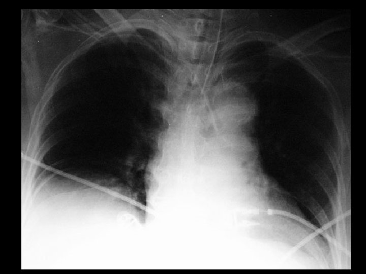
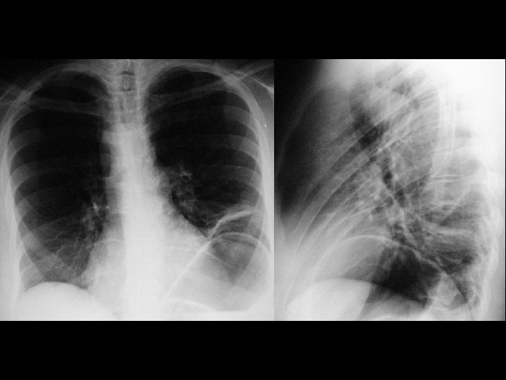
Tension pneumothorax • Findings: – hyperlucent right hemithorax – inverted right hemidiaphragm – leftward mediastinal shift • ddx: unilateral lucent lung) – Swyer-James syndrome – air-trapping from FB – oligemia (massive PE)
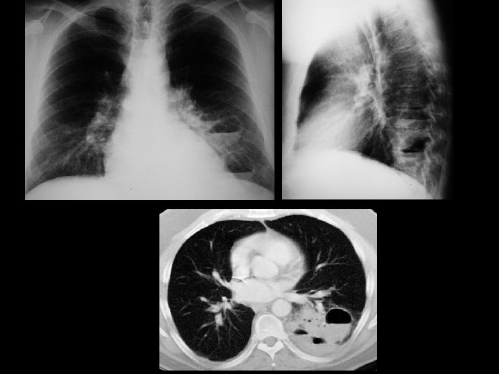

Pulmonary sequestration • Findings: – LLL consolidation with multiple cavities containing gas-fluid levels • ddx: – necrotizing pneumonia – bronchogenic carcinoma with necrosis
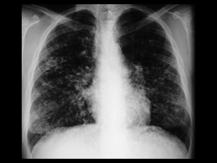

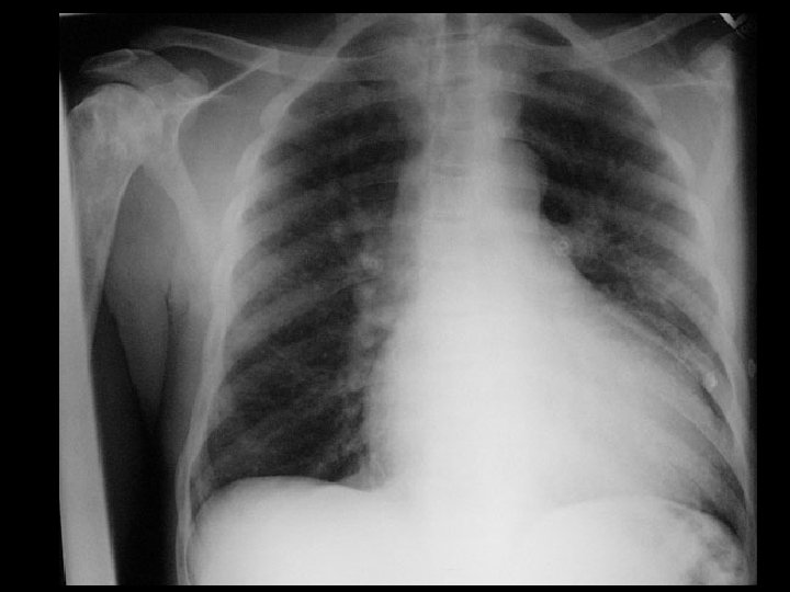
Cystic fibrosis • Findings: – hyperinflated lungs with diffuse, bilateral bronchiectasis • ddx: – immotile cilia syndrome – congenital immunodeficiency and recurrent infections

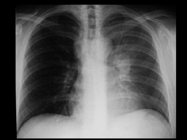
A 1 -antitrypsin • Findings: – Extremely hyperexpanded lungs with a basal predominance – enlarged central pulmonary arteries • ddx: – ritalin toxicity

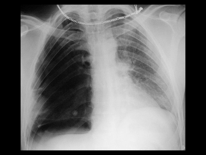
Left upper lobe collapse & central bronchogenic CA • Findings: – “veil-like” opacity of the left hemithorax – central left hilar mass – subtle signs of left-sided volume loss • ddx: – NONE! – This is an Aunt Minnie!
- Slides: 88