
Medical Complication in Pregnancy Associate Professor Doctor Atiwut









- Slides: 126
Medical Complication in Pregnancy Associate Professor Doctor Atiwut Kamudhamas Department of Obstetrics and Gynecology Faculty of Medicine Thammasat University
Contents • • Gestational diabetes mellitus Iron deficiency anemia Thalassemia Cardiovascular disease
Gestational Diabetes mellitus
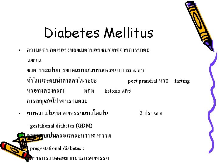
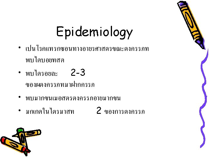
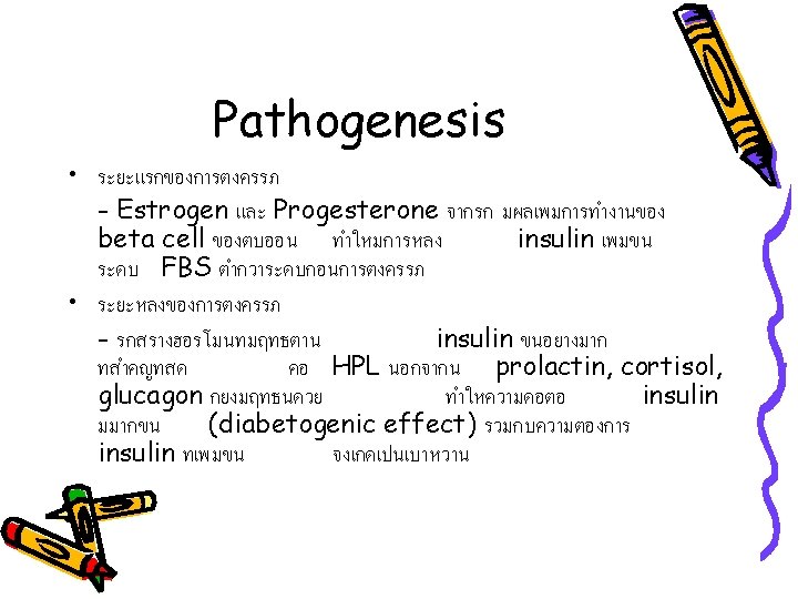
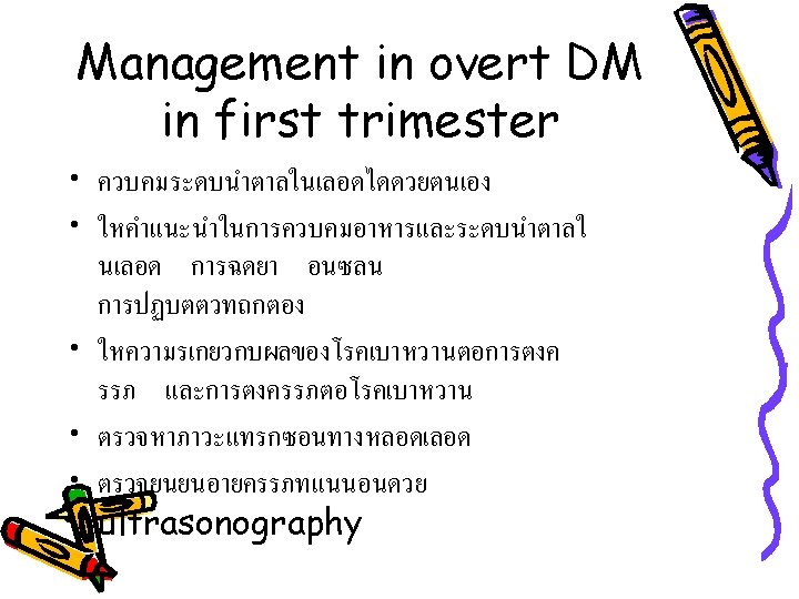
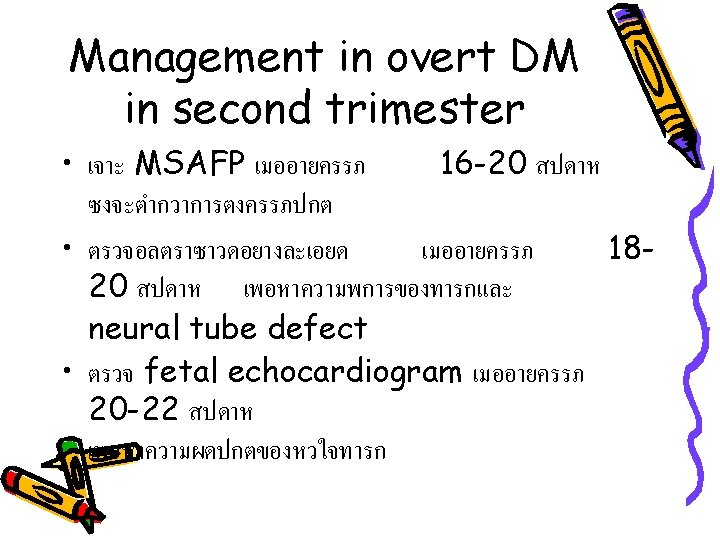
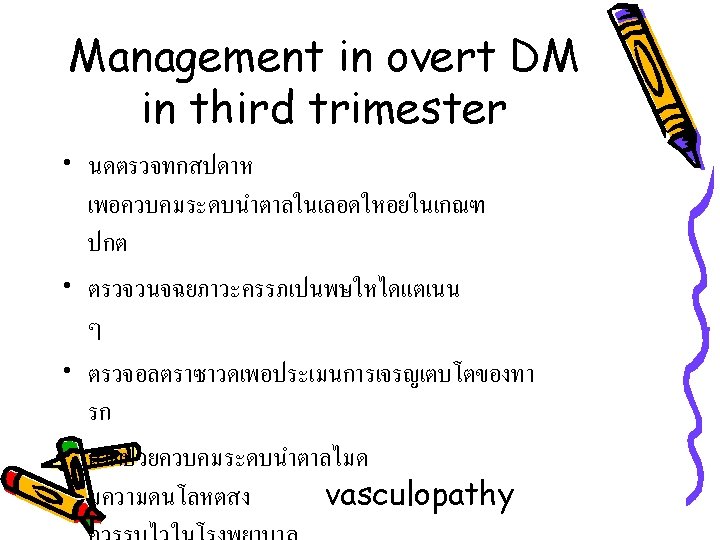
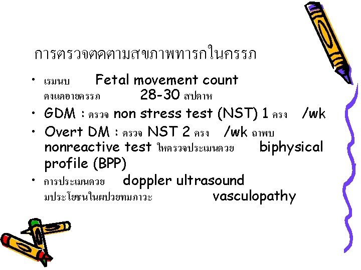
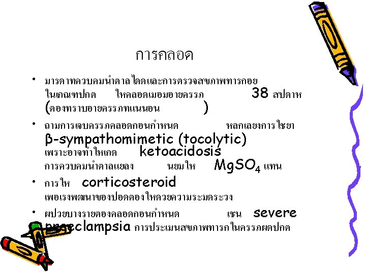
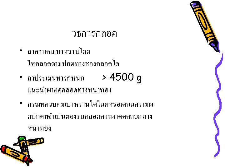
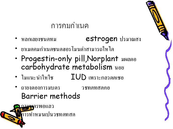
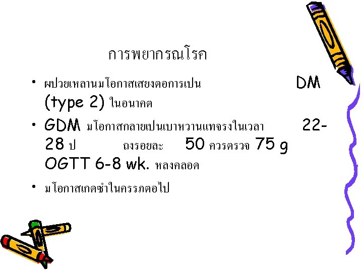
Outline • • Definition Epidemiology Pathogenesis Classification Screening test Risk Assessment for Detecting GDM Management in GDM
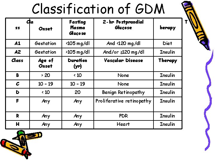
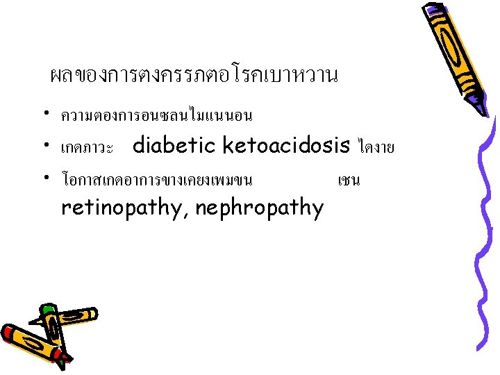
Classification of GDM Onset Fasting Plasma Glucose 2 -hr Postprandial Glucose herapy A 1 Gestation <105 mg/dl And <120 mg/dl Diet A 2 Gestation >105 mg/dl And/or ≥ 120 mg/dl Insulin Class Age of Onset Duration (yr) Vascular Disease Therapy B > 20 < 10 None Insulin C 10 – 19 None Insulin D < 10 20 Benign Retinopathy Insulin F Any Proliferative retinopathy Insulin R Any PDR Insulin H Any Heart Insulin ss Cla T
GDM • Class A 1 (chemical DM) พบเปนสวนใหญ • Class A 2 (overt DM) ม fasting hyperglycemia
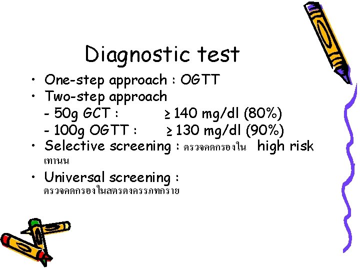
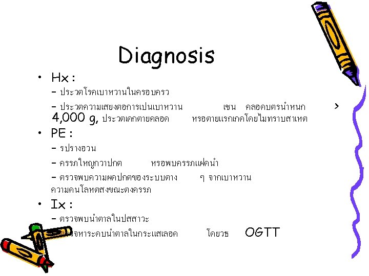
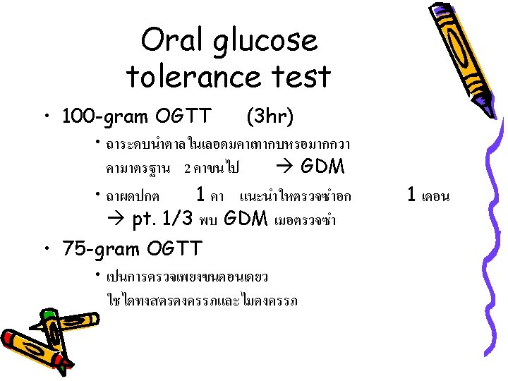
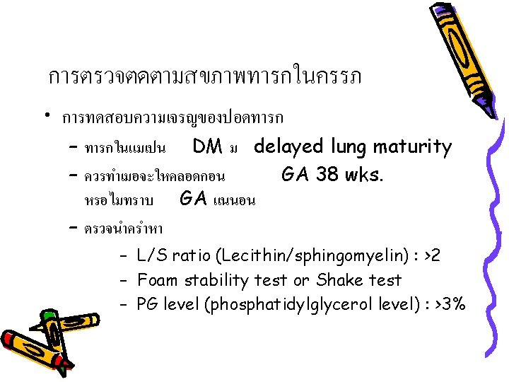
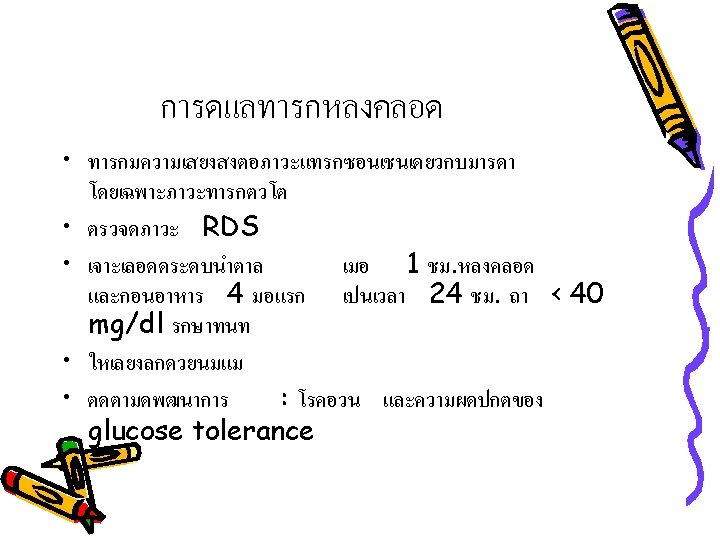
Diagnostic test • One-step approach : OGTT • Two-step approach - 50 g GCT : ≥ 140 mg/dl (80%) - 100 g OGTT : ≥ 130 mg/dl (90%) • Selective screening : ตรวจคดกรองใน high risk เทานน • Universal screening : ตรวจคดกรองในสตรตงครรภทกราย
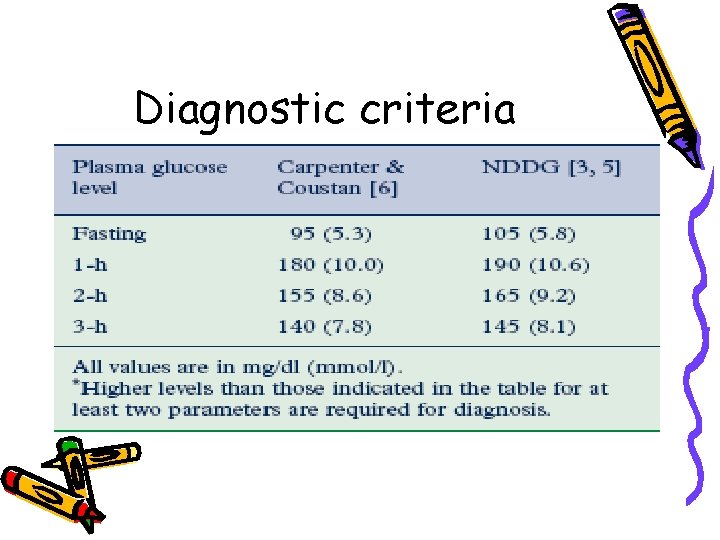
Diagnostic criteria
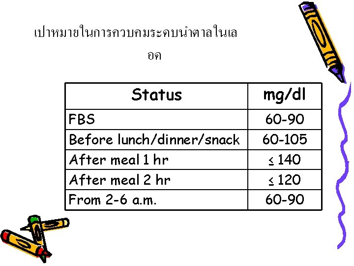
เปาหมายในการควบคมระดบนำตาลในเล อด Status mg/dl FBS Before lunch/dinner/snack After meal 1 hr After meal 2 hr From 2 -6 a. m. 60 -90 60 -105 ≤ 140 ≤ 120 60 -90
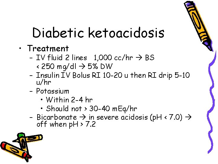
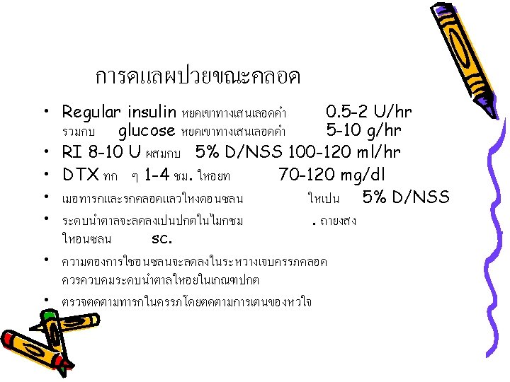
Diabetic ketoacidosis • Treatment – IV fluid 2 lines 1, 000 cc/hr BS < 250 mg/dl 5% DW – Insulin IV Bolus RI 10 -20 u then RI drip 5 -10 u/hr – Potassium • Within 2 -4 hr • Should not > 30 -40 m. Eq/hr – Bicarbonate in severe acidosis (p. H < 7. 0) off when p. H > 7. 2
Iron deficiency anemia
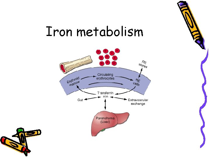
Iron metabolism
Iron contents in the body • Ferritin Storage form • Transferrin Transporting form • Hemoglobin, Myoglobin, Fe-containing enzymes
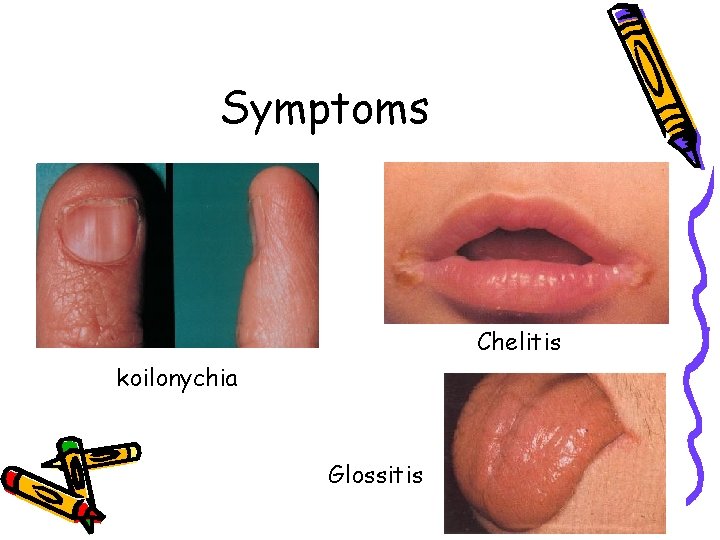
Iron-deficiency Anemia • Most common cause of anemia during pregnancy 75% • Signs : Fatique, Dyspnea, Headache, PICA • Symptoms : Pallor, Glossitis, Chelitis, Koilonychia
Symptoms Chelitis koilonychia Glossitis
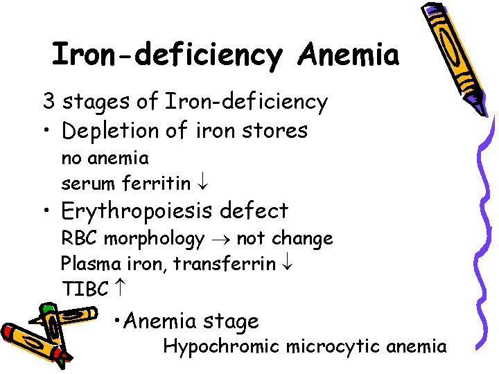
Iron-deficiency Anemia 3 stages of Iron-deficiency • Depletion of iron stores no anemia serum ferritin • Erythropoiesis defect RBC morphology not change Plasma iron, transferrin TIBC • Anemia stage Hypochromic microcytic anemia
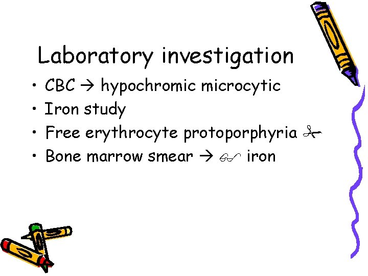
Laboratory investigation • • CBC hypochromic microcytic Iron study Free erythrocyte protoporphyria Bone marrow smear iron
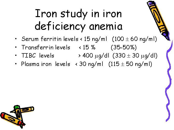
Iron study in iron deficiency anemia • • Serum ferritin levels < 15 ng/ml (100 60 ng/ml) Transferrin levels < 15 % (35 -50%) TIBC levels > 400 g/dl (330 30 g/dl) Plasma iron levels < 30 ng/ml (115 50 ng/ml)
Treatment • Oral administration • Parenteral administration
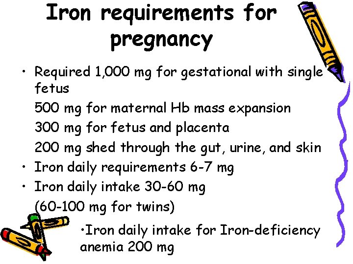
Iron requirements for pregnancy • Required 1, 000 mg for gestational with single fetus 500 mg for maternal Hb mass expansion 300 mg for fetus and placenta 200 mg shed through the gut, urine, and skin • Iron daily requirements 6 -7 mg • Iron daily intake 30 -60 mg (60 -100 mg for twins) • Iron daily intake for Iron-deficiency anemia 200 mg
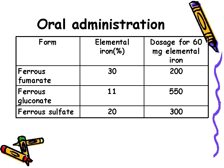
Oral administration Form Elemental iron(%) Ferrous fumarate 30 Dosage for 60 mg elemental iron 200 Ferrous gluconate 11 550 Ferrous sulfate 20 300
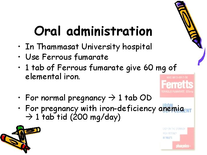
Oral administration • In Thammasat University hospital • Use Ferrous fumarate • 1 tab of Ferrous fumarate give 60 mg of elemental iron. • For normal pregnancy 1 tab OD • For pregnancy with iron-deficiency anemia 1 tab tid (200 mg/day)
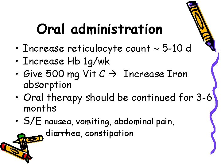
Oral administration • Increase reticulocyte count 5 -10 d • Increase Hb 1 g/wk • Give 500 mg Vit C Increase Iron absorption • Oral therapy should be continued for 3 -6 months • S/E nausea, vomiting, abdominal pain, diarrhea, constipation
Parenteral administration • IV or IM • Treat for woman can’t take oral iron preparations - malabsorption syndrome - intolerance to S/E • Hb < 8. 5 g/dl
Thalassemia
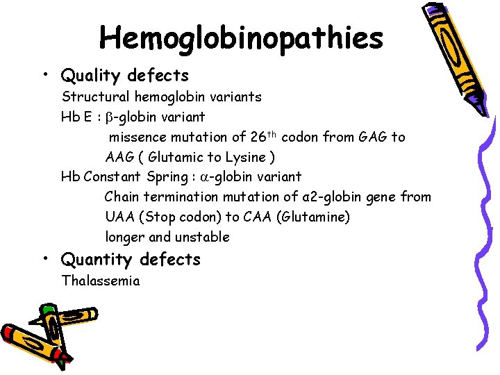
Hemoglobinopathies • Quality defects Structural hemoglobin variants Hb E : -globin variant missence mutation of 26 th codon from GAG to AAG ( Glutamic to Lysine ) Hb Constant Spring : -globin variant Chain termination mutation of α 2 -globin gene from UAA (Stop codon) to CAA (Glutamine) longer and unstable • Quantity defects Thalassemia
Definition • Thalassemia is caused by globin gene mutation which are characterized by impaired production of one or more of the normal globin peptide chains. • Abnormal synthesis rates may result in ineffective erythropoiesis, hemolysis, and varying degrees of anemia.
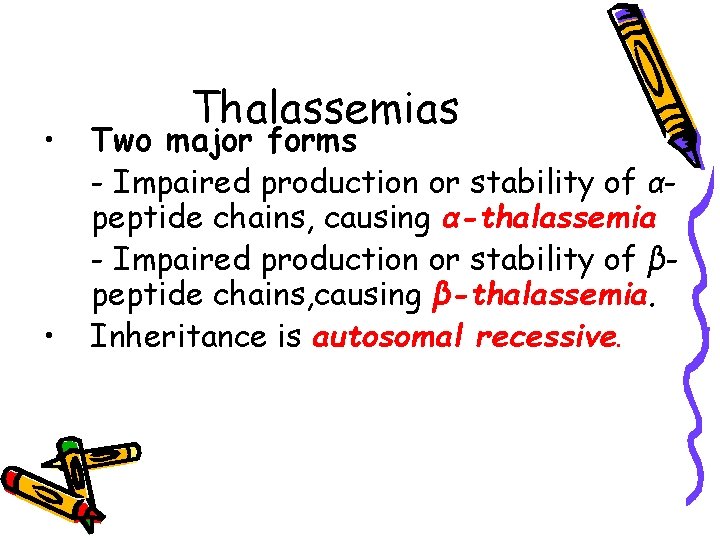
• • Thalassemias Two major forms - Impaired production or stability of αpeptide chains, causing α-thalassemia - Impaired production or stability of βpeptide chains, causing β-thalassemia. Inheritance is autosomal recessive.
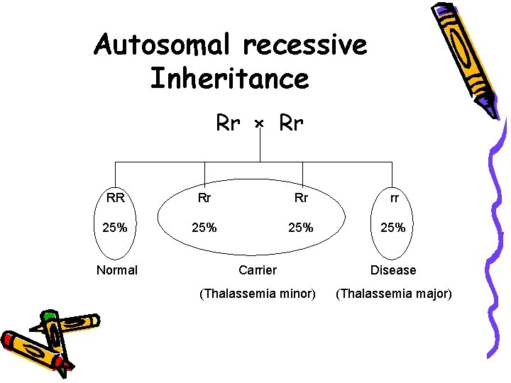
Autosomal recessive Inheritance Rr × Rr RR Rr Rr rr 25% 25% Normal Carrier Disease (Thalassemia minor) (Thalassemia major)
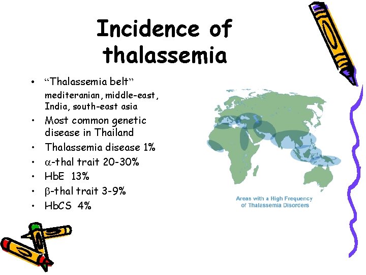
Incidence of thalassemia • “Thalassemia belt” mediteranian, middle-east, India, south-east asia • Most common genetic disease in Thailand • Thalassemia disease 1% • -thal trait 20 -30% • Hb. E 13% • -thal trait 3 -9% • Hb. CS 4%
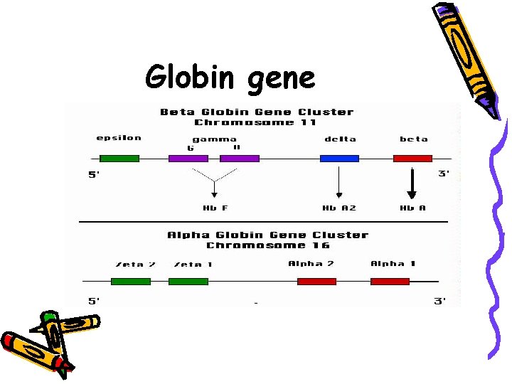
Globin gene
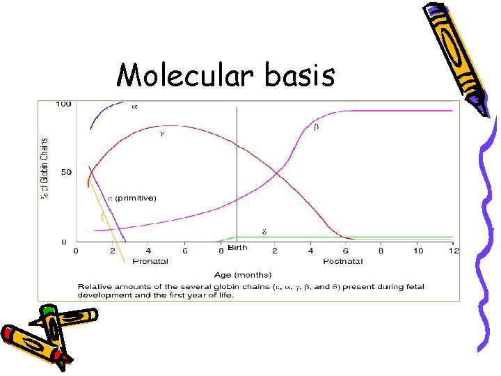
Molecular basis ζ
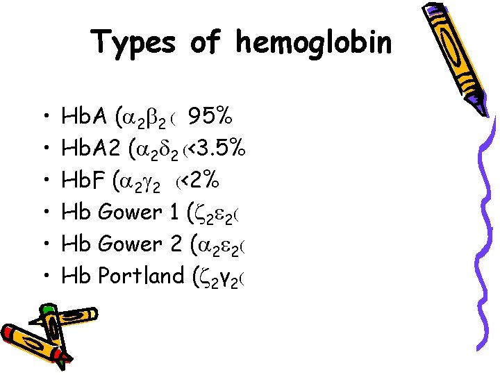
Types of hemoglobin • • • Hb. A ( 2 2 ( 95% Hb. A 2 ( 2 2 (<3. 5% Hb. F ( 2 2 (<2% Hb Gower 1 ( 2 2( Hb Gower 2 ( 2 2( Hb Portland ( 2γ 2(
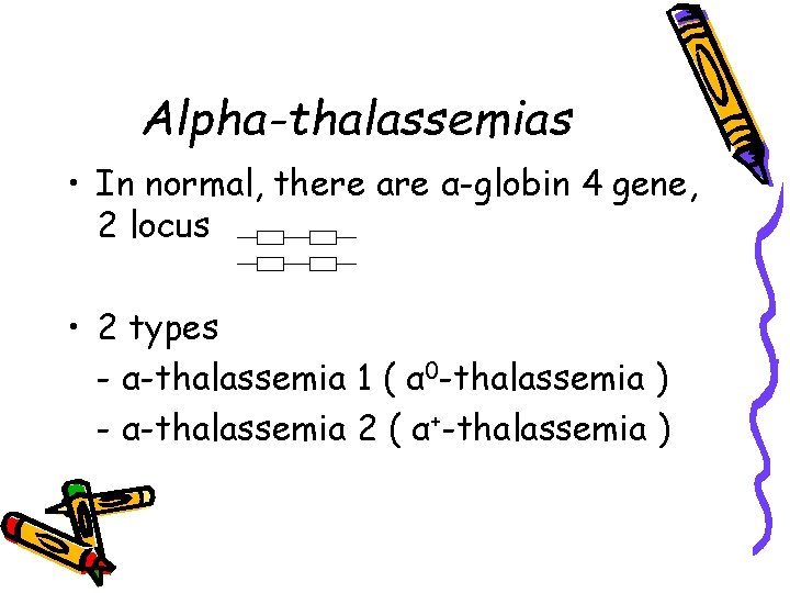
Alpha-thalassemias • In normal, there are α-globin 4 gene, 2 locus • 2 types - α-thalassemia 1 ( α 0 -thalassemia ) - α-thalassemia 2 ( α+-thalassemia )
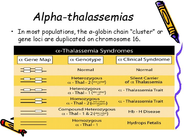
Alpha-thalassemias • In most populations, the α-globin chain “cluster” or gene loci are duplicated on chromosome 16.
-Thalassemia • 0 - Thalassemia : β 0 thal , no -globin protein • +- Thalassemia : β+thal , some -globin protein • Mechanism of gene mutation Point mutation
Most common Thalassemia in Thailand 1. 2. 3. 4. 5. Hb Bart’s hydrops fetalis Homozygous -thalassemia/Hb. E Hb H disease Homozygous Hb Constant Spring
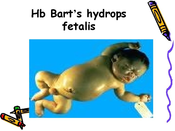
Hb Bart’s hydrops fetalis • Deletion of all four -globin chain genes (--/--) characterizes homozygous α-thalassemia 1 • Genetic from maternal and paternal (--/ ( • Without -globin chains hemoglobin Bart ( 4 ( • Fetus is affected Severe anemia, lack of O 2, heart failure, hydrops fetalis • Maternal complications : Preeclampsia, Pre and Post-partum hemorrhage, dystonia • Fetal blood sampling found Hb Bart’s (γ 4) (Normal fetus : Hb. F (α 2γ 2) )
Hb Bart’s hydrops fetalis
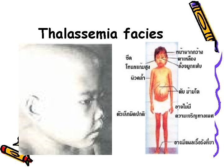
Homozygous -Thalassemia • • -thalassemia major or Cooley anemia At birth Hb. F ↑ no symptom -globin precipitate into RBC lysis lead to severe anemia (Hb 3 -4 g/dl), Hepatosplenomegaly, jaundice, slow growth and bone change (Thalassemia facies) • Blood transfusion + Iron chelator • Complication : congestive heart failure cirrhosis DM Sterility
-thalassemia/Hb. E • Variation in degree of anemia ( moderate to severe ) • Hb 2. 5 - 12. 5 g/d. L • Chronic anemia • Thalassemia facies • Hepatosplenomegaly • Complication : Respiratory tract infections Congestive heart failure Epilepsy Amenorrhea Infertile
Hb. H disease • Mild to moderate severe. • Not necessary to have blood transfusion. • If patients have infection, acute hemolysis and anemia may be occurred rapidly.
Homozygous Hb Constant Spring • Mild severe • Not necessary to have blood transfusion
Thalassemia facies
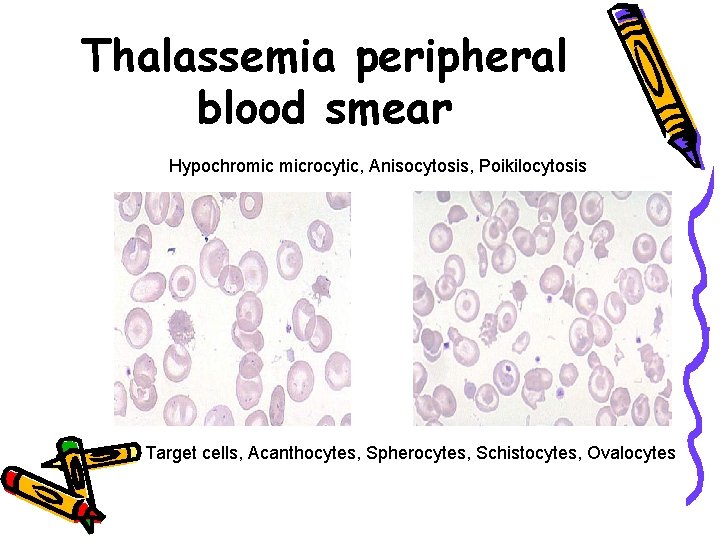
Thalassemia peripheral blood smear Hypochromic microcytic, Anisocytosis, Poikilocytosis Target cells, Acanthocytes, Spherocytes, Schistocytes, Ovalocytes
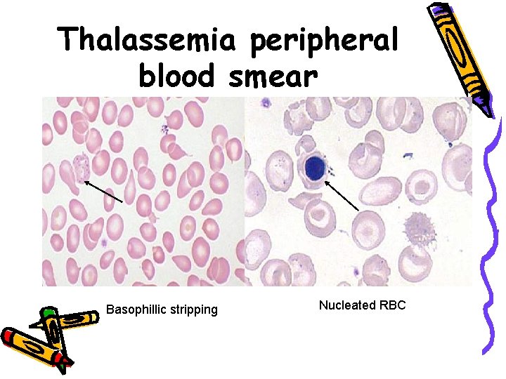
Thalassemia peripheral blood smear Basophillic stripping Nucleated RBC
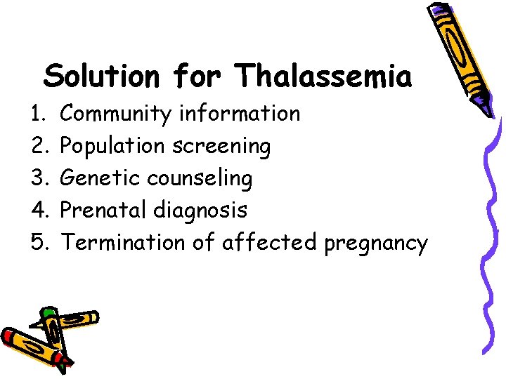
Solution for Thalassemia 1. 2. 3. 4. 5. Community information Population screening Genetic counseling Prenatal diagnosis Termination of affected pregnancy
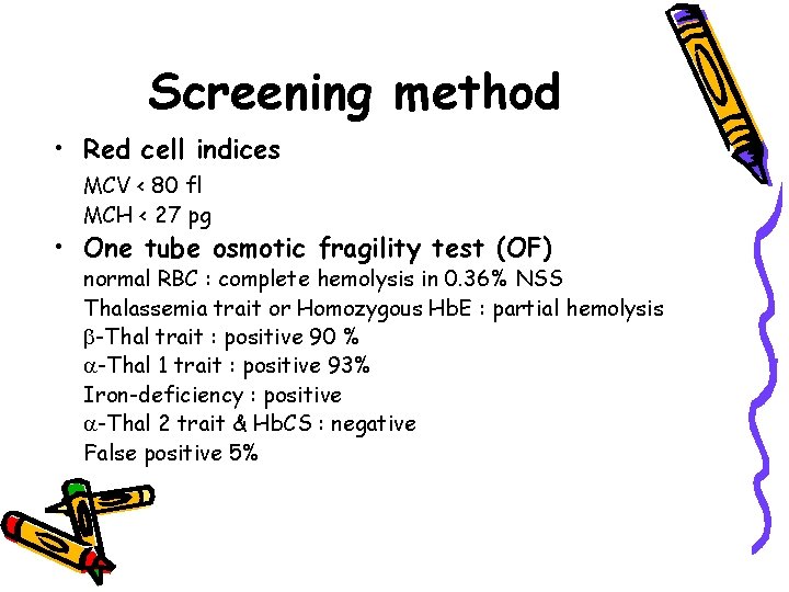
Screening method • Red cell indices MCV < 80 fl MCH < 27 pg • One tube osmotic fragility test (OF) normal RBC : complete hemolysis in 0. 36% NSS Thalassemia trait or Homozygous Hb. E : partial hemolysis -Thal trait : positive 90 % -Thal 1 trait : positive 93% Iron-deficiency : positive -Thal 2 trait & Hb. CS : negative False positive 5%
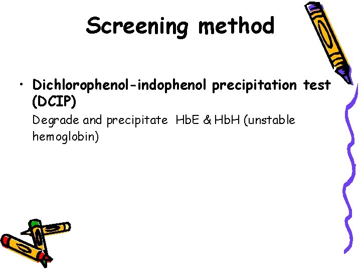
Screening method • Dichlorophenol-indophenol precipitation test (DCIP) Degrade and precipitate Hb. E & Hb. H (unstable hemoglobin)
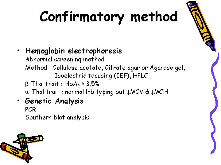
Confirmatory method • Hemoglobin electrophoresis Abnormal screening method Method : Cellulose acetate, Citrate agar or Agarose gel, Isoelectric focusing (IEF), HPLC -Thal trait : Hb. A 2 > 3. 5% -Thal trait : normal Hb typing but ↓MCV & ↓MCH • Genetic Analysis PCR Southern blot analysis
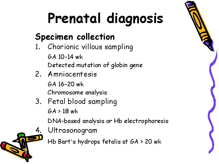
Prenatal diagnosis Specimen collection 1. Chorionic villous sampling GA 10 -14 wk Detected mutation of globin gene 2. Amniocentesis GA 16 -20 wk Chromosome analysis 3. Fetal blood sampling GA > 18 wk DNA-based analysis or Hb electrophoresis 4. Ultrasonogram Hb Bart’s hydrops fetalis at GA > 20 wk
Perinatal care • • • Maintain Hb level between 7 – 10 g/dl Blood transfusion Iron chelating Folate supplement Terminate in hydrops fetalis Fetal health assessment
Cardiovascular disease in pregnancy
Outline • Physiological conditions with heart disease in pregnancy • Diagnosis of heart disease • General management
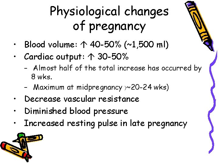
Physiological changes of pregnancy • Blood volume: 40 -50% (~1, 500 ml) • Cardiac output: 30 -50% – Almost half of the total increase has occurred by 8 wks. – Maximum at midpregnancy )~20 -24 wks) • Decrease vascular resistance • Diminished blood pressure • Increased resting pulse in late pregnancy
Effect of pregnancy to heart disease • • Problem on diagnosis Increase severity of heart disease Recurrence of rheumatic fever Cardiomyopathy
Effect of heart disease to pregnancy • • Spontaneous abortion, preterm Increased rate of DFIU Increased IUGR Increased rate of congenital heart disease in child • Increased rate of therapeutic abortion
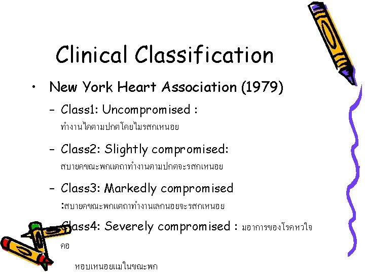
Diagnosis of heart disease • Diagnostic studies • Clinical classification
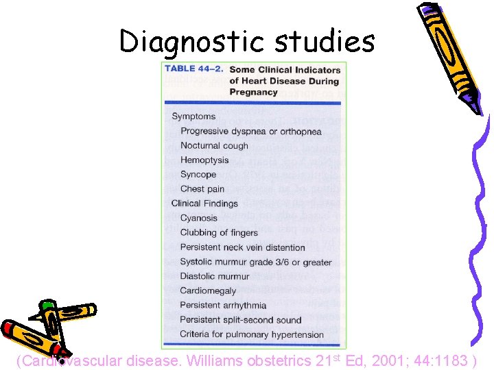
Diagnostic studies (Cardiovascular disease. Williams obstetrics 21 st Ed, 2001; 44: 1183 )
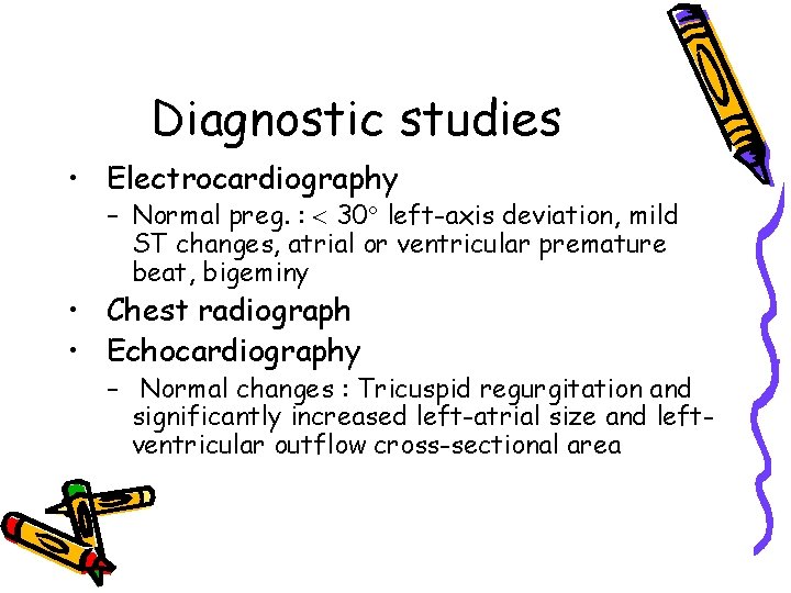
Diagnostic studies • Electrocardiography – Normal preg. : 30 left-axis deviation, mild ST changes, atrial or ventricular premature beat, bigeminy • Chest radiograph • Echocardiography – Normal changes : Tricuspid regurgitation and significantly increased left-atrial size and leftventricular outflow cross-sectional area
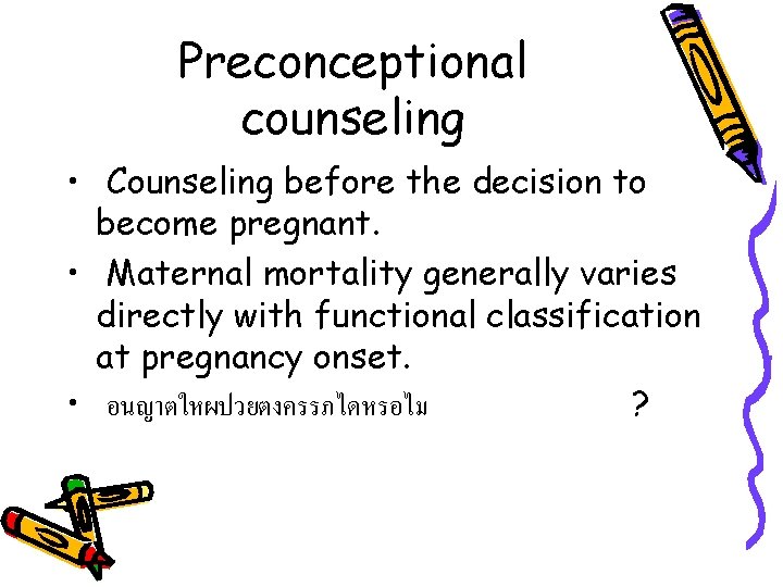
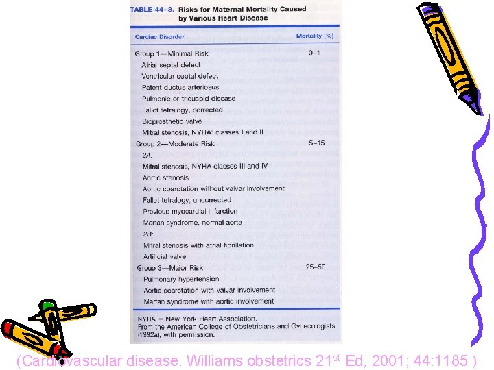
Preconceptional counseling • Counseling before the decision to become pregnant. • Maternal mortality generally varies directly with functional classification at pregnancy onset. • อนญาตใหผปวยตงครรภไดหรอไม ?
(Cardiovascular disease. Williams obstetrics 21 st Ed, 2001; 44: 1185 )
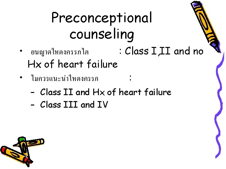
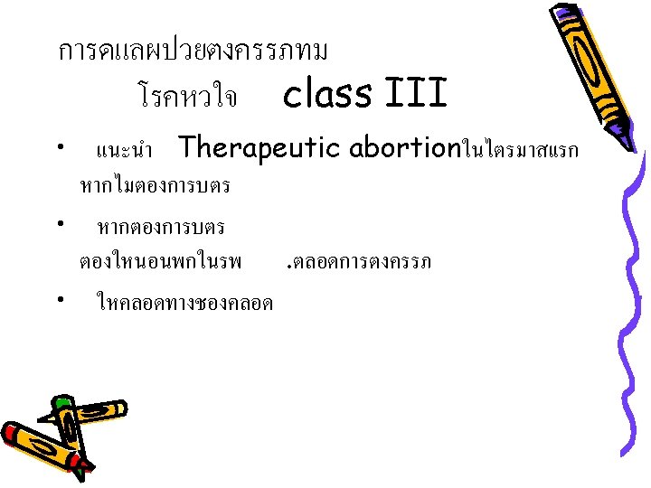
Preconceptional counseling • อนญาตใหตงครรภได : Class I, II and no Hx of heart failure • ไมควรแนะนำใหตงครรภ : – Class II and Hx of heart failure – Class III and IV
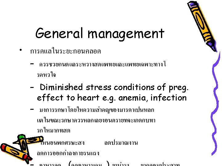
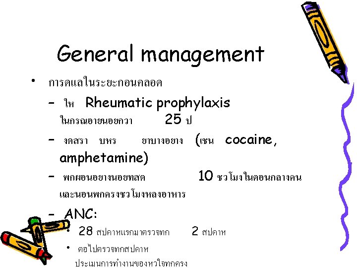
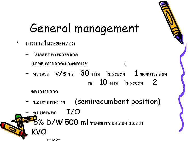
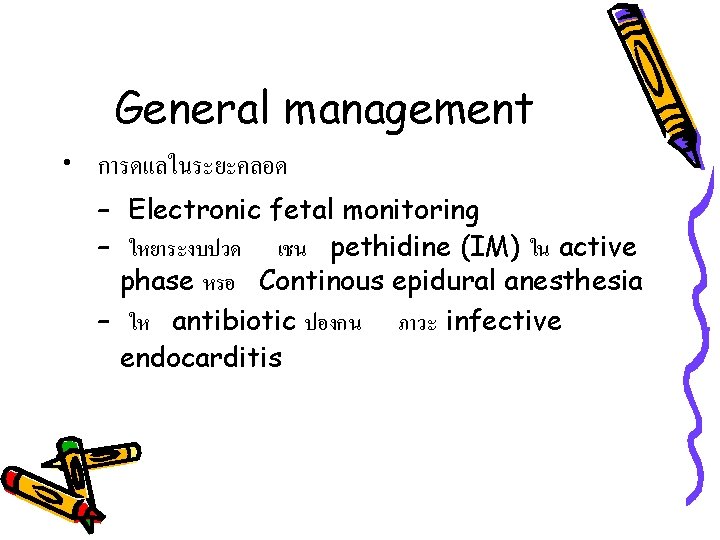
General management • การดแลในระยะคลอด – Electronic fetal monitoring – ใหยาระงบปวด เชน pethidine (IM) ใน active phase หรอ Continous epidural anesthesia – ให antibiotic ปองกน ภาวะ infective endocarditis
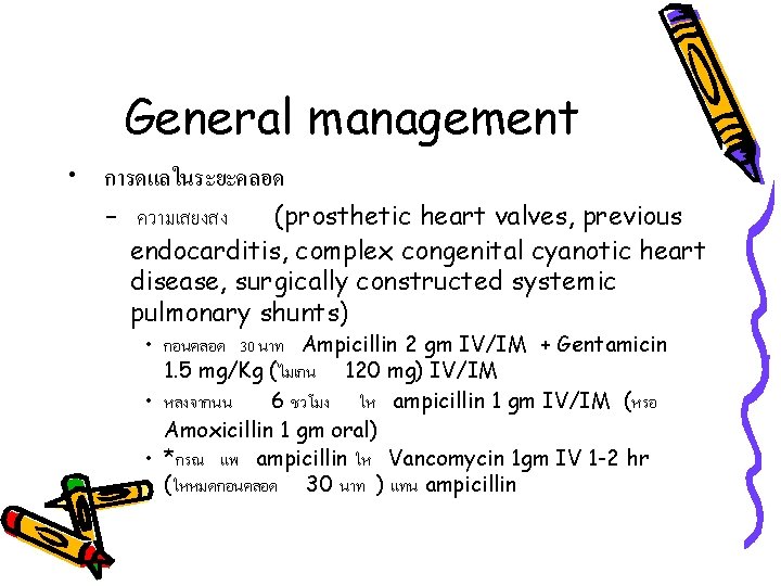
General management • การดแลในระยะคลอด – ความเสยงสง (prosthetic heart valves, previous endocarditis, complex congenital cyanotic heart disease, surgically constructed systemic pulmonary shunts) • กอนคลอด 30 นาท Ampicillin 2 gm IV/IM + Gentamicin 1. 5 mg/Kg (ไมเกน 120 mg) IV/IM • หลงจากนน 6 ชวโมง ให ampicillin 1 gm IV/IM (หรอ Amoxicillin 1 gm oral) • *กรณ แพ ampicillin ให Vancomycin 1 gm IV 1 -2 hr (ใหหมดกอนคลอด 30 นาท ) แทน ampicillin
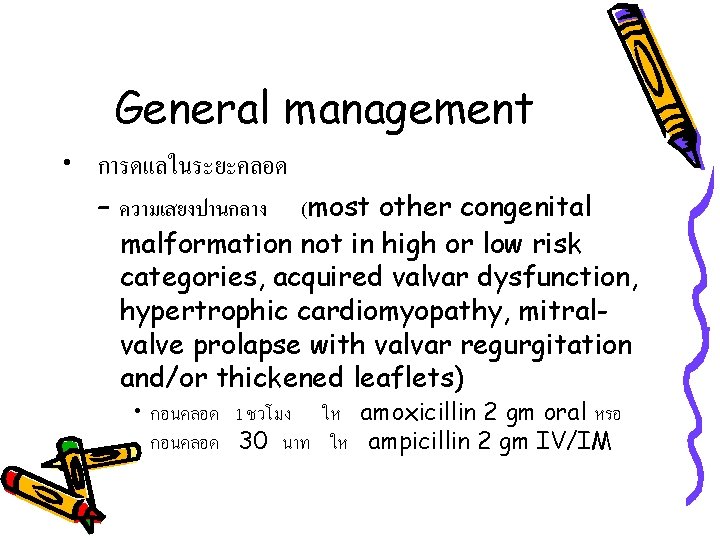
General management • การดแลในระยะคลอด – ความเสยงปานกลาง (most other congenital malformation not in high or low risk categories, acquired valvar dysfunction, hypertrophic cardiomyopathy, mitralvalve prolapse with valvar regurgitation and/or thickened leaflets) • กอนคลอด 1 ชวโมง ให amoxicillin 2 gm oral หรอ กอนคลอด 30 นาท ให ampicillin 2 gm IV/IM
General management • การดแลในระยะคลอด – ความเสยงตำ (ASD, surgically corrected lesions without prosthesis (ASD, VSD, PDA), coronary artery disease with previous bypass surgery, mitral-valve prolapse without regurgitation, physiological murmurs, previous rheumatic fever without valvar dysfunction, pacemakers) • ไมให antibiotic prophylaxis
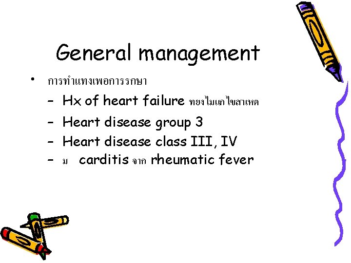
General management • การทำแทงเพอการรกษา – Hx of heart failure ทยงไมแกไขสาเหต – Heart disease group 3 – Heart disease class III, IV – ม carditis จาก rheumatic fever
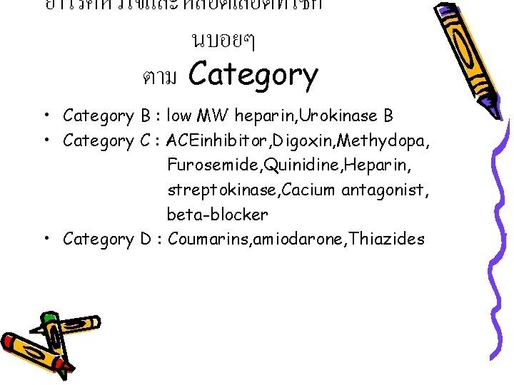
ยาโรคหวใจและหลอดเลอดทใชก นบอยๆ ตาม Category • Category B : low MW heparin, Urokinase B • Category C : ACEinhibitor, Digoxin, Methydopa, Furosemide, Quinidine, Heparin, streptokinase, Cacium antagonist, beta-blocker • Category D : Coumarins, amiodarone, Thiazides
References • ธระ ทองสง, ชเนนทร วนาภรกษ , บรรณาธการ. สตศาสตร. พมพครงท 4. เชยงใหมฯ : พ บ ฟอเรน บคส , 2541. • Cunningham FG, Gant NF, Lenovo KJ, Gilstrap III LC, Hauth JC, Wenstrom KD, eds. William obstertrics. 22 nd ed. New York : Mc. Graw-Hill, Inc. , 2005.
Thank you for your attention