Shoulder Region Dr S Nishan Silva MBBS Why
")
Shoulder Region Dr. S. Nishan Silva (MBBS)

Why? ? ?


Shoulder Anatomy Bones Clavicle Scapula Humerus Shoulder vs Shoulder Girdle

Shoulder Anatomy Joints Sternoclavicular

Shoulder Anatomy Joints Sternoclavicular Acromioclavicular

Distal Clavicle � Coracoclavicular ligaments “Suspensory ligaments of the upper extremity” Two components: ▪ Trapezoid ▪ Conoid Stronger than AC ligaments Provide vertical stability to AC joint

Shoulder Anatomy Joints Sternoclavicular Acromioclavicular Glenohumeral

Glenohumeral Joint Most common dislocated joint Lacks bony stability Composed of: Fibrous capsule Ligaments Surrounding muscles Glenoid labrum

Shoulder Anatomy Ligaments Acromioclavicular Joint ▪ Acromioclavicular Ligament

Shoulder Anatomy Ligaments Glenohumeral Joint ▪ Glenohumeral ligaments ▪ Superior ▪ Middle ▪ Inferior

Glenohumeral joint 25% humeral head surface in contact with glenoid Humeral head coverage increased to 75% with glenoid labrum

Shoulder Anatomy Cartilage Glenoid labrum

Glenoid Labrum

Radiographic Anatomy

Shoulder Anatomy Shoulder Girdle Muscles Trapezius

Major muscles of the trunk Trapezius Origin: superior nuchal line, external occipital protuberance, ligamentum nuchae and spinous processes of seventh cervical and all thoracic vertebrae Insertion: lateral third of clavicle, acromion, and spine of scapulartery Acton: upper fibers elevate scapula, lower fibers depress scapula; if scapula is fixed, one side acting along, draws head toward the same side, and turn face to opposite side; both sides together, draw head directly backward

Major muscles of the trunk Latissimus dorsi Origin: spinous processes of lower six thoracic and all lumbar vertebrae, median sacral crest, and posterior part of iliac crest. Insertion: floor of intertubercular groove of humerus. Action: trunk fixed, extends, adducts and medially rotates arm ; arm fixed, elevates trunk.

Shoulder Anatomy Shoulder Girdle Muscles Trapezius Serratus Anterior

Serratus Anterior

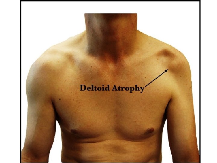
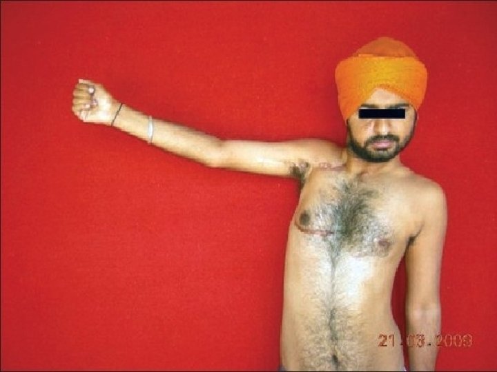
Winged Scapula

Shoulder Anatomy Glenohumeral Muscles Rotator Cuff ▪ ▪ Suprispinatus Infraspinatus Teres Minor Subscapularis

Rotator cuff �Subscapularis �Supraspinatus �Infraspinatus �Teres minor

Rotator cuff muscles � Supraspinatus, infraspinatus, teres minor, subscapularis � Form cuff around humeral head � Keep humeral head within joint (counteract deltoid) � Abduction, external rotation, internal rotation

Movement of RC Muscles � Subscapularis is an internal rotator of the arm. � Supraspinatus assists the deltoid in abducting the arm, with its greatest contribution being the initiation of abduction. � Infraspinatus and teres minor muscles both externally rotate the arm.

Supraspinatus “empty can sign”

Teres major Origin: dorsal surface of inferior angle of scapula Insertion: crest of lesser tubercle of humerus Action: medially rotates and adducts arm

Shoulder Anatomy Glenohumeral Muscles Latissimus Dorsi Pectoralis Major

Pectoralis major Origin: medial half of clavicle, sternum, 1 th-6 th costal cartilages. Insertion: crest of greater tubercle of humerus. Action: flexes, adducts and rotates arm medially; arm fixed, elevates trunk; elevates ribs 1 -6, aidding in forced inspiration.

Shoulder Anatomy Glenohumeral Muscles Latissimus Dorsi Pectoralis Major Deltoid

Major muscles of upper limb Deltoid Origin: lateral third of clavicle, acromion, and spine of scapula Insertion: deltoid tuberosity of humerus Action: abducts,flexes and medically rotates, extends, and laterally rotates arm

Shoulder Anatomy Glenohumeral Muscles Latissimus Dorsi Pectoralis Major Deltoid Biceps

Biceps strength testing Arms outstretched with palms up at level of shoulder Forced supination of hand with elbow flexed at 90 degrees

Shoulder Anatomy Glenohumeral Muscles Latissimus Dorsi Pectoralis Major Deltoid Biceps Triceps

Shoulder Anatomy Other structures Brachial Plexus Brachial Artery

Brachial plexus Formation: Five roots: formed by anterior rami of C 5 -C 8 and T 1 spinal nerves, roots C 5~C 7 give rise to long thoracic n. Three trunks The upper trunk is formed by the joining of root C 4, C 5, C 6. The middle trunk is the continuation of root C 7. The lower trunk is formed by the joining of root C 8 and T 1. Six divisions: above clavicle, trunks form anterior and posterior divisions Three cords: below clavicle, divisions form three cords that surround the second portion of axillary a.

Position: passes through the scalene fissure to posterosuperior of subclavian artery, then enters the axilla to form lateral, medial and posterior cords Main branches Lateral cord Musculocutaneous n. Lateral root to median n. Medial cord Medial root to median n. Ulnar n. Medial brachial cutaneous n. Medial antebrachial cutaneous n.

Posterior cord radial n. axillary n. thoracodorsal n.

Axillary artery Continuation of subclavian artery at lateral border of first rib Becomes brachial artery at lower border of teres major Divided into three parts by overlying pectoralis minor First portion, above muscle-gives rise to thoracoacromial a. Second portion, behind muscle-gives rise to lateral thoracic a. Third portion, below muscle-gives rise to subscapular a. anterior and posterior humeral circumflex a. ; the former then divides into throcodorsal a. and circumflex scapular a.

Axillary a. Thoracoacromial a. Lateral pectoral n. Musculocutaneous n. Medial antebrachial cutaneous n. Median n. Ulnar n. Medial brachial cutaneous n. Intercostobrachial n. Thoracodorsal n. & a. Long thoracic n. & lateral thoracic a.

Integrated Action
 Corachobrachialis Deltoid (anterior) Name")
Practice Name the muscles for Horizontal Adduction Pect Major (both) Corachobrachialis Deltoid (anterior) Name the muscles for Horizontal Abduction Deltoid (post) Infraspinatus Teres minor Lats

Practice List the muscles that do flexion of the shoulder Coracobrachialis Pectoralis major (upper to 60°) Anterior Deltoid • List the muscles that do extension of the shoulder • Latissimus dorsi • Teres major • Posterior deltoid • Pectoralis major (lower fibers to neutral)

Practice List the muscles that do adduction of the adduction shoulder Pectoralis major (lower and upper below 90°) Coracobrachialis Latissimus dorsi Teres major List the muscles that do abduction of the shoulder • Deltoid (all sections) • Supraspinatus • Pectoralis major (upper past 90°)

Practice List the muscles that do internal rotation of the shoulder Subscapularis Latissimus dorsi Teres major Anterior deltoid Pect. major • List the muscles that do external rotation of the shoulder • Infraspinatus • Teres minor • Posterior deltoid

Name the muscle. Coracobrachialis Name the action Adduction of the shoulder Also, flexion and hor. add.

Name the muscle. Subscapularis Name the action Internal rotation of the shoulder

Name the muscle. Deltoid Name the action Abduction of shoulder

Name the muscle. Infraspinatus Name the action External rotation

Name the muscle. Name the action Teres Major Adduction of scapula

Name the muscle. Teres Minor Name the action if the humerus move directly to the posterior Extension of the shoulder

Name the muscle. Supraspinatus Name the action Abduction of the shoulder

Coracobrachialis Subscapularis Pect. Major Deltoid

Supraspinatus Teres Major Infraspinatus Teres Minor

What position are her shoulders in? Flexion

What position is his right shoulder in? Horizontal Abduction and External Rotation

What rotation action is his shoulder performing as he continues to through the ball? Internal Rotation

What position are her shoulders in? Flexion

What position are his shoulders in? Horizontal abduction or Extension

Position of their shoulders? 1. Flexion 2. Extension

What is the position of shoulders? Extension

Major Muscles of the Shoulder Pectoralis major Latissimus dorsi Push-ups Chinning Pull-ups Robe climb Bench press Dips on parallel bars Throwing Pullover exercises Tennis serve Pulldown exercises Rowing
 = ? Abduction Deltoid Supraspinatus")
Shoulder action = ? Shoulder muscle(s) = ? Abduction Deltoid Supraspinatus
 = ? Flexion Ant Deltoid Upper Pect Major")
Shoulder action = ? Shoulder muscle(s) = ? Flexion Ant Deltoid Upper Pect Major Coracobrach.
 = ? Horizontal Add. Ant. Deltoid Pect. Major")
Shoulder action = ? Shoulder muscle(s) = ? Horizontal Add. Ant. Deltoid Pect. Major (both) Coracobrachialis
 = ? Horizontal Abduction Latissimus Dorsi Post. Deltoid")
Shoulder action = ? Shoulder muscle(s) = ? Horizontal Abduction Latissimus Dorsi Post. Deltoid Teres Minor Infraspinatus
 = ? Adduction Pect. Major (both) Coracobrachialis Latissimus")
Shoulder action = ? Shoulder muscle(s) = ? Adduction Pect. Major (both) Coracobrachialis Latissimus Dorsi Teres Major
 = ? Horizontal Add Ant. Deltoid Pect. Major")
Shoulder action = ? Shoulder muscle(s) = ? Horizontal Add Ant. Deltoid Pect. Major (both) Coracobrachialis
 = ? Extension Lats Post. Deltoid Infraspinatus Teres")
Shoulder action = ? Shoulder muscle(s) = ? Extension Lats Post. Deltoid Infraspinatus Teres Major Pectoralis Major (lower) Teres minor
 = ? External Rotation Infrspinatus Teres Minor Post.")
Shoulder action = ? Shoulder muscle(s) = ? External Rotation Infrspinatus Teres Minor Post. Deltoid

Name a shoulder muscle isolated with the following exercises. �Side arm dumbbell raises �Deltoid �Push-ups �Pectoralis major �Rowing and pull-overs �Latissimus dorsi

What is the action to the left? What muscles perform that action? Internal Rotation External Rotation Internal Rotation Subscapularis, Ant. Deltoid, Pect, Major, Lats. And Teres Major
")
Rotator Cuff Exercises External Rotation Internal Rotation External Rotation Abduction (to work the supraspinatus)

Shoulder Dysfunction and Injury �Factors Predisposing to Shoulder Pain Instability of glenohumeral joint Weakness in scapular stabilizing muscles Previous injury (dislocation of glenohumeral joint, separation of AC joint) Hypomobility of cervical or thoracic spine Postural dysfunction Muscle imbalances
 � Differentiation of Shoulder Pain Active inflammation: pain that")
Shoulder Dysfunction and Injury (cont’d) � Differentiation of Shoulder Pain Active inflammation: pain that occurs or increases at night Irritation of a sensory nerve root: sharp pain, numbing, & tingling in a dermatome Rotator cuff injury: pain at lateral portion of upper arm, painful limitation when elevating arm overhead Bicipital tendinitis: well-localized pain at anterior portion of head of humerus & aggravation with Speed’s test Adhesive capsulitis: stiffness in shoulder, dramatic loss of arm motion (especially external rotation)
 �Differentiation of Shoulder Pain Impingement: pain over anterior humerus,")
Shoulder Dysfunction and Injury (cont’d) �Differentiation of Shoulder Pain Impingement: pain over anterior humerus, loss of internal rotation, & painful Neer’s test Instability: clunking in shoulder with active circumduction & excessive joint play in passive motion test for glenohumeral joint Pain originating in glenohumeral joint: rarely felt at joint, but over lateral brachial region
 �Characteristics of Shoulder Pain (vs. neck pain) Elicited or")
Shoulder Dysfunction and Injury (cont’d) �Characteristics of Shoulder Pain (vs. neck pain) Elicited or increased from active shoulder motion & relieved by rest Isometric challenge will be painful with localized lesion Painless weakness in arm & shoulder muscles from motor nerve root problem in cervical spine
 �Common Dysfunctions and Injuries of the Shoulder Rotator cuff")
Shoulder Dysfunction and Injury (cont’d) �Common Dysfunctions and Injuries of the Shoulder Rotator cuff tendinitis (supraspinatus tendinitis) Infraspinatus tendinitis Subscapularis tendinitis Adhesive capsulitis (frozen shoulder) Impingement syndrome Instability syndrome of the glenohumeral joint
 � Common Dysfunctions and Injuries of the Shoulder Bicipital")
Shoulder Dysfunction and Injury (cont’d) � Common Dysfunctions and Injuries of the Shoulder Bicipital tendinitis Subacromial (subdeltoid) bursitis Acromioclavicular ligament sprain Suprascapular nerve entrapment Costoclavicular syndrome (part of thoracic outlet syndrome) Pectoralis minor syndrome (part of thoracic outlet syndrome)

Impringement Syndrome Abduction/adduction Painful arc of abduction – sensitive, not specific Impringement of inflammed subacromial bursae / biceps tendon etc underneath

Anterior shoulder dislocation �Fig :

Shoulder - summary
![Name Description Muscles Scapularretraction [7] (aka scapular adduction) The scapula is moved posteriorly and](http://slidetodoc.com/presentation_image/e690ba448d24eabd0a602e412eb4c8ad/image-88.jpg "Name Description Muscles Scapularretraction [7] (aka scapular adduction) The scapula is moved posteriorly and")
Name Description Muscles Scapularretraction [7] (aka scapular adduction) The scapula is moved posteriorly and medially along the back, moving the arm and shoulder joint posteriorly. Retracting both rhomboideus major, minor, and trapezius scapulae gives a sensation of "squeezing the shoulder blades together. " Scapularprotraction[7](aka scapular abduction) The opposite motion of scapular retraction. The scapula is moved anteriorly and laterally along the back, moving the arm and shoulder joint anteriorly. If both scapulae are protracted, the serratus anterior (prime mover), pectoralis minor and major scapulae are separated and the pectoralis major muscles are squeezed together. Scapularelevation [8] The scapula is raised in a shrugging motion. Scapulardepression [8] The scapula is lowered from elevation. The scapulae may be pectoralis minor, lower fibers of the trapezius, subclavius, depressed so that the angle formed by the neck and shoulders is latissimus dorsi obtuse, giving the appearance of "slumped" shoulders. Arm abduction [9] Arm abduction occurs when the arms are held at the sides, parallel to the length of the torso, and are then raised in the plane of the torso. This movement may be broken down into True abduction: supraspinatus (first 15 degrees), deltoid; two parts: True abduction of the arm, which takes the humerus Upward rotation: trapezius, serratus anterior from parallel to the spine to perpendicular; and upward rotation of the scapula, which raises the humerus above the shoulders until it points straight upwards. Arm adduction[10] Downward rotation: pectoralis minor, pectoralis major, Arm adduction is the opposite motion of arm abduction. It can subclavius, latissimus dorsi (same as scapular depression, with be broken down into two parts: downward rotation of the pec major replacing lower fibers of trapezius); True Adduction: scapula and true adduction of the arm. same as downward rotation with addition of teres major and the lowest fibers of the deltoid Arm flexion [11] Arm extension[11] The humerus is rotated out of the plane of the torso so that it points forward (anteriorly). The humerus is rotated out of the plane of the torso so that it points backwards (posteriorly) levator scapulae, the upper fibers of the trapezius pectoralis major, coracobrachialis, biceps brachii, anterior fibers of deltoid. latissimus dorsi and teres major, long head of triceps, posterior fibers of the deltoid Medial rotation of the arm [12] Medial rotation of the arm is most easily observed when the elbow is held at a 90 -degree angle and the fingers are extended subscapularis, latissimus dorsi, teres major, pectoralis major, so they are parallel to the ground. Medial rotation occurs when anterior fibers of deltoid the arm is rotated at the shoulder so that the fingers change from pointing straight forward to pointing across the body. Lateral rotationof the arm[12] The opposite of medial rotation of the arm. Armcircumduction[13] Movement of the shoulder in a circular motion so that if the elbow and fingers are fully extended the subject draws a circle in pectoralis major, subscapularis, coracobrachialis, biceps brachii, the air lateral to the body. In circumduction, the arm is not lifted supraspinatus, deltoid, latissimus dorsi, teres major and minor, above parallel to the ground so that "circle" that is drawn is infraspinatus, long head of triceps flattened on top. infraspinatus and teres minor, posterior fibers of deltoid


- Slides: 90