DISEASES OF THE HEART ENDOCARDIUM Dr Eman MS

 is an auto-immune collagen disease characterized by")


. C.")
, or subacute bacterial endocarditis (SBE). n Etiology:")


. vegetations are denoted by")


, or acute bacterial endocarditis (ABE) is a part")


. B. Sometimes")


- Slides: 85
DISEASES OF THE HEART ENDOCARDIUM Dr Eman MS Muhammad
RHEUMATIC FEVER Definition: n Rheumatic fever (RF) is an auto-immune collagen disease characterized by an inflammatory changes in the fibrous tissue of different body structures specially the heart, joints, subcutaneous tissue, blood vessels, and CNS. n RF accounts for 40 % of all heart diseases and 90 % of heart diseases in children. n Its incidence has fallen as a result of n
Etiology: n RF is a febrile illness follows a streptococcal pharyngitis and/or tonsillitis caused by group A of ß- hemolytic streptococci. n RF develops after a latent period of 2 -4 weeks (after recovery from infection). n Only a small number of patients with streptococcal infection develop an attack of RF. n The bacteria are neither found in the lesions nor in the blood stream. n
RF tends to recur after subsequent attacks of streptococcal pharyngitis and each recurrence ↑the risk of serious valvular diseases. n Treatment of streptococcal pharyngitis does not abolish the risk of subsequent RF. n Long term administration of penicillin prevents further attacks of streptococcal pharyngitis and of subsequent RF. n Heart failure (HF) may occur during acute illness and is occasionally fatal. n In large majority of cases the myocardial function usually recovers completely. n
Age incidence: n Common in children and young adults, between 5 -20 years, rare before the age of 3 years. n Girls are affected more than boys. n There is a strong individual predisposition. n Predisposing factors: n Overcrowding, damp weather, poor housing and low socioeconomic conditions. n These factors predispose to upper respiratory tract infections with subsequent development of RF. n
Pathogenesis: Rheumatic fever is a systemic disease affecting the connective tissue around arterioles. n Streptococci pyogenes and myocardium shares common antigens. n Two mechanisms are involved in RF development: 1. Type II hypersensitivity reaction caused by the antibodies which the immune system generates against strept antigens. n
An autoimmune reaction triggered by these antibodies that may cross-react with the similar antigen of the heart muscle, and arteries → tissue destruction. This crossreactivity is termed molecular mimicry (an autoimmune reaction), and leads to RF. n Similar antibodies occur after uncomplicated streptococci infections and in patients who develop poststreptococcal glomerulonephritis but not RF. n Rheumatic valvular lesions may also involve a cell-mediated immune reaction (type IV hypersensitivity reaction) as these lesions 2.
Acute rheumatic fever Changes in the heart: n Although all the three layers of the heart are affected in acute RF (pancarditis) the severity of their individual involvement varies greatly. n Characteristic Aschoff bodies can be seen on light microscopy. n The larger macrophages may become Anitschkow cells or Aschoff giant cells. n These lesions can be found in any layer of n
Pericarditis: n A serofibrinous exudative pericardial inflammation with effusion and fibrin deposition on both pericardial surfaces. n Sometimes a thick layer of fibrin is formed giving them a rough shaggy appearance which has been compared with that observed when two generously buttered pieces of bread are pressed together and then pulled apart (bread and butter appearance). n Pericarditis usually resolves without sequelae. n
Myocarditis: n In acute fatal cases the myocardium is flabby and the ventricles especially the left, are dilated. n Characteristic Aschoff bodies; pathognomonic of RF are scattered throughout the myocardium being numerous in the left atrium and ventricle. n They develop as tinny just visible pale foci in the connective tissue septa of the myocardium. n Microscopically: n Aschoff bodies are composed of swollen n
The larger macrophages may become Anitschkow cells or Aschoff giant cells with two or three nuclei or a single convoluted nucleus. n With time the Aschoff bodies subside and healing by fibrosis occurs, leaving minute scars in the CT of the myocardium. n
Aschoff’s body
Endocarditis: n In acute RF, the most prominent endocardial lesion consists of thrombotic vegetations forming a line of multiple, fine, adherent, grey pink, firm, nodular deposits 1 -3 mm on the surface of the valve cusps. n They form mainly on the line of contact between the cusps when the valves close. n Similar vegetations form on the chordae tendinae. n Aschoff bodies develop and are particularly numerous in the posterior wall of the left atrium. n
Microscopically: n Involvement of the endocardium typically results in fibrinoid necrosis and wart formation along the lines of closure of the left -sided valves of the heart. n There is also an inflammatory edema, infiltration by polymorph, lymphocytes, macrophages and focal fibrinoid necrosis. n Focal ulceration occurs and acute thrombotic vegetation are formed in these areas. n There is an ingrowth of capillaries from the base of the valves, followed by organization of vegetations and diffuse thickening of the n
Organization of vegetations results in fibrous union between adjacent cusp margins → valve stenosis. n Organization of vegetations on the chordae tendinae leading to their becoming matted together → shortening of the chordae and distortion of the cusps → valve incompetence → regurgitation. n With recurrent acute attacks, injury of the valves becomes more severe and further fibrous thickening and deformity results. n If patients die in acute illness, all valves are inflamed, but permanent distortion are n
Rheumatic vegetations
Chronic rheumatic heart diseases: Following recovery from RF the function of the myocardium usually returns to normal, although minute fibrous scars mark the site of healed Aschoff bodies. n Organization of fibrous peicarditis results in fibrous peicardial adhesions or even obliteration of the sac, but the fibrous tissue is seldom thick enough to impair cardiac function. n
Injury to the valves commonly causes permanent deformity resulting in stenosis or incompetence or a combination of both defects. n In 30% of RF the valve lesions results in heart failure. n In chronic RF both the mitral and aortic valves are affected in 50% of cases, and the mitral valve alone in about 25% of cases. n The mitral, aortic and tricuspid valves are involved in about 15% of cases, while involvement of all 4 valves or of the aortic valve alone is rare. n
Changes in other tissues in RF: Joints: n Fleeting arthritis; one joint is affected after the other, occurs specially in large joints. n Joint cavity shows serous exudate; joint effusion. n Synovial, capsular and pericapsular tissues (tendons and their sheaths) show congestion, edema, cellular infiltrate of histiocytes, lymphocytes, plasma cells and scanty Aschoff bodies. n Articular cartilage is not affected, so with n
Subcutaneous nodules: n It develops over bony prominences or tendons of the arms and legs, elbow, wrist, spine, occipital protuberance in severe cases. n They are most commonly found on the extensor surfaces of the elbow and overlying the ulna. n The nodules are between 1 and 2 cm in diameter, painless and resemble enlarged Aschoff bodies. n
SUBCUTANEOUS NODULES
Brain: n Edema, thrombosis, hemorrhage and perivascular round cell infiltration occur in the basal ganglia and cerebral cortex. n It causes sudden, involuntary, irregular, purposeless, rapid movements particularly of the extremities called “Sydenham's chorea” with muscular in-coordination and weakness. n
Skin: n Erythematous skin rashes may occur; “erythema marginatum”. n It is a long-lasting reddish rash that begins on the trunk or arms as macules, which spread outward and clear in the middle to form rings, which continue to spread and coalesce with other rings. n This rash typically spares the face and is made worse with heat. n
ERYTHEMA MARGINATUM
Pleura and Peritoneum: n They show serofibrinous inflammation. n Blood vessels: n Aschoff’s nodules form in the media and adventitia of the large arteries. n
Clinical features of acute RF: n RF develops usually 2 -4 weeks after streptococcocal sore throat infection. n Symptoms are fever, tachycardia, malaise, arthralgia flitting from one joint to another. n The affected joints are sometimes swollen. n The most serious effect at this stage is rheumatic myocarditis → various degrees of acute HF. n Signs of acute pericarditis appear later. n The valvular lesions are undetectable at this stage. n Secondary mitral incompetence due to n
Chorea may develop during or apart from acute illness due to cerebral irritability. n Skin rashes and subcutaneous nodules may develop during or apart from acute illness too. n There is no specific test for RF. n A raised ESR, elevated C-reactive protein, and high ASOT are usually present. n Anemia and slight leucocytosis. n
n Causes of death: A. Heart failure: from active carditis or secondary to chronic rheumatic valvulitis. B. Embolic manifestation: from atrial or vascular thrombosis. It causes death in 20 -35% of cases. C. Bacterial endocarditis: causes death in 10 -15% of cases. D. Pneumonia: in congested lungs.
ENDOCARDITIS n n n Definition: Inflammation of the valvular endocardium. Types: 1. Infective endocarditis (bacterial endocarditis): A. Acute infective endocarditis or acute bacterial endocarditis (AIE or ABE). B. Subacute infective endocarditis or subacute bacterial endocarditis (SIE or SBE).
2. Non-infective endocarditis: A. Rheumatic endocarditis. B. Atypical verrucous endocarditis (Libman. Sacks endocarditis). C. Non infective thrombotic endocarditis (Terminal endocarditis)
SUBACUTE INFECTIVE ENDOCARDITIS Subacute infective endocarditis (SIE), or subacute bacterial endocarditis (SBE). n Etiology: n It is caused by bacteria relatively of low virulence. n The most important is “viridans” group of αhemolytic streptococci. n It forms part of the normal flora of the mouth and pharynx. n It is followed by steptococcus bovis, and n
Pathogenesis: n The organisms enter the blood stream from dental sepsis and throat infections during mastication, dental extraction and tonsillectomy (bacteremia). n They invade valves predisposed to infection by: a) Chronic rheumatic valvulitis. b) Congenital malformations as bicuspid aortic valve, pulmonary stenosis, septal defects. . . etc. n These predisposing factors cause roughness of the cusps upon which minute platelet n
The bacteria lodge in the thrombi, multiply and invade the cusps. n This causes an inflammatory response, with recruitment and destruction of collagen. n Subacute bacterial endocarditis can be considered a form of Type III hypersensitivity reaction. n
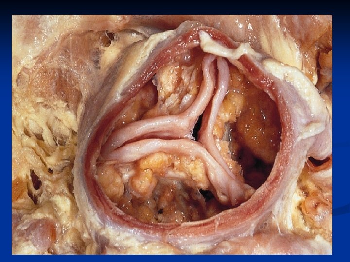
Pathological lesions: n The vegetations are friable, soft, bulky, polypoid, and easily detached. n They are shaggy, grey reddish, or brown in color, projecting from the surface of the valve cusps. n They usually develop on the contact surface of a cusp, where they may be patchy or continuous. n Spread from the aortic valve to the mitral valve, and vice versa is common. n The micro-organisms invade the cusp tissue and cause necrosis, which may lead to cusp n
The lesion may spread from the mitral valve to Mc. Callum’s patch in the left atrium, and from the base of the valve into the adjacent myocardium or into the root of the aorta. n Microscopically: n The vegetations consists mostly of platelets and fibrin and contain colonies of microorganisms. n Polymorphs are usually scanty. n The underlying cusp is vascularized, thickened, and fibrosed and may have areas of necroses. n It is infiltrated by polymorphs, macrophages n
Eradication of infection followed by gradual repair of the damaged cusps. n Organization of vegetations, results in irregular scarring and distortion of the cusps, fibrous thickening and matting of the chordae tendinae. n The end result may be indistinguishable from chronic rheumatic valvulitis, but usually the cusps are more irregularly thickened and calcified. n
Endocarditis of the mitral valve (subacute, caused by Streptococcus viridens). vegetations are denoted by arrows.
Histological appearance of vegetation of endocarditis with extensive acute inflammatory cells and fibrin.
n n Complications and effects: I. Embolic manifestations: Emboli from detached parts of the vegetations, leads to aseptic infarctions in the spleen, kidneys and brain because of the low virulence of the organisms.
Renal lesions: in the form of: a. Renal infarctions. b. Acute diffuse glomerulonephritis. c. Focal embolic glomerulonephritis: due to sensitization of the glomerular capillaries to the circulating organism. Only some of the glomeruli are affected and is focal within the tuft. This leads to diffuse petechial hemorrhage appear on the surface of the kidneys and hence the name “flea-bitten kidney”. II. n n
Mycotic aneurysms: The cerebral, superior mesentric, renal and limb arteries may show areas of aneurysmal dilatations due to lodgment of a low grade infected embolus. III. n
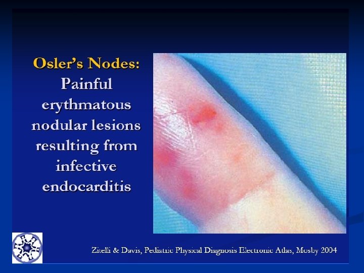
IV. Skin n n lesions: Splinter hemorrhage and Osler’s nodes. These appear as small raised painful nodules, mainly in the extremities. They were thought to be embolic but now regarded as toxic or allergic vasculitis. Clubbing of the fingers
Infective endocarditis: splinter hemorrhage of nail bed
Clinical features and course: n SIE often starts insidiously with fever, malaise and mild anemia. n Petechiae usually appear in small number in the skin, mucous membrane and retina. n Splinter hemorrhages are seen under the nail; they are probably embolic. n The spleen is usually enlarged and palpable. n Cardiac murmurs may be present from previous heart disease or may develop or change in quality as the infective lesions progress. n
Causes of death: a. The commonest cause of death in SIE is HF. b. Other major causes of death are embolic phenomena and renal failure. n Investigations: n Blood culture to detect the organism. n The blood sample is best taken at the height of fever. n Blood picture: shows progressive anemia, slight polymorphnuclear leucocytosis, sometimes with monocytosis. n High ESR. n
ACUTE INFECTIVE ENDOCARDITIS This is much more severe infection than SIE, and without effective treatment it is fatal within few days or weeks. n The commonest causal organism is steptococcal hemolyticus, staphylococcal aureus, pneumococci, and gonococci. n In immunodeficiency states less virulent bacteria, including those responsible for SIE can cause acute infective endocarditis. n
Pathogenesis: n Acute infective endocarditis (AIE), or acute bacterial endocarditis (ABE) is a part of a septicemic condition. n Bacteria reach the heart by blood stream from a septic focus in the body as puerperal sepsis, acute osteomyelitis, carbuncle, pneumonia or gonococcal infections. n They attack the healthy valves usually in cases with poor resistance. n
Pathological features: n The vegetations resemble those of SIE, but tend to be larger and localized to one part of the valve. n They consists mainly of fibrin containing large clusters of bacteria surrounded by PNL. n Virulent organisms rapidly invade the cusps → suppuration and necrosis. n It may cause cusp “aneurysms” or complete rupture. n The vegetations commonly extend to and cause rupture of the chordae tendinae. n
Spread of infection from the aortic valve to the adjacent aorta may cause aneurysm or rupture. n The organisms may also spread into the myocardium causing abscesses and necrosis. n The mitral and aortic valves are more commonly involved. n The tricuspid valve is often affected in cases of gonorrhea and drug addicts. n
Complications: n Local: n Ulceration, perforation or rupture of cusps or chordae tendinae may occur. n General: n Mainly septic emboli form distant pyemic abscesses and septic infarcts. n
Clinical features and course: n Clinical features are those of severe bacterial infection. n Blood culture is nearly always positive. n There are cardiac murmurs which may change rapidly as valve cusps are destroyed. n Septicemia may develop and embolic fragments may produce septic infarcts “pyemic abscesses” in the brain, kidneys lung and spleen. n
Causes of death: n Acute HF due to combination of toxemia, destruction of the affected valves and septic myocarditis. n Even with intensive treatment the mortality is over 50%. n
VALVULAR DISEASES
NORMAL HEART
Diseases of mitral valve Mitral stenosis: n Etiology: n Occurs in young and middle age due to: A. Healed rheumatic valvulitis; the commonest cause. B. Healed mild SBE. C. Rare causes as SLE, rheumatoid arthritis. n Women are affected more often than men. n In two thirds of cases the aortic valve is involved. I.
Gross picture: n The cusps are thickened, rigid and fused. n Their surfaces are irregular due to fibrosis and calcification. n Thickening and rigidity → a combination of stenosis and incompetence. n The mitral orifice is narrowed and slit-like “Button hole” and the valve looks like a funnel when viewed from the atrial surface. n The cordae tendinae are fibrosed, thick and short. n The papillary muscles show hypertrophy. n Pure stenosis results when the fused cusps n
Microscopically: n The cusps become thickened, distorted, vascularized throughout. n It consists of dense fibrous tissue which may be infiltrated by lymphocytes and plasma cells. n In some cases the cusps may be irregularly calcified. n Effects: n Left atial hypertrophy is very limited. n It follows by left atrial enlargement and dilatation. n Atrial fibrillation (AF) is common, but the n
Thrombus formation in the LA; almost always accompanies AF. n Systemic embolism → cerebral infarction. n Initially, the raised left atrial pressure is sufficient to drive normal blood volume into the left ventricle. n During exercise the requirements for increasing blood flow cannot be met. n With severe stenosis even the resting blood flow is decreased. n Dyspnea and persistent cough result from pulmonary congestion and edema. n Mild hemoptysis due to engorged pulmonary n
Attacks of acute pulmonary edema are brought on by exercise and paroxysmal nocturnal dyspnea occurs during night. n Increased pulmonary venous pressure → ↑ vascular tone and pressure. n This reduces the danger of pulmonary edema, but restricts pulmonary blood flow. n The resulting pulmonary hypertension leads to right ventricular hypertrophy, and failure with generalized venous congestion and edema. n
Complications: A. Atrial thrombosis → cerebral embolism. B. Hemorrhagic infarctions of the lung caused by thrombosis in pulmonary arteries or by emboli from the right atrium or leg veins. C. SIE complicates mitral stenosis caused by chronic rheumatic valvulitis. n Causes of death: n Pulmonary hypertension and right sided heart failure (RSHF). n
Mitral stenosis with commissural fusion
II. n n n Mitral incompetence: Occurs alone or in association with mitral stenosis in young and middle age. Etiology: A. Rheumatic valvulitis is the commonest cause. B. SIE. C. Dilatation of the LV as a result of anemia, hypertension, healed myocardial infarction and aortic incompetence causing stretching of the mitral ring and mitral incompetence. Post-inflammatory scarring of the mitral valve
Fibrosis of the cusps, and thickening and rigidity of the chordae, if severe may hold the cusps firmly in a partially opened position. n Increased rigidity of the cusps and fusion along part of their free margins often results in a combination of stenosis and incompetence. n Pure incompetence is unusual, n Sudden mitral incompetence may develop due to perforation of the cusps or rupture of the affected chordae. n
Effects: n Incompetence of the mitral valve → regurgitation of blood into the left atrium during ventricular systole. n The left atrium becomes distended but the additional volume of blood passes freely into the left ventricle during ventricular diastole. n Stretching of the LV by the extra volume load results in more forcible contraction. n If mitral incompetence develops gradually, there is time for LV to undergo hypertrophy and this enables it to eject the normal amount of blood into the aorta. n
The LV becomes handicapped by the mitral leak; its maximal effective stroke volume is ↓ → diminished exercise tolerance; fatigue and weakness becomes the presenting symptoms. n The ↑ volume load → left ventricular failure (LVF) accumulation of the residual blood → ↑ pressure in the left atrium → pulmonary congestion and edema. n In cases of combined mitral stenosis and incompetence pulmonary congestion and edema → pulmonary hypertension, right ventricular failure (RVF), then; congested n
n Causes of death: A. Mostly of left sided heart failure (LSHF). B. Sometimes pulmonary hypertension and RSHF.
Diseases of aortic valve I. n n Aortic incompetence: Etiology: A. Rheumatic valvulitis; the most important cause → fibrous thickening and contraction of the cusps. It is combined with stenosis and in most cases the mitral valve is also affected. B. Syphilitic aortitis and syphilitic valvulitis. This ↑ the work load of the LV which at each contraction must expel both normal stroke volume and the amount of blood regurgitated
Effects: n The ↑ volume of blood which has to be expelled by the LV results in raised systolic blood pressure. n During diastole the regurgitation of blood → rapid fall to abnormally low pressure. n The pulse pressure is thus ↑ giving a collapsing (water hummer) pulse “Corrigan’s sign”. n In response to ↑ volume and pressure loads, the LV hypertrophies and dilates. n A state of compensation may last for many years. n
Angina pectoris is always a feature, but arrhythmias and sudden death are less common. n Lastly there is left ventricular dilatation and failure. n n Causes of death: A. LSHF. B. Arrhythmias
Aortic regurgitation
II. n Aortic stenosis: Etiology: A. Rheumatic valvulitis. The cusps are thickened, vascularized, rigid and partly adherent. Stenosis is usually combined with incompetence, and in 90% of cases the mitral valve is also affected. B. SIE. C. Congenital. D. Calcific aortic stenosis. It is usually combined with normal mitral valve and little if any aortic incompetence. This is an exaggeration of changes occurring with old
Effects: n Reduction of valve orifice by over 50% ↑ the resistance to ejection of blood into the aorta → left ventricular hypertrophy which is concentric. n In most patients this maintains an adequate cardiac output for many years. n There is severe ↑ in the left ventricular pressure to overcome the resistance of the stenotic valve. n Since the pressure in the aorta is not ↑ and may fall below normal during ventricular diastole, coronary perfusion pressure is ↓. n
These factors ↑ pressure load and ↓ coronary circulation, together with left ventricular hypertrophy, predispose to angina pectoris which occurs sometimes in absence of coronary atheroma. n Fainting is a common symptom, perhaps due to cerebral insufficiency and/or transient arrhythmias triggered by ↑ workload on the LV. n n Causes of death: A. About 15% of patients with aortic stenosis dies suddenly due to ventricular fibrillation
Aortic stenosis
Diseases of tricuspid valve I. n n Tricuspid stenosis: In 15% of cases of post-rheumatic valve diseases, the mitral, and aortic valves are affected. Etiology: A. Chronic rheumatic valvulitis. B. Carcinoid syndrome. C. Congenital.
II. n Tricuspid incompetence: Etiology: A. Functional in cases of mitral stenosis; it is the most common cause. B. Chronic rheumatic valvulitis. In 15% of cases of post-rheumatic valve diseases, the mitral, aortic valves are affected. C. Infective endocarditis; it is the most common cause of tricuspid incompetence due to intravenous drug abuse. D. Stenosis may also result from congenital malformation of the cusps.
The changes are similar to that occurs in the mitral valve, but usually less severe, giving rise to stenosis or combined stenosis and incompetence. n Carcinoid syndrome causes tricuspid stenosis, either pure or combined stenosis and incompetence. n Pure incompetence is a feature of RSHF due to dilatation of the valve ring. n
Effects: n Tricuspid stenosis, incompetence or a combination of the two have similar effects. n Pressure rises in the right atrium, which dilates. n The central venous pressure (CVP) ↑, and systemic venous congestion occurs with the development of “cardiac edema”. n When associated with mitral stenosis or LVF, the tricuspid lesions tend to ↓ the degree of pulmonary venous congestion and pulmonary hypertension by limiting the volume of blood reaching the lung and left side of the heart. n
Diseases of pulmonary valve I. n n Pulmonary stenosis: Causes: A. Congenital. B. Rheumatic valvulitis. C. Infective endocarditis. D. Carcinoid syndrome. Pulmonary stenosis is a feature of carcinoid syndrome but occurs more commonly as a congenital malformation.
n Effects: A. Right ventricular hypertrophy. B. RSHF. C. Pulmonary oligemia and cyanosis.
Pulmonary incompetence: n Causes: A. Functional in cases of mitral stenosis. B. Congenital. C. Infective endocarditis. D. Carcinoid syndrome. n More often it is due to pulmonary hypertension, with dilatation of the pulmonary artery and valve. n In carcinoid syndrome it may accompany stenosis. n It also occurs as a congenital anomaly or rarely due to infective endocarditis. II.
Effects: n The mechanical effects are not serious unless there is pulmonary hypertension and they are: A. Right ventricular hypertrophy and dilatation. B. RSHF. C. Pulmonary hypertension. n The cause of death in tricuspid and/or pulmonary valve diseases is: RSHF. n
Thank you