Plant Pathology and Plant Diseases Anne Marte Tronsmo


- Slides: 18

Plant Pathology and Plant Diseases © Anne Marte Tronsmo, David B. Collinge, Annika Djurle, Lisa Munk, Jonathan Yuen and Arne Tronsmo 2020 TEACHING MATERIALS

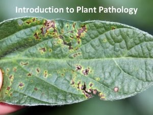
Fig. 9. 1 A-B Examples of symptoms. (A) Rhytisma acerinum causing tar spot on acer. (B) Canker of apple tree caused by Neonectria ditissima. (A, B, © L. Munk. ) TEACHING MATERIALS Plant Pathology and Plant Diseases © Anne Marte Tronsmo, David B. Collinge, Annika Djurle, Lisa Munk, Jonathan Yuen and Arne Tronsmo 2020

Fig. 9. 1 C-D Examples of symptoms. (C) Colletotrichum gloeosporioides causing bitter rot on apple. (D) Aphanomyces cochlioides causing dry rot of sugar beet. (C: © L. Munk; D: © A. Djurle. ) TEACHING MATERIALS Plant Pathology and Plant Diseases © Anne Marte Tronsmo, David B. Collinge, Annika Djurle, Lisa Munk, Jonathan Yuen and Arne Tronsmo 2020

Fig. 9. 1 E-F Examples of visible signs of disease. (E) Smut spores (teliospores) of Ustilago maydis. (F) Mycelium and sclerotia of Sclerotinia sclerotiorum. (© L. Munk. ) TEACHING MATERIALS Plant Pathology and Plant Diseases © Anne Marte Tronsmo, David B. Collinge, Annika Djurle, Lisa Munk, Jonathan Yuen and Arne Tronsmo 2020

Fig. 9. 2 A-B Examples of visible signs of disease. (A) Conidia of Monilia fructigena causing brown rot on apple. (B) Conidia and chasmothecia of Erysiphe trifolii causing powdery mildew on lupin. . (© L. Munk). TEACHING MATERIALS Plant Pathology and Plant Diseases © Anne Marte Tronsmo, David B. Collinge, Annika Djurle, Lisa Munk, Jonathan Yuen and Arne Tronsmo 2020

Fig. 9. 2 C-D Examples of visible signs of disease. (C) Aecia of Uromyces beticola causing beet rust. (D) Uredinia of Puccinia striiformis causing yellow rust on wheat. (© L. Munk). TEACHING MATERIALS Plant Pathology and Plant Diseases © Anne Marte Tronsmo, David B. Collinge, Annika Djurle, Lisa Munk, Jonathan Yuen and Arne Tronsmo 2020

Fig. 9. 2 E-F Examples of visible signs of disease. (E) Smut spores (teliospores) of Ustilago maydis. (F) Mycelium and sclerotia of Sclerotinia sclerotiorum. (© L. Munk. ) TEACHING MATERIALS Plant Pathology and Plant Diseases © Anne Marte Tronsmo, David B. Collinge, Annika Djurle, Lisa Munk, Jonathan Yuen and Arne Tronsmo 2020

Fig. 9. 2 G-H Examples of visible signs of disease. (G) Sporangia of Albugo candida causing white rust on shepherd’s purse. (H) Sporangia of Peronospora viciae causing downy mildew on faba bean. (© L. Munk. ) TEACHING MATERIALS Plant Pathology and Plant Diseases © Anne Marte Tronsmo, David B. Collinge, Annika Djurle, Lisa Munk, Jonathan Yuen and Arne Tronsmo 2020

Fig. 9. 3 A-B Examples of abiotic disorders. (A) Hail damage on oilseed rape. (B) Frost damage on a shoot of grapevine. (A: © SLU; B: © L. Munk. ) TEACHING MATERIALS Plant Pathology and Plant Diseases © Anne Marte Tronsmo, David B. Collinge, Annika Djurle, Lisa Munk, Jonathan Yuen and Arne Tronsmo 2020

Fig. 9. 3 C-D Examples of abiotic disorders. (C) Ice damage on golf course. (D) Kale with leaf drop due to sudden change in temperature. (C: © A. Tronsmo. D: © L. Munk. ) TEACHING MATERIALS Plant Pathology and Plant Diseases © Anne Marte Tronsmo, David B. Collinge, Annika Djurle, Lisa Munk, Jonathan Yuen and Arne Tronsmo 2020

Fig. 9. 3 E-F Examples of abiotic disorders. (E) Iron deficiency. (F) Boron deficiency in celeriac. (E: T. Krogstad, © NMBU F: I. Aasen, © NMBU. ) TEACHING MATERIALS Plant Pathology and Plant Diseases © Anne Marte Tronsmo, David B. Collinge, Annika Djurle, Lisa Munk, Jonathan Yuen and Arne Tronsmo 2020

Fig. 9. 4 A compound microscope with phasecontrast. 1, Eye pieces; 2, rotating revolver with 4 objectives; 3, stage; 4, condenser; 5, ring for shift between light field, phase-contrast and dark field; 6, adjustment knob for the aperture; 7, adjustment knob for condenser; 8, coarse and fine adjustment knob; 9, lamp; 10, power switch; 11, light control; 12, brightness adjustment. (© A. Tronsmo) TEACHING MATERIALS Plant Pathology and Plant Diseases © Anne Marte Tronsmo, David B. Collinge, Annika Djurle, Lisa Munk, Jonathan Yuen and Arne Tronsmo 2020

Fig. 9. 5 A-B Examples of fungal and fungal-like structures. (A) Spores of Fusarium sp. (B) A conidiophore and conidia of Botrytis cinerea. (E) Mature and immature teliospores of Phragmidium sp. (F) Sporangiophores and a sporangium of Peronospora viciae on pea. (© L. Munk). TEACHING MATERIALS Plant Pathology and Plant Diseases © Anne Marte Tronsmo, David B. Collinge, Annika Djurle, Lisa Munk, Jonathan Yuen and Arne Tronsmo 2020

Fig. 9. 5 C-D Examples of fungal and fungal-like structures. (C) Ascospores of Venturia inaequalis. (D) Chasmothecium – ascocarp of the fungus causing powdery mildew on oak, Erysiphe alphitoides. (© L. Munk. ) TEACHING MATERIALS Plant Pathology and Plant Diseases © Anne Marte Tronsmo, David B. Collinge, Annika Djurle, Lisa Munk, Jonathan Yuen and Arne Tronsmo 2020

Fig. 9. 5 E-F Examples of fungal and fungal-like structures. (E) Mature and immature teliospores of Phragmidium sp. (F) Sporangiophores and a sporangium of Peronospora viciae on pea. . (© L. Munk). TEACHING MATERIALS Plant Pathology and Plant Diseases © Anne Marte Tronsmo, David B. Collinge, Annika Djurle, Lisa Munk, Jonathan Yuen and Arne Tronsmo 2020

Fig. 9. 6 A scheme for diagnosis following isolation of the pathogen. (© L. Munk. ) TEACHING MATERIALS Plant Pathology and Plant Diseases © Anne Marte Tronsmo, David B. Collinge, Annika Djurle, Lisa Munk, Jonathan Yuen and Arne Tronsmo 2020

Fig. 9. 7 DNA sequencing for identification of microorganisms: (A) Scheme for isolating DNA and PCR amplification of specific DNA sequences from infected plant tissue. (B) Schematic structure of eukaryotic ribosomal genes illustrating the internal transcribed spacer (ITS) sequences. (© D. B. Collinge). TEACHING MATERIALS Plant Pathology and Plant Diseases © Anne Marte Tronsmo, David B. Collinge, Annika Djurle, Lisa Munk, Jonathan Yuen and Arne Tronsmo 2020

Fig. 9. 8 Lateral flow device – field kit. (© L. Munk. ) TEACHING MATERIALS Plant Pathology and Plant Diseases © Anne Marte Tronsmo, David B. Collinge, Annika Djurle, Lisa Munk, Jonathan Yuen and Arne Tronsmo 2020
 Tronsmo plant pathology and plant diseases download
 Tronsmo plant pathology and plant diseases download
 Tronsmo plant pathology and plant diseases download
 Conclusion of plant diseases
 Plant pathology
 Plant pathology
 Plant pathology
 Plant pathology
 Plant pathology
 Disease cycle
 Plant pathology
 Plant pathology
 Biological nematicides
 Plant pathology
 Funo verde
 Saturno leyenda
 Planetas metalicos de la alquimia
 Marte henriksen
 Didicou
