THE EAR The ear is the organ of


- Slides: 119
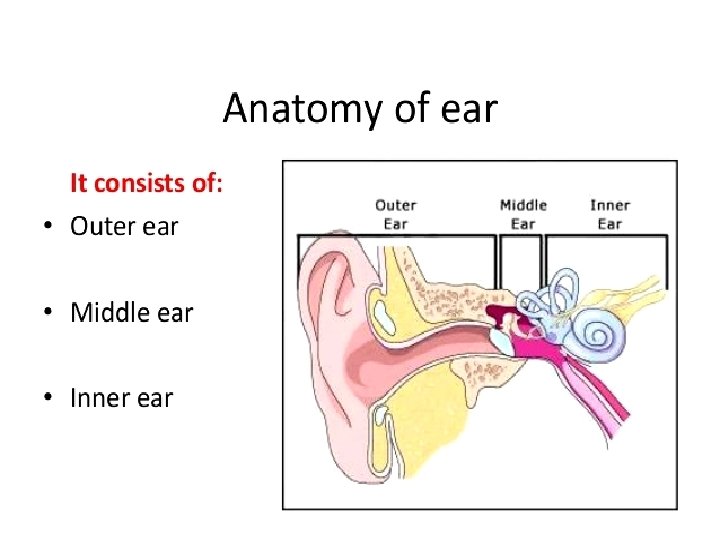
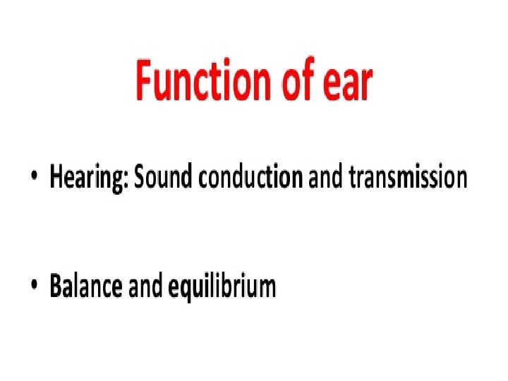
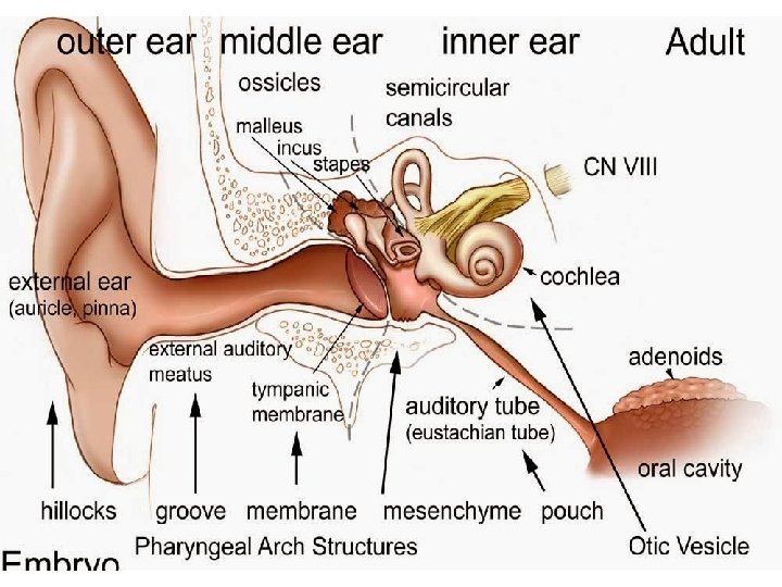
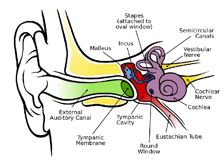
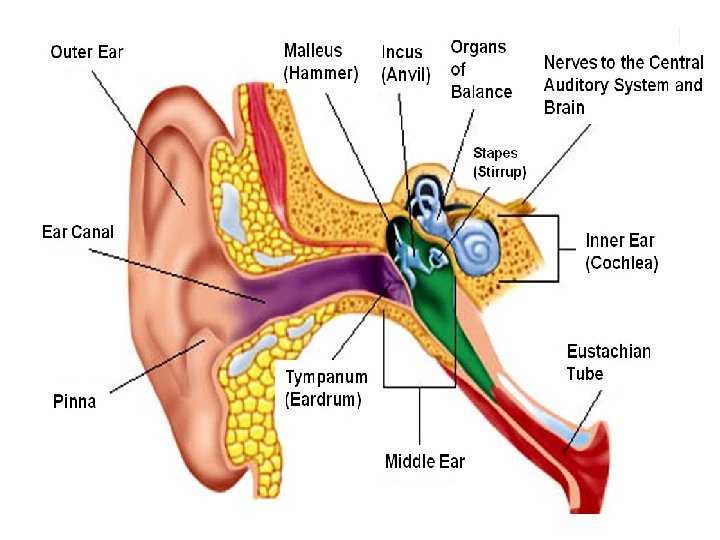
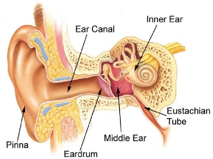
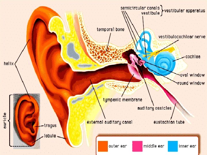
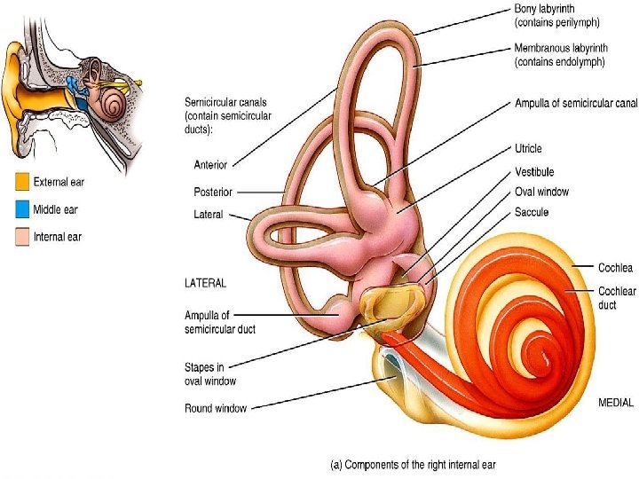
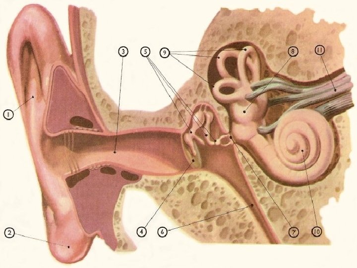
THE EAR • The ear is the organ of hearing and equilibrium. It is formed of 3 parts : • The external ear. • The middle ear or the tympanic cavity. • The internal ear or the labyrinth.
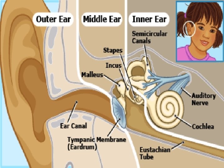
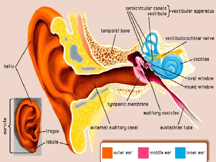
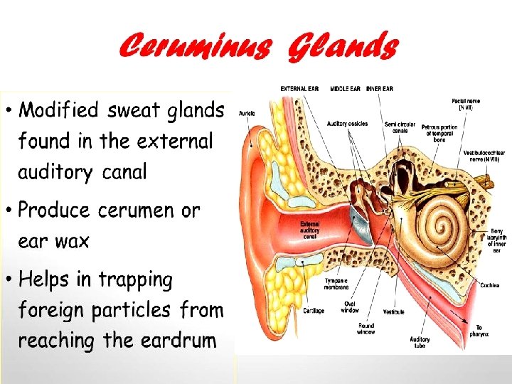
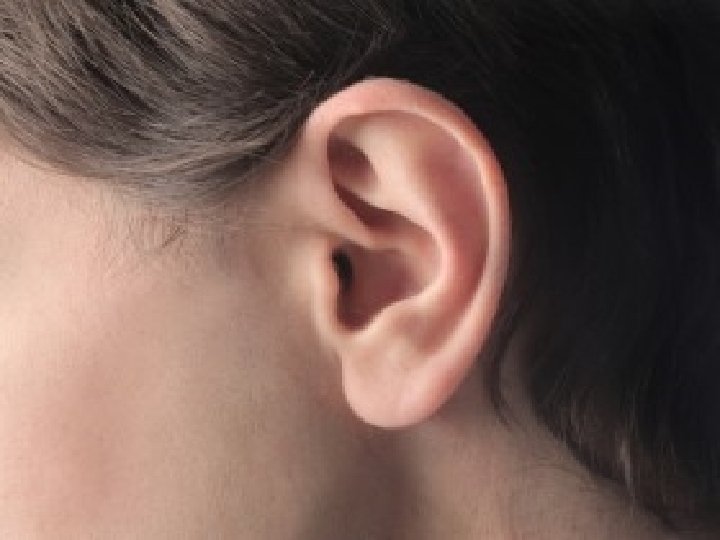
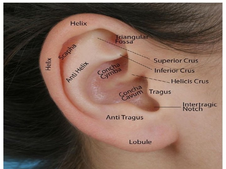
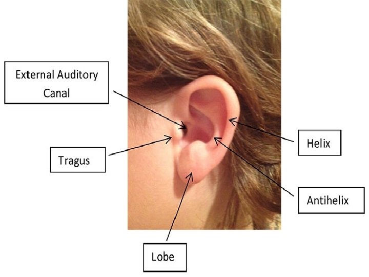
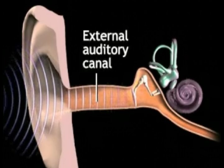
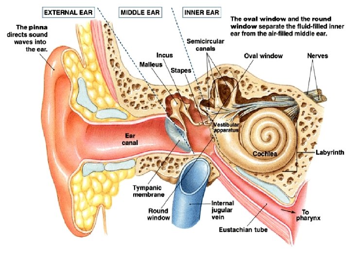
The External Ear Consists of : • The Ear Pinna : It is formed of elastic cartilage covered on both sides by skin. • The External Auditory Meatus : Its outer part is formed of elastic cartilage while its inner part is formed of bone. It is covered with skin rich in ceruminous glands which secrete the cerumen (wax of the ear ). Hairs are also present in the external ear. The hairs and cerumen secretion have a protective function.
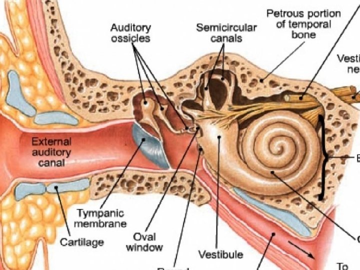
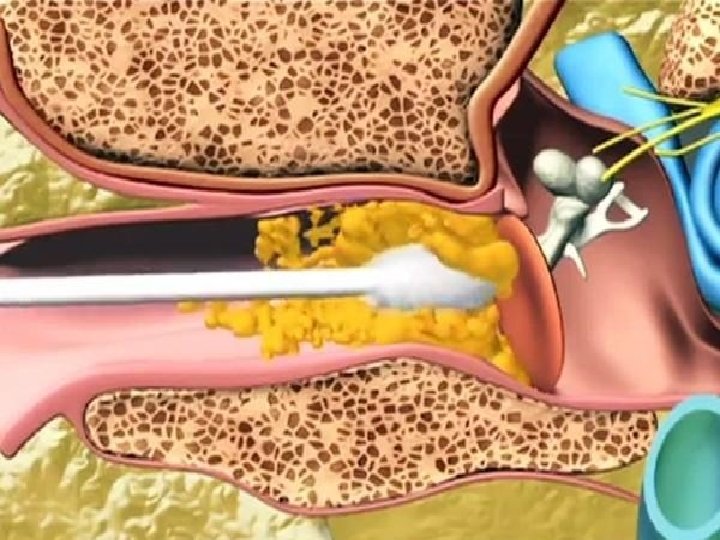
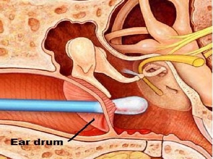
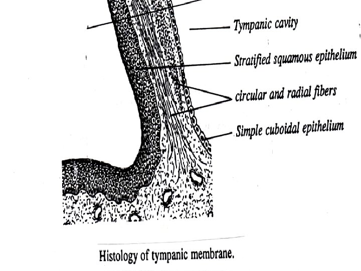
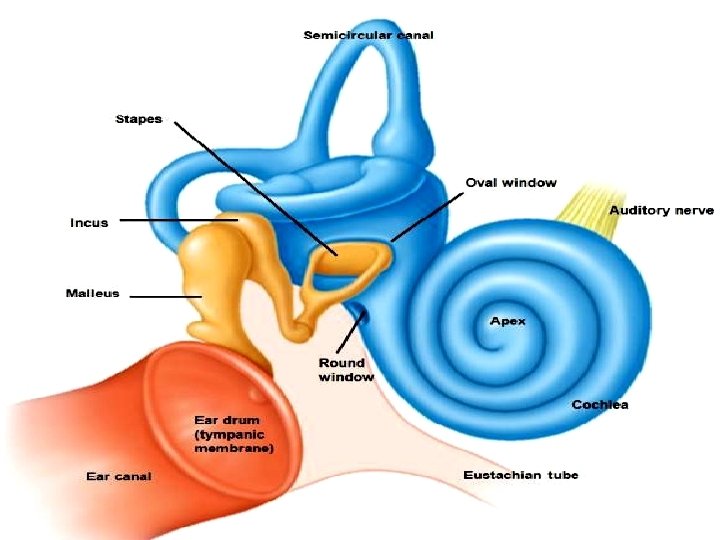
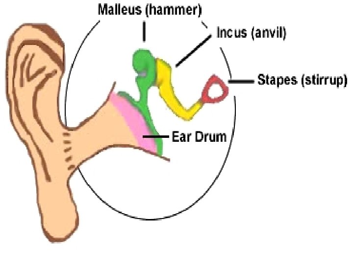
The Tympanic Membrane or Eardrum • It is an oval fibroelastic membrane which forms a partition between the external and middle car. • The tympanic membrane is formed of the following layers from outside inwards : - • Stratified squamous epithelium having no papillae. In the centre of the drum, the epithelium is thin formed only of 2 layers of cells. Mitosis is present only in the cells near the margin.
• Radially arranged collagenous bundles. • Circularly arranged collagenous bundles. • Simple cuboidal epithelium covers the inner surface of the drum. • N. B. : The upper part of the drum is thin and flaccid and is called Schrapnell's membrane. • Functions of the tympanic membrane : It transmits the vibrations of sounds to the ossicles of the middle ear.
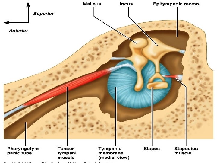
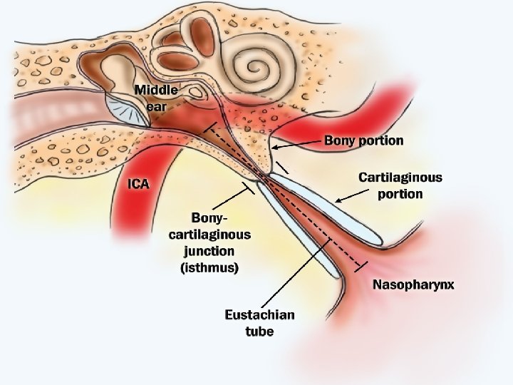
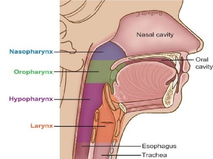
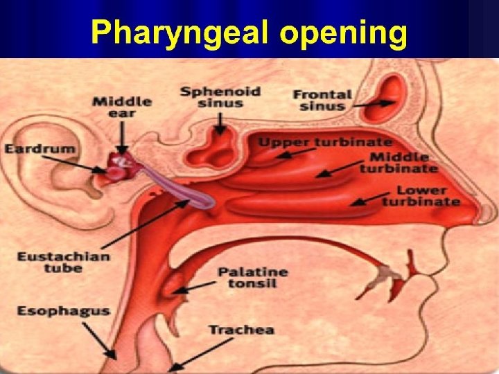
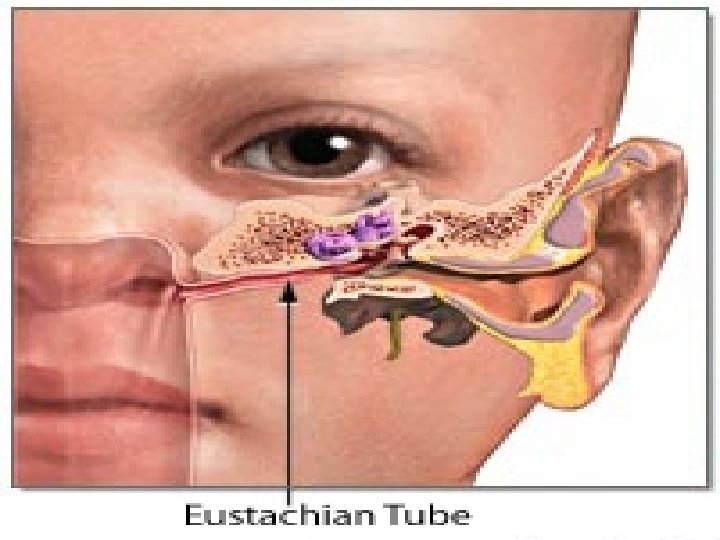
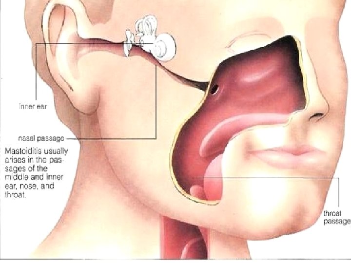
THE MIDDLE or THE TYMPANIC CAVITY • It is a cavity in the temporal bone, connected anteriorly with the nasopharynx by the Eustachian tube and connected posterorly with the air cavities in the mastoid process.
• The middle ear is lined with simple cubical epithelium with no basement membrane. • The middle ear contains • 3 bony ossicles, two muscles and the chorda tympani nerve
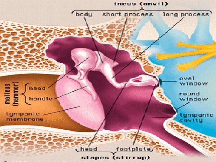
The Three Bony Ossicles Of The Middle Ear • The bony ossicles are : • the malleus, the incus and the stapes. They articulate with each other by synovial joints. They are formed of spongy bone and are covered with periosteum. Outside the periosteum there is a layer of C. T. covered with simple cubical epithelium.
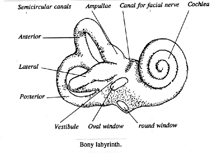
• The three ossicles transmit the vibrations of sounds from the tympanic membrane to the oval window which is present in the medial wall of the middle ear.
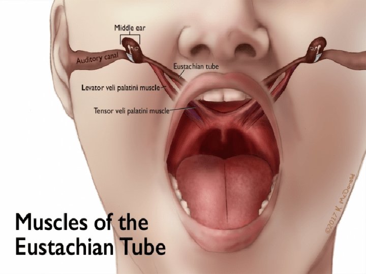
The Muscles Of The Middle Ear • The Two Muscles Which Are Attached To The Ossicles Are : • The tensor tympani muscle which is attached to the malleus. It makes the tympanic membrane tense and ready for vibrations. • The stapedius muscule which is attached to the stapes protecting the internal ear from highly pitched sounds. • There are two windows in the medial wall of the middle ear which are :
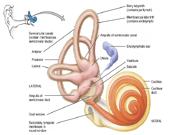
• The Oval Window : It is closed by the annular ligament which is attached to the foot plate of the stapes. Through the oval window, sound vibrations are conducted to the perilymph of the vestibule of the internal ear. • The Round Window : It is closed by an elastic membrane called secondary tympanic membrane which separates the middle ear from the internal ear.
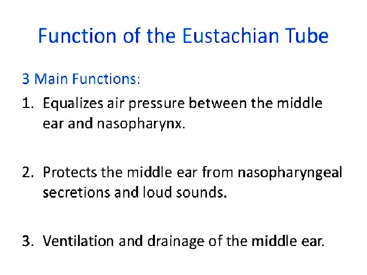
The Eustachian or Pharyngotympanic Tube : • The tube connects the middle ear with the nasopharynx. • The anterior ⅔ of its wall is formed of elastic cartilage and is lined with pseudostratified columnar ciliated epithelium with goblet cells. • The posterior ⅓ of its wall is formed of bone and is lined with simple columnar ciliated epithelium. • The walls of the Eustachian tube open during swallowing, thus the pressure in the middle ear is balanced with the atmospheric pressure.
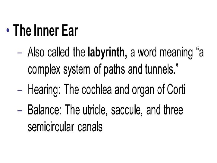
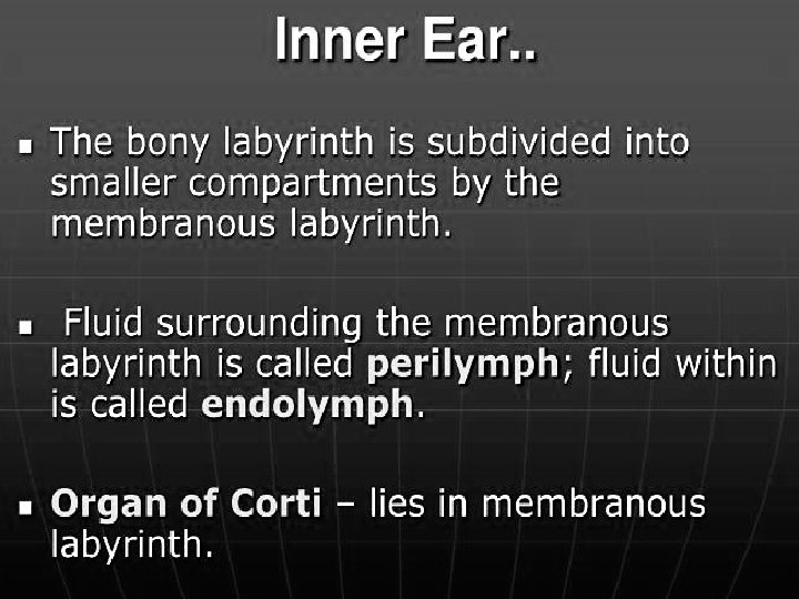
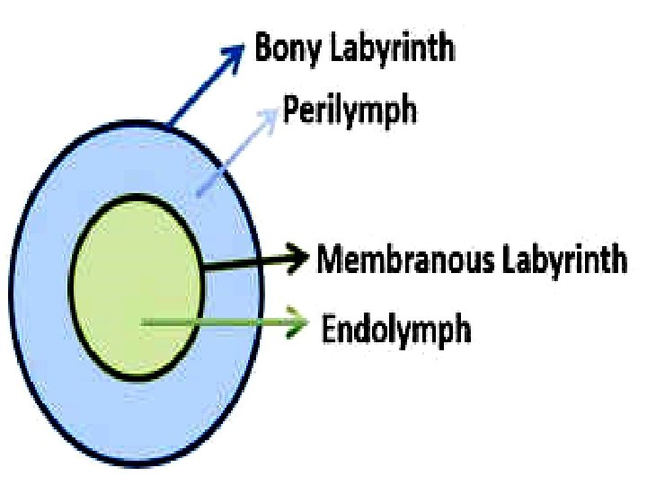
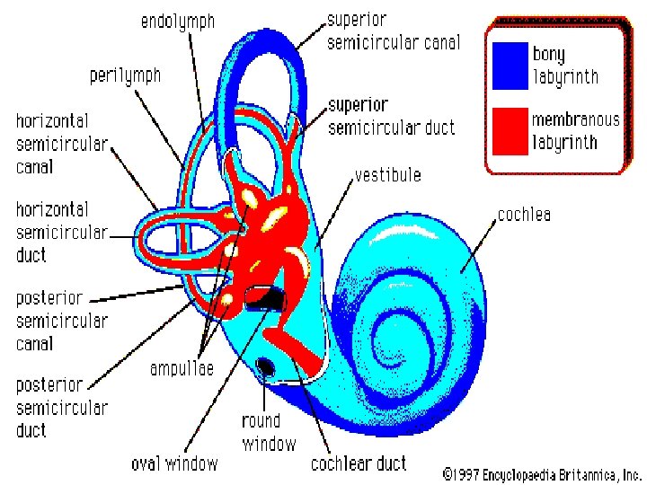
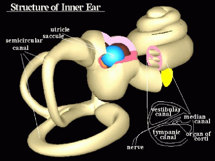
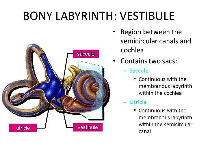
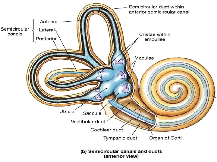
• THE INTERNAL EAR or LABYRINTH • The internal ear is present in the temporal bone. It is concerned with hearing and balance. • The internal ear consists of : A-Bony Labyrinth which is formed of bone, it is filled with a fluid called perilymph and it communicates with the subarachnoid space of the brain meninges. The Bony Labyrinth Consists Of : -
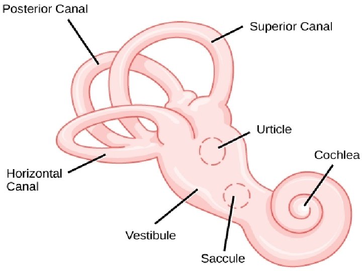
• The Cochlea. • The three Semicircular Canals. • The vestibule which contains the utricle and saccule.
B-Membranous labyrinth which are the membranous sacs and canals, present in the cavities of the bony labyrinth.
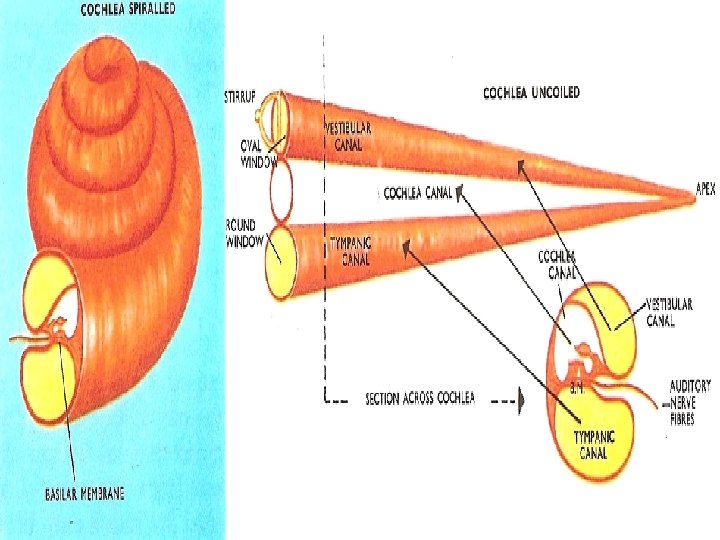
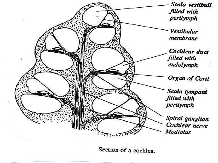
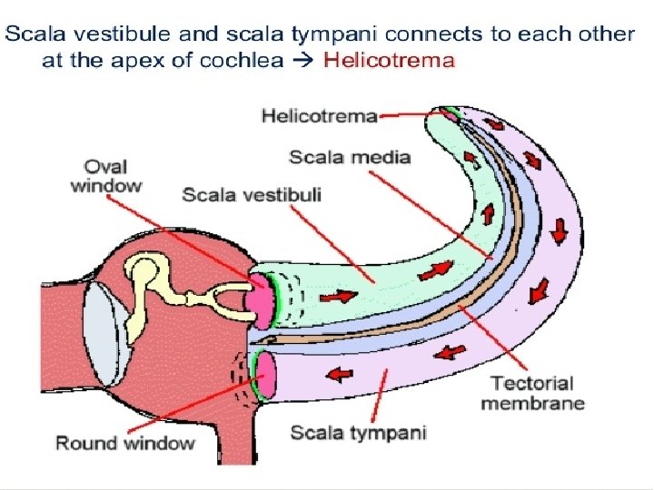
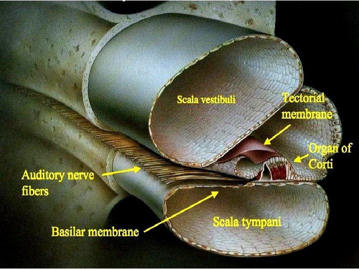
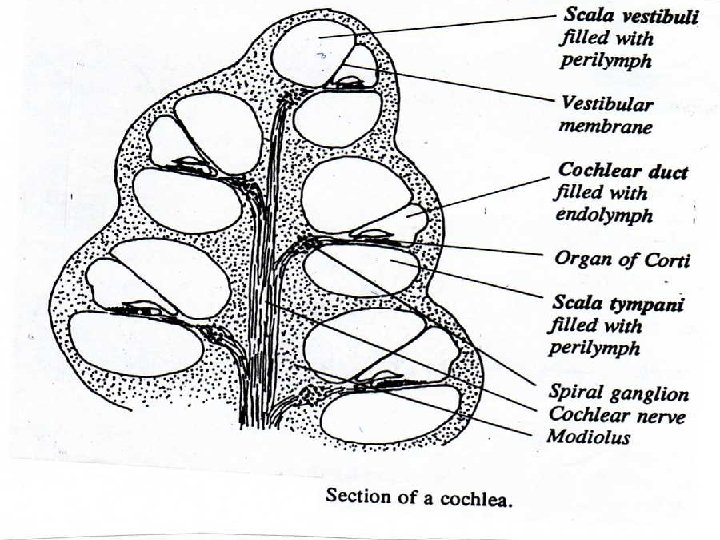
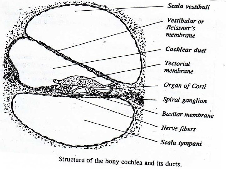
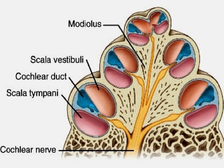
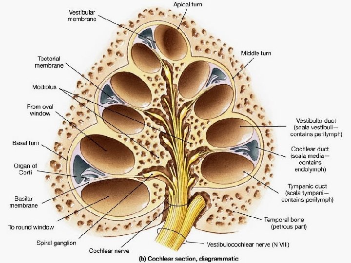
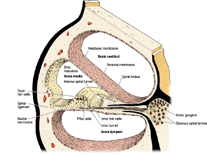
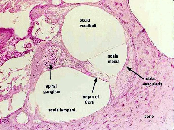
THE COCHLEA • It is a bony coiled tube which is conical in shape. It wounds and runs about 2½ turns in a spiral form around a central bony axis called the modiolus. The modiolus is a spongy bone containing nerve fibres, blood capillaries and spiral ganglia. • The turns of the cochlea around the modiolus form 5 bony cavities at lateral sides of the modiolus. • Each cavity is divided into 3 compartments by 2 membranes : The vestibular membrane and the basilar membrane.
The 3 compartments are : • The scala vestibuli. It is present in the upper part of the cavity. It communicates with the vestibule. • The scala tympani which is present at the lower part of the cavity. It communicates with the tympanic cavity through the round window.
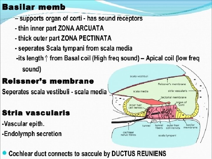
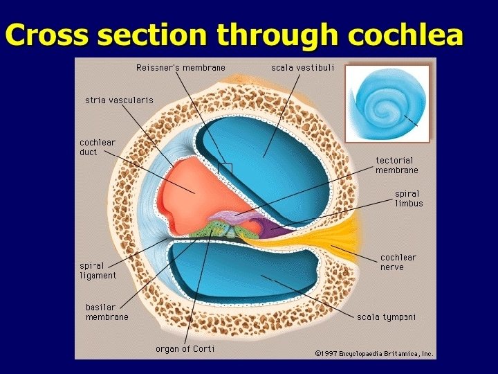
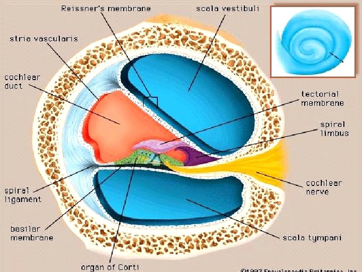
• The cochlear duct present inbetween the scala vestibuli and scala tympani. It represents the membranous part of the cochlea. It contains endolymph. • The cochlear duct : It is a triangular duct. The vestibular and the basilar membranes form its two sides. The stria vascularis which is formed of vascular C. T. forms the base of the cochlear duct.
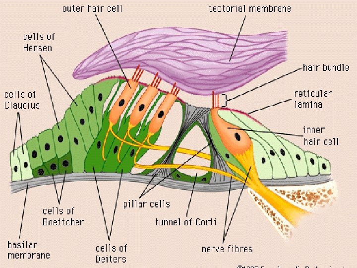
• The vestibular or Reissner's membrane is formed of C. T. covered with simple squamous epithelium. It separates the cochlear duct from the scala vestibuli. • The stria vascularis is formed of vascular C. T. covered with pseudostratified columnar epithelium. The cells of the stria secrete the endolymph which is present in the cochlear duct. • The basilar membrane is formed of elastic and collagenous C. T. fibres covered with simple cubical epithelium. organ of corti is present on this membrane.
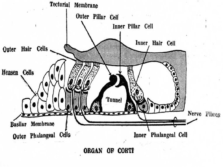
THE ORGAN OF CORTI • It is a neuro-epithelial structure found on the upper surface of the basilar membrane. It is covered with a gelatinous membrane called tectorial membrane. • The organ of Corti as any neuroepithellal structure is formed of :
• a-Sensory hair cells. • b-Supporting cells which are: Pillar cells, phalangeal cells and Hensen cells: • In the centre of the cells of organ of Corti there is a triangular space called the tunnel. The tunnel is bounded by outer and inner pillar cells
• The outer and inner pillar cells. The inner pillar cell has a concave upper end, while the outer pillar cell has a rounded upper end. The hair cells are present outer to and inner to these pillar cells.
• The phalangeal cells : These are tall columnar cells. They serve as supporting cells to the hair cells. They have concavities to hold the hair cells. • The Hensen cells : They are supporting cells which are present outer to the phalangeal cells.
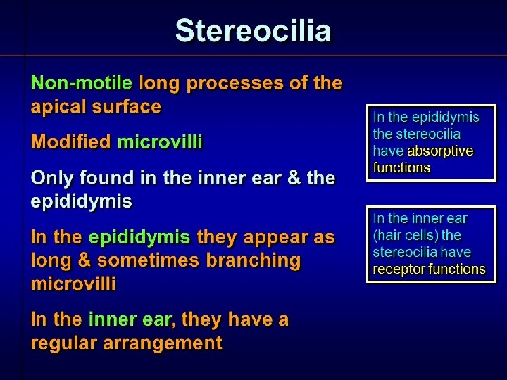
The Sensory Hair Cells In The Organ Of Corti. • There Are Two Types Of Hair Cells : • Outer hair cells arranged in 3 to 5 rows. • Inner hair cells arranged in one row. The bases of these cells do not rest on the basilar membrane, but they are present on cup-like processes of the phalangeal cells. • The free surface of each hair cell has hair-like processes. These processes are stereocilia (non motile cilia).
• The basal part of each cell is surrounded by synaptic ribbon of nerve endings. These nerve fibres are the dendrites of the bipolar nerve cells of the spiral ganglia in the internal ear. The axons of these bipolar nerve cells form the cochlear nerve which passes first in the modiolus of the cochlea.
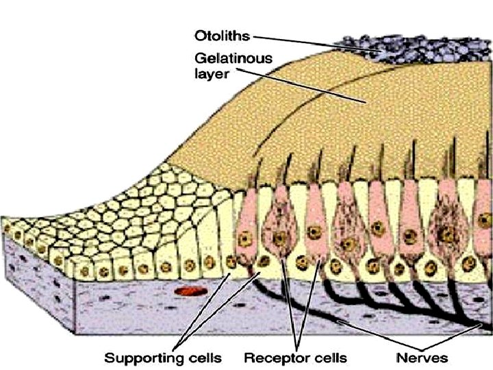
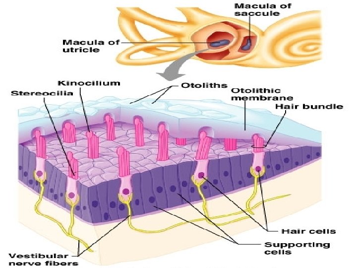
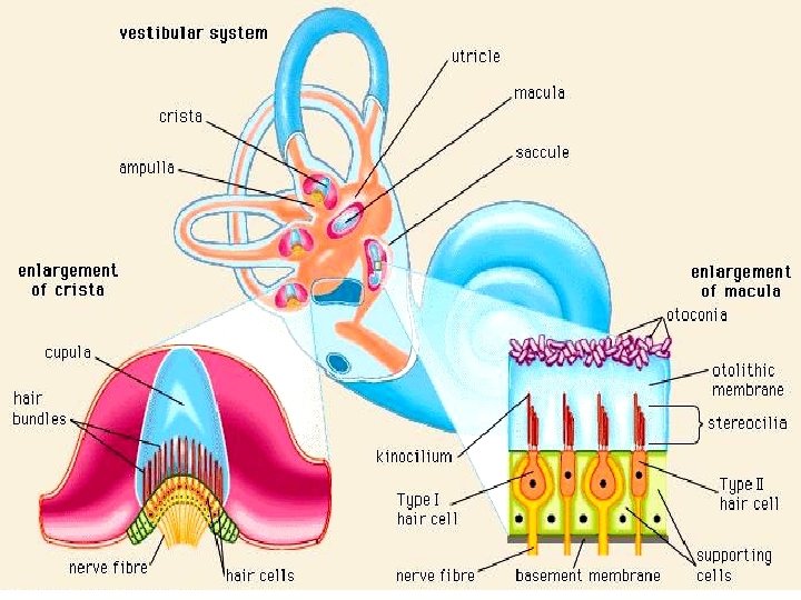
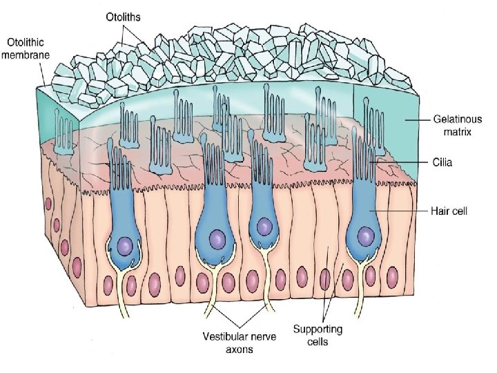
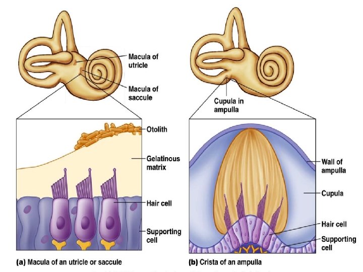
The bony vestibule is filled with perilymph. It contains two sacs of the membranous labyrinth, the utricle and saccule which are filled with endolymph. The utricle and saccule sacs are lined by simple squamous mesothelium. Each sac contains a flat plaque-like sensory ending called a macula.
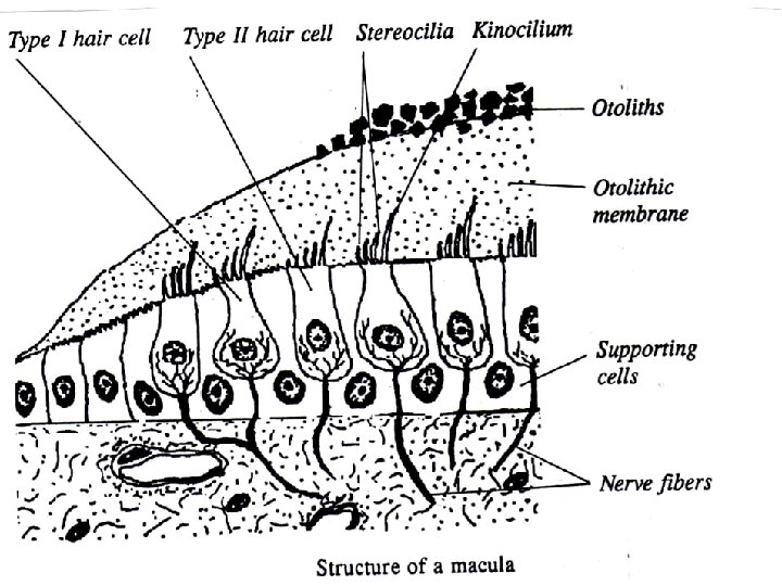
Structure Of The Macula : • The macula is formed of 2 main types of cells resting on a clear basement membrane. The cells are covered with a thick gelatinous glycoprotein layer. The surface of this layer is rich in calcium carbonate crystals, known as Otolith.
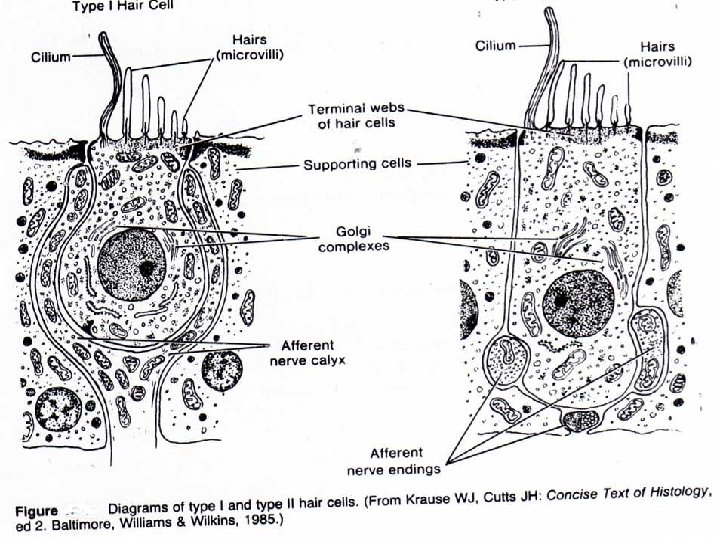
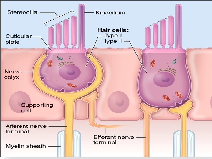
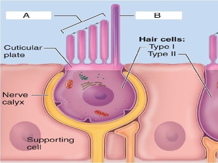
The Macula Contains These Cells 1. The Hair Cells Of the Macula Are Of Two types – Type I hair cells are flask-shaped cells. They are surrounded by afferent nerve endings. Their free surfaces have stereocilia (non-motile cilia). – Type II hair cells are columnar cells. They are surrounded by afferent and efferent nerve endings. Their free surfaces have stereocilia
• 2. The Supporting Cells In The Macula : They are columnar cells supporting the hair cells. Their free surfaces are covered with stereocilia. • Functions : The saccule is responsible for the sense of gravity and linear acceleration. The utricle is the organ of balance which regulates the position of the head in relation to the gravity.
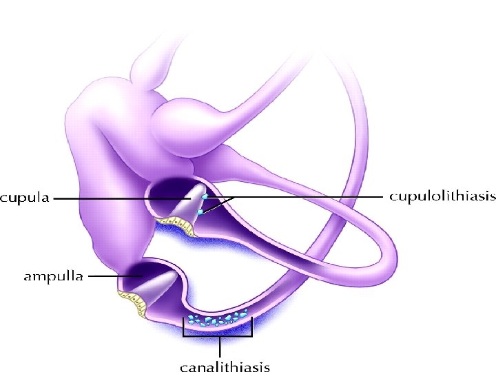
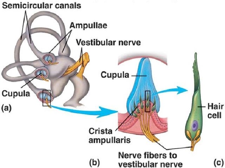
• The Semicircular Canals • Each semicircular canal has one expanded end called ampulla. The 3 canals have 5 openings into the utricle because the non expanded ends of the superior and posterior canals have a common opening in the utricle.
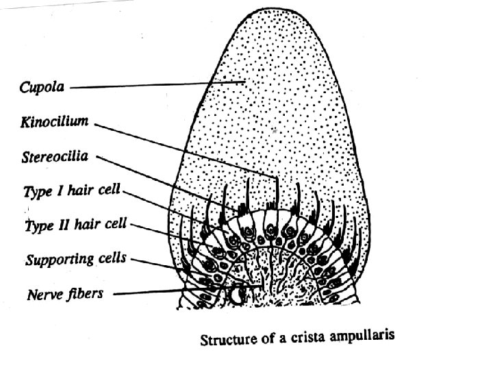
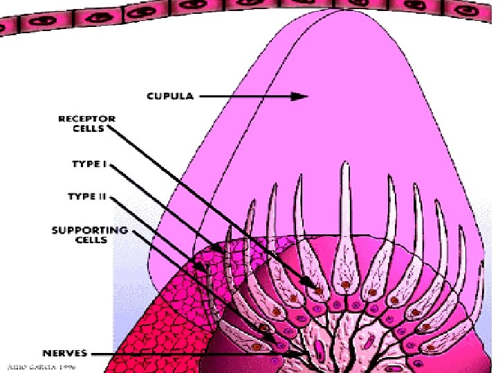
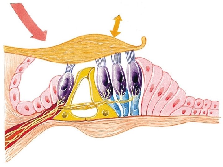
• The three bony semicircular canals contain the membranous labyrinth which takes the same shape of the bony part. This membranous labyrinth contains a neuro-epithelial structure which is pressent in the ampulla and is called crista ampularis.
Each crista ampularis is formed of two types of cells : – Hair cells which are of two types; flask-shaped cells and columnar cells covered by stereocilia. – Supporting cells which are columnar cells. • On the surface of the crista ampularis, there is a gelationus thick membrane called Copula. It has no calcium carbonate crystals on its surface. • Functions of the crista ampularis : • It is stimulated by the movement of endolymph following angular acceleration of the head. It regulates the position of the body.