Chapter 26 Male reproductive system Primary secondary sex


 deferens Thick muscular tube that extends from tail of epididymis to the")


 head, 2) mid piece and 3) tail. – Head")


- Slides: 23
Chapter 26 Male reproductive system
Primary & secondary sex organs Testes are the primary sex organ of the male reproductive system. They manufacture and store spermatozoa for fertilization of ova and reproduction of the species. Produce the sex cells or gametes = spermatozoa in males Secondary or accessory sex organs include: – The scrotum, epididymis, ductus (vas) deferens, urethra, ejaculatory duct, prostate gland, bulbourethral gland, seminal vesicles, and penis.
Male reproductive organs
Scrotum Divided pouch of skin that contains the testes and is suspended outside of the body where it keeps the temp ~ 3. 5 degrees cooler and prevents the sperm from dying. Covered with stratified squamous epithelium and sparse pubic hairs it is divided in the middle by a septum that separates the two testicles. Externally the septum is marked by a seam the perineal raphe which extends from the inferior surface of the penis, around the scrotum to the margin of the anus. In cold weather the scrotum can retract by contraction of the cremaster muscle (sk. m. ) to bring the testicles up closer to the body for warmth. The cremaster muscle is an extension of the ext. oblique muscles. The dartos muscle, a thin layer of smooth muscle in the superficial fascia, causes the scrotum to wrinkle and thicken the skin in order to preserve heat in the cold, as well.
Scrotum The scrotum is richly supplied by many sensory and motor nerve fibers It is also highly vascularized by the testicular artery to provide nourishment and oxygen to the spermatogenic apparatus necessary for production of sperm. The pampiniform plexus is a network of veins that surround the testicular artery and serve as a heat exchanger to cool the blood in the testicular artery and allow for production of live sperm.
Testes
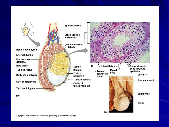
Testes Location: Contained within the scrotum inferior and outside of the abdominal cavity at the root of and posterior to the penis. Structure: Enclosed in a serous sac tunica vaginalis tunica albuginea “white coat”, fibrous outer capsule of the testes. Septal extensions of the t. albuginea form lobules – compartments; contain 1 -4 seminiferous tubules – tightly coiled “sperm factories” lined with germinal epithelium and sustentacular or Sertoli cells. The “S” cells protect the germinal epithelium and form a blood-testes-barrier to prevent the immune system antibodies from attacking the sperm septum – divides testis into numerous wedge shaped compartments that arise from tunica albuginea. interstitial cells (Cells of Leydig) – produce androgenic hormones (testosterone, etc. ); sourround seminiferous tubules and secrete fluid into surrounding interstitial fluid.
Testes
Spermatic ducts Consists of those ducts that carry sperm from the testes to the urethra. Includes the efferent ductules, epididymis, ductus deferens and ejaculatory duct. Efferent ductules are 12 small ducts that carry sperm from the seminiferous tubules to the epididymis. They are lined with clusters of ciliated cells that help the sperm move along.
Epididymis Coiled tube ~ 20 feet long on posterior surface of testicles. Serve as storage sites and place of maturation of the sperm produced in the seminiferous tubules. Has a head, body and tail and is lined with pseudostratified columnar epithelium covered with stereocilia. Upon entering the epididymis it may take up to 20 days for sperm to mature and become motile and capable of fertilizing an ova. Sperm are stored in the epididymis for 20 days before they are phagocytized and destroyed by surrounding epithelial cells. At the time of ejaculation, the sperm are expelled from the tail of the epididymis into the ductus deferens. Sperm do not begin swimming until ejaculated into the vagina. They are expelled from the sperm ducts by intense peristaltic contractions.
Ductus (vas) deferens Thick muscular tube that extends from tail of epididymis to the ejaculatory duct. ~ 45 cm long and 2. 5 mm in diameter. Runs superiorly through the inguinal canal and spermatic cord up the anterior wall of pelvic cavity over and behind the urinary bladder and turns anteriorly to join the duct of the seminal vesicles which form the ejaculatory duct. Propels sperm from epididymis to ejaculatory duct by peristaltic contractions. Lined with pseudostratified columnar epithelium.
Ejaculatory duct A short segment of spermatic duct system the is formed by the convergence of the ductus deferens and seminal vesicle duct. It is ~ 2 cm long and passes into the prostate gland where it empties into the urethra.
Accessory glands of male reproductive system Includes three sets of accessory glands: – The seminal vesicles – The prostate gland – The bulbourethral (Cowper) glands
Seminal vesicles Paired glands ~ 5 cm long and dorsal to the urinary bladder. Internally they are a complex labyrinth of ducts Produce a yellowish secretion that is ~ 60% of the semen volume.
Prostate gland A bulbous gland situated at the base of the urinary bladder and consists of smooth muscle and connective tissue. Composed of 30 -50 tubuloacinar glands that empty into the urethra. Milky secretion is ~ 30% of ejaculate volume.
Bulbourethral gland Two brownish spherical glands ~ 1 cm diameter. They are located just superior to the bulb of the penis and their duct empties into the penile urethra. Their secretion neutralizes the residual acidic urine in the urethra and serves a pre-ejaculate to lubricate the penis for easy insertion into the vagina.
Semen Is a complex mixture of sperm and glandular secretions released upon ejaculation. Typical volume is ~ 2 to 5 ml of ejaculate. Sperm count is 50 to 120 million/ ml. of semen. It consists of: – ~ 10% spermatozoa – 30% prostatic fluids – 60% seminal fluids – High in fructose and enzymes to neutralize vaginal secretions and bacteria.
Spermatozoa Three distinct regions: 1) head, 2) mid piece and 3) tail. – Head is a flattened oval containing chromosomes. Tip contains Acrosomal cap with enzymes that will penetrate the ova membrane. – Mid piece is joined to head by a short neck and contains centrioles and mitochondria. – Tail is a flagellum that propels the spermatozoa through the female reproductive tract.
Spermatozoa
Urethra Consists of three regions in males – Prostatic urethra – Membranous urethra – Penile “spongy” urethra
Penis Tubular organ that contains the distal portion of the urethra and introduces semen into the vagina during sexual intercourse. Internal half is the root and external half is a shaft and glans. Together the penis and scrotum constitute the external genitalia of males. The skin of the penis is loose and slides distally over the glans to form a “foreskin” or prepuce. Internally the penis consists of 3 long cylindrical bodies of erectile tissue. These erectile bodies fill with blood during arousal and engorge the penis creating an erection. The erectile bodies are: a single corpus spongiosum, a paired corpus cavernosa.
Penile structure Root = bulb + crus Shaft = body + glans Erectile tissue: Corpus cavernosa Corpus spongiosum Vascular supply: Internal pudendal art. Nerve supply: Parasym. → erection Sym. → ejaculation