EAR THE ORGAN OF HEARING INTRODUCTION The ear


 b) External ear: Collect")

Composed of elastic Cartilage covered by")


- Slides: 45
EAR THE ORGAN OF HEARING
INTRODUCTION • The ear is the organ for collection, conduction, modification, amplification and analysis of sound reaching it. It not only receives sound, but also aids in balance and body position. • Ears are responsive to extremes of sound frequencies from 20 to 20, 000 Hz
EAR
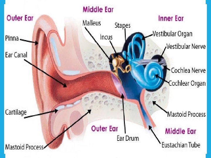
PARTS OF EAR • Three parts 1. 2. 3. a) b) External ear: Collect and conduct the sound wave to the tympanic membrane Middle ear: Filled with air, Intensifies the force of sound vibration Internal ear: consists of. Organ of corti- concerned with hearing Vestibule & semicircular duct- concerned with balancing
EXTERNAL EAR
EXTERNAL EAR • Consists of- 1. Auricle ( pinna)Composed of elastic Cartilage covered by skin 2. External acoustic meatus- 2 parts- i. Lateral 1/3 rd-cartilage of pinna ii. Medial 2/3 is bony
PINNA
EXTERNAL EAR • Lobule- It hangs below the anti tragus, no cartilage , skin covers fibro fatty tissue. • External acoustic meatus- lined by true skin. The skin surrounding the external acoustic meatus contains glands that produce wax (cerumen). The ear canal ends at the external surface of the tympanic membrane • Inflammation of this meatus is extreme painful as skin is adherent to the underlying structure.
Middle EAR
MIDDLE EAR • The middle ear is an air-filled cavity between outer & inner ear, contained within the petrous part of temporal bone and separated from external ear by tympanic membrane
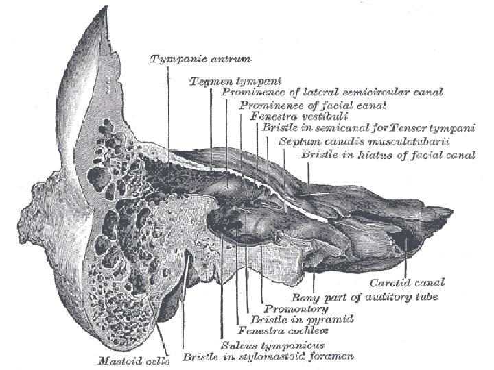
BOUNDARIES OF MIDDLE EAR • Roof- is formed by tegmen tympani • Floor – formed by a thin plate of bone separating the cavity From the jugular bulb.
BOUNDARIES OF MIDDLE • Anterior wall – EAR presents canal for tensor tympani muscle, orifice for auditory tube, carotid canal. • Posterior wall – presents aditus to the mastoid antrum and canal for the facial nerve
MIDDLE EAR
BOUNDARIES OF MIDDLE EAR • Medial wall – separates the cavity from the inner ear and presentsv. Promontory – a rounded elevation. v. Fenestra vestibule- lies above and behind the promontory v. Fenesta cochlea - lies below and behind the promontory
BOUNDARIES OF MIDDLE EAR • Lateral wall – is formed entirely by the tympanic membrane or ear drum which receives the sound waves and conduct it to three ossicles as vibration
CONTENTS OF MIDDLE • Three ossicles- EAR malleus, incus and stapes • Muscles- tensor tympani and stapedius • Nerves- facial, Glossopharyngeal, greater and lesser petrosal nerves • Arteries- internal carotid, branch of maxillary, middle meningeal artery • Ligaments • Air
TYMPANIC MEMBRANE • It is a pearly grey oval membrane. • Parts: Malleolar fold divides the membrane into two parts 1. Pars flaccid- small triangular area above the fold, ruptures more 2. Pars tensa- below the fold where handle of the malleus is attached.
STRUCTURE OF TM it has three layers • Outer cuticular layer- lined by keratinize stratified squamous epithelium • Intermediate fibrous- handle of the malleus is attached here • Inner mucous- lined by ciliated columnar epithelium • Surgical Incision of TM is called Myringotomy
TYMPANIC MEMBRANE • Artery supply: - Maxillary artery and posterior auricular artery. • Nerve supply: Auriculo temporal, glossopharyngeal and vagus nerve • Development: • Outer layer- from ectoderm of 1 st brachial cleft • Intermediate layer- from mesoderm of 1 st brachial cleft • Inner layer- from endoderm of 1 st pharyngeal pouch
EAR OSSICLES
EAR OSSICLES • The three ossicles transmit sound from the tympanic membrane to the inner ear. The handle of malleus is connected to the tympanic membrane. The incus is the bridge between the malleus and stapes. The stapes connects to the oval window by its footplate. The ossicles help in amplification of sound waves by nearly thirty times.
MUSCLES OF MIDDLE EAR • The movement of the ossicles is stiffened by two muscles. 1. The stapedius muscle, is connected to the stapes; 2. The tensor tympani muscle is connected to the base of the malleus. These muscles exert damping effect of sound vibration to protect the inner ear from loud sounds.
MASTOID ANTRUM The mastoid antrum is an air space in the petrous part and communicating posteriorly with the mastoid air cells and anteriorly with the middle ear. These air spaces function as sound receptor; provide voice resonance, acts as acoustic insulation. The mastoid air cell system is a major contributor to middle ear inflammatory diseases.
INNER EAR
INTRODUCTION • The inner ear consists of an outer bony and inner membranous labyrinths. This contains the sensory organs for balance and motion, namely the vestibules of the ear (utricle and saccule), and the semicircular canals. This also contains the sensory organ for hearing, the cochlea.
INNER EAR- at a glance Bony labyrinth Membranous labyrinth Function Cochlear duct Contains organ of corti vestibule Saccule & utricle Concerned with static equilibrium Semicircular canal Semicircular duct Concerned with kinetic equilibrium
BONY AND MEMBRANOUS LABYRINTH
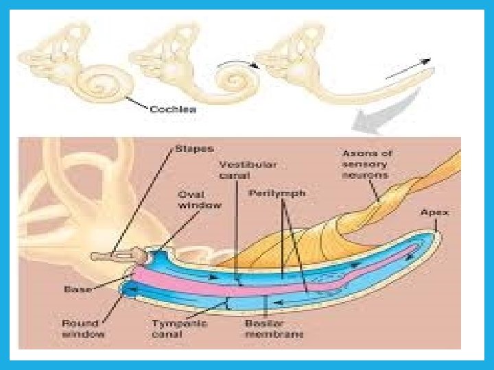
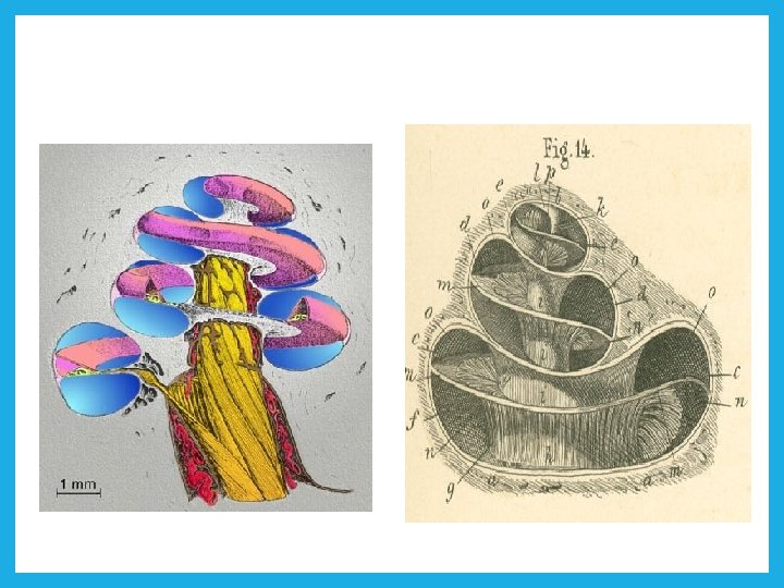
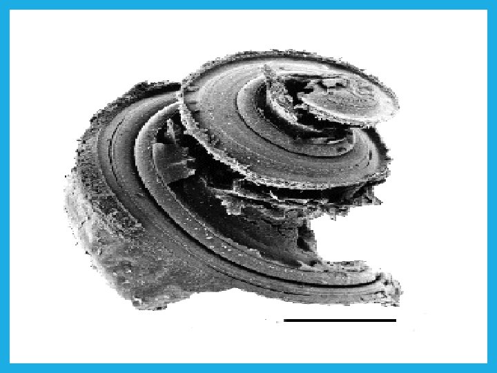
COCHLEA • The cochlea is conical in shape, resembles of a common snail. Its apex known copula and directed forward and base lies posteriorly at the bottom of the internal acoustic meatus. The cochlear canal is arranged spirally.
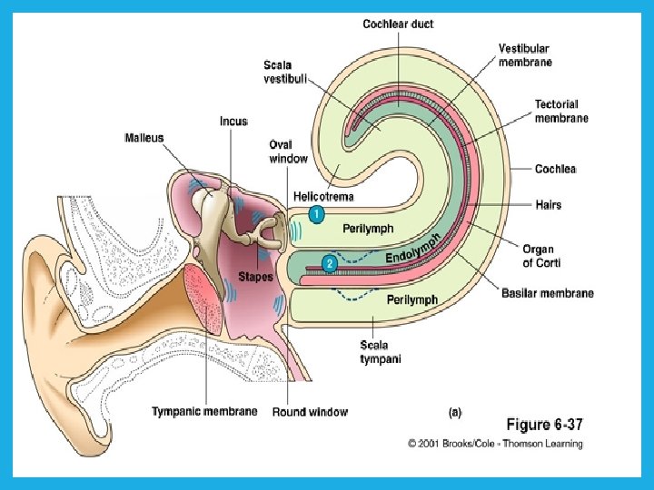
COCHLEAR DUCT • Cochlear duct is the membranous labyrinth within bony spiral cochlea. Inside, from the wall of cochlea two membrane arise vestibular & basilar membrane divides the cochlea into • 1. scala vestibule 2. cochlear duct 3. scala tympany v Two scala contain perilymph and cochlear duct contain endolymph.
COCHLEAR DUCT
COCHLEAR DUCT
VESTIBULE- SACCULE & UTRICLE & SSEMICIRCULAR CANAL - DUCT • Vestibule intervenes between the cochlea in front and semicircular canal behind. By an oblique ridge it is divided into two areas which lodges saccule and utricle • 5 Semicircular canal contains Semicircular duct
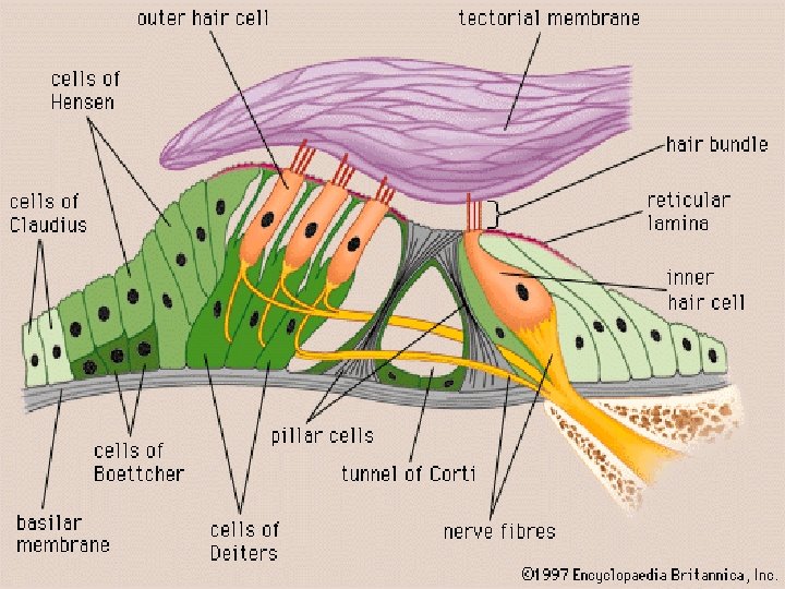
SPIRAL ORGAN OF CORTI • It contains special auditory receptors , rest on basilar membrane of cochlear duct. Organ of corti contains hair cells, which respond to different sound frequency. The most characteristic feature of hair cells is the presence of array of sterocilia. The afferent fibers of cochlear nerve come in direct synaptic contact with the hair cells. Between these cells following supporting cells are found: – Pillar (rod) cells, Deiter’s cells and Hensen’s cells
SPIRAL ORGAN OF CORTI
TRACING OF THE SOUND WAVE
INTERNAL ACOUSTIC MEATUS SHOWING THE NERVES
THANK YOU