The Middle Ear Middle ear Anatomy Air containing


 Mesotympanum (middle) Hypotympanum (lower).")


 implies a permanent abnormality of the pars tensa")


. • keratinizing epithelium has")


")
")


")


")


 • Scanty thin otorrhea (Foul smell) • Complications")
 • Take any cultures if")

 �MRI in case of suspicion of complications.")
 o Facial paralysis o Labyrinthitis. �Intracranial complications:")


- Slides: 78
The Middle Ear
Middle ear Anatomy Air containing cavity Lined with mucous membrane Containing auditory ossicles Lies in the petrous part of the temporal bone. Surfaces: • Roof: tegmen tympani (tegmental roof). • Floor: jugular bulb. • Anterior wall (carotid wall): auditory tube & tube for tensor tympani muscle. • Posterior wall (mastoid wall): aditus to mastoid antrum. • Lateral wall (membranous wall): tympanic membrane. • Medial wall: facial nerve, promontory, fenestra vestibuli, fenestra cochleae. • •
Middle ear Anatomy Middle ear cleft contains: 1. Middle ear cavity 2. Attic 3. Eustachian tube 4. Aditus to mastoid antrum 5. Mastoid antrum 6. Mastoid air cells
THE TYMPANIC CAVITY Divided into three compartments: Epitympanum (upper) Mesotympanum (middle) Hypotympanum (lower).
THE CONTENTS OF THE TYMPANIC CAVITY • The tympanic cavity contains the – Ossicles – two muscles (tensor tympani, stapedius) – the chorda tympani (facial nerve branch) – the tympanic plexus. Chorda tympani: branch of the facial nerve, Carries taste sensation from the anterior 2/3 of the tongue & sends secretomotor fibers to the submandibular gland Tensor tympani : is supplied by • Stapedius muscle: is supplied by facial Ø mandibular nerve
The ossicles • • The ossicles are the malleus, incus and stapes that form a semi -rigid bony chain for conducting sound. The malleus is the most lateral and is attached to the tympanic membrane, whereas the stapes is attached to the oval window.
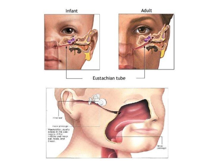
THE EUSTACHIAN TUBE It links the middle ear with the nasopharynx. Length = 36 mm (reached by the age of 7). It runs downwards from the middle ear at 45° and is turned forwards and medially. Medial two-thirds cartilaginous part. Lateral one-third bony. Its narrowest portion is called the isthmus, where the diameter is only 0. 5 mm or less. It ventilates the middle ear and equalizes air pressure on both sides of tympanic membrane
Otitis media with effusion
Otitis media with effusion • Non purulent effusion in the middle ear that may be either mucoid or serous. • Symptoms usually involve hearing loss or aural fullness but typically do not involve pain or fever.
• Pathogenesis: 2 main mechanisms: 1. Malfunctioning of eustachian tube: Eustachian tube fails to aerate the middle ear and is also unable to drain the fluid. 2. Increased secretory activity of middle ear mucosa: Biopsies of middle ear mucosa in these cases have confirmed increase in number of mucus or serous-secreting cells.
• Aetiology: 1. Malfunctioning of the Eustachian tube: like in: – – – Adenoid hypertrophy Chronic rhinosinusitis Chronic tonsillitis Benign and malignant tumours of the nasopharynx: this should be always excluded in unilateral otitis media with effusion in an adult. Palatal defects e. g cleft palate 2. Allergy 3. Unresolved otitis media 4. Viral infections
eustachian tube dysfunction absorption and/or diffusion of nitrogen and oxygen into the middle ear mucosal cells negative pressure develops within the middle ear transudate from the mucosa Non-sterile Non-infectious effusion
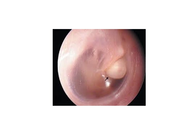
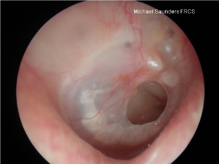
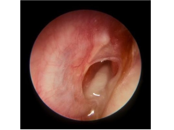
• Clinical features: – Symptoms: hearing loss, delayed and defective speech, mild earache – Otoscopic findings: TM is dull and opaque with loss of light reflex. It may appear yellow, grey or bluish in colour. TM may appear retracted or slightly bulging. Fluid level or air bubbles may appear. Mobility of the TM is restricted. – Hearing tests: audiometry: conductive hearing loss. Tympanometry: flat curve (type B)
• Treatment : – Medical: can be used for up to 12 weeks, e. g decongestents, antiallergic measures, antibiotics, middle ear aeration (Valsalva manoeuvre, chewing gum, otovent®)
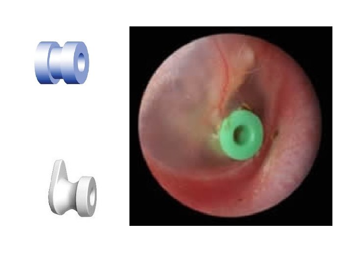
• Treatment: – Surgical: when medical treatment fails, persistent fluid for more than 12 weeks. 1. 2. 3. 4. Myringotomy and aspiration of fluid Grommet (ventilation tube) insertion Tympanotomy or cortical mastoidectomy Surgical treatment of the causative factor: adenoidectomy, tonsillectomy…
Acute otitis media
Acute otitis media • An acute inflammation of middle ear by pyogenic organisms. Here, middle ear implies middle ear cleft, i. e. eustachian tube, middle ear, attic, aditus, antrum and mastoid air cells • More common especially in infants and children of lower socioeconomic group. Typically, the disease follows viral infection of upper respiratory tract but soon the pyogenic organisms invade the middle ear.
• Epidemiology • 70% of all children experience one or more attacks of AOM before their second birthday. • Children aged 6 -11 months appear particularly susceptible to AOM. • most common affliction necessitating medical therapy for children younger than 5 years.
• Routes of infection: 1. 2. 3. Via the Eustachian tube: the most common route, Eustachian tube in infants and young children is shorter, wider and more horizontal and thus may account for higher incidence of infections in this age group. Breast or bottle feeding in a young infant in horizontal position may force fluids through the tube into the middle ear and hence the need to keep the infant propped up with head a little higher. Via the External Ear: through TM perforation Blood borne: uncommon route
• Predisposing factors: 1. Recurrent attacks of common cold, upper respiratory tract infections and exanthematous fevers like measles, diphtheria or whooping cough. 2. Infections of tonsils and adenoids. 3. Chronic rhinitis and sinusitis. 4. Nasal allergy. 5. Tumours of nasopharynx, packing of nose or nasopharynx for epistaxis. 6. Cleft palate.
• Bacteriorlogy: Most common organisms in infants and young children are Streptococcus pneumoniae (30%), Haemophilus influenzae (20%) and Moraxella catarrhalis (12%).
• Viral infections that attack and damage mucosal linings of respiratory tracts may facilitate the ability of the bacteria to become pathogenic in the nasopharynx, eustachian tube, and middle ear cleft. The infection is usually of viral origin
Inflammation in the nasopharynx extends to the medial end of the eustachian tube stasis and inflammation alter the pressure within the middle ear. Stasis also permits pathogenic bacteria to colonize the normally sterile middle ear space
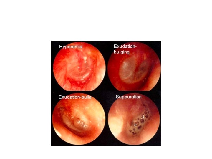
Stages of AOM • • a. Stage of Hyperemia b. Stage of Exudation c. Stage of Suppuration f. Stage of Resolution • d. Stage of Coalescence and Surgical Mastoiditis • e. Stage of Complication
Diagnosis • Signs and symptoms • Although the history of AOM varies with age, a number of constant features manifest during the otitis-prone years, including the following: • Neonates: Irritability or feeding difficulties may be the only indication of a septic focus • Older children: This age group begins to demonstrate a consistent presence of fever and otalgia, or ear tugging • Older children and adults: Hearing loss becomes a constant feature of AOM
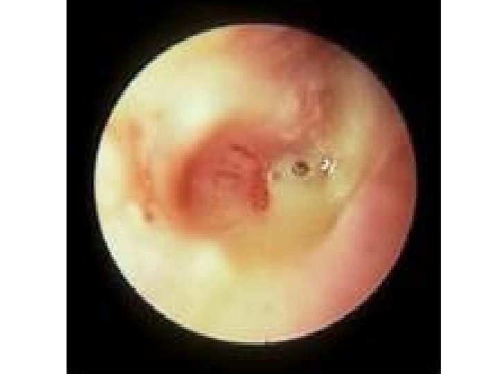
• Otoscopy: – inflammation – Bulging – Perforated tympanic membrane (most frequently in posterior or inferior quadrants).
• • Management : Medical : Amoxicillin is the antibiotic of choice Allergy to penicillin: Trimethoprimsulfamethoxazole , cefixim , macrolides.
• Surgical : – Tympanocentesis – Myringotomy with insertion of a ventilating tube in case of recurrent acute otitis media
Complications • • • • Chronic suppurative otitis media Postauricular abscess Facial nerve paresis Labyrinthitis Labyrinthine fistula Mastoiditis most common complication Temporal abscess Petrositis Intracranial abscess Meningitis Otitic hydrocephalus Sigmoid sinus thrombosis Encephalocele Cerebrospinal fluid (CSF) leak
Acute mastoiditis Management • IV antibiotics • Grommet insertion • Cortical mastoidectomy
Chronic otitis media and Cholesteatoma
INTRODUCTION • Chronic otitis media (COM) implies a permanent abnormality of the pars tensa or flaccida. • Chronic suppurative otitis media (CSOM) is a perforated tympanic membrane with persistent drainage from the middle ear (ie, lasting >6 -12 wk).
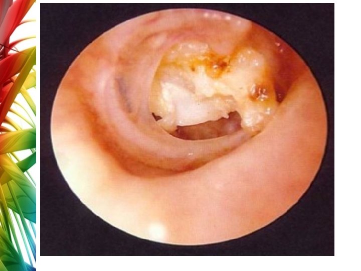
Cholesteatoma • “skin in the wrong place. ” • It is an epidermal inclusion cyst of the middle ear or mastoid That contains the desquamated debris (principally keratin) from their keratinizing, squamous epithelial lining. • Consists of two parts: 1. 2. the matrix, which is made up of keratinizing squamous epithelium resting on a thin stroma of fibrous tissues a central white mass, consisting of keratin debris produced by the matrix
Cholesteatoma • CLASSIFICATION OF CHOLESTEATOMA 1. Congenital 2. Acquired, primary: It is called primary as there is no history of previous otitis media or a pre-existing perforation. Most commonly due to midlle ear negative pressure and formation of attic retraction pocket. 3. Acquired, secondary: there is already a pre-existing perforation in pars tensa, more associated with marginal perforations.
Attic cholesteatoma
• A typical attic retraction cholesteatoma (primary acquired cholesteatoma). • keratinizing epithelium has migrated through a perforation into the middle ear (secondary acquired cholesteatoma) • Behind or within an intact tympanic membrane (congenital cholesteatoma)
Bone erosion in cholesteatoma 1. Enzymatic induced dissolution of bone 2. Pressure necrosis: not widely accepted these days.
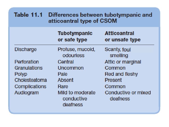
Chronic Suppurative Otitis Media CSOM • a long-standing infection of a part or whole of the middle ear cleft characterized by ear discharge and a permanent perforation. • Types: 1. Tubotympanic: Also called the safe or benign type; it is associated with a central perforation. There is no risk of serious complications. 2. Atticoantral: Also called unsafe or dangerous type; it is associated with an attic or a marginal perforation. The disease is often associated with a bone eroding process such as cholesteatoma, granulations or osteitis. Risk of complications is high in this variety.
Chronic suppurative otitis media without cholesteatoma (tubotympanic disease)
Chronic Suppurative Otitis Media without Cholesteatoma (Tubo-tympanic Disease)
Clinical presentation Otorrhea Hearing loss
• Otorrhea • Profuse, intermittent, mucoid drainage
• Hearing loss : – conductive hearing loss ü 50 to 60 % ümoderate to severe in up to two-thirds. ü low frequencies.
Clinical presentation �Otalgia is uncommon �Complications(Vertigo , facial palsy, …. )
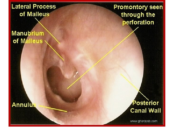
Physical examination : • Inspection. • Otoscopic examination. EAC aural polyps and granulati on tissue Perforation middle ear mucosa • fistula test. • inspection of the nasopharynx • tuning fork bone erosion and ossicular disruption
• AUDIOMETRIC TESTING : • Esp. if ear dry, prior to any surgical intervention. • Degree of hearing loss the extent of the middle ear disease. • Perforations of the TM 15 to 20 Db • ossicular chain damage 30 and 50 d. B.
• RADIOGRAPHIC STUDIES : • Temporal bone CT : § Indications: -Unresponsive to treatment -Cholesteatoma -Suspected complications
INITIAL THERAPY Aural toilet empiric topical antibiotics Ciprofloxacin otic solution
• Surgery : o Otorrhea that persists for longer than 6 weeks despite antibiotic use o Radiographic evidence of chronic mastoiditis, such as coalescent mastoiditis o Conductive hearing loss.
Complications �occur because the infection spreads through natural or pathologic bony dehiscences and vascular channels. �Intratemporal complications: • • Mastoiditis Petrositis (Gradenigo syndrome: VI cranial nerve palsy, ear discharge, severe otalgia or retro orbital pain)) Facial paralysis Labyrinthitis. � Intracranial complications: • • lateral sinus thrombophlebitis Meningitis • intracranial abscess. �Sequelae include hearing loss, acquired cholesteatoma, and tympanosclerosis
Chronic Otitis Media with Cholesteatoma (Attico-Antral Disease)
Epidemiology � 13 -50 per 100, 000 of the population �In US, the overall incidence is 7 per 100, 000 children per year in children �Both males and females are affected, with a ratio of 3: 2 �Increase incidence in poor socioeconomic standards , poor nutrition , lack of health education and increase smoking. �More aggressive in children
Bacteriology • Mixed aerobic and Anaerobic
Diagnosis • The diagnosis of aural cholesteatoma is made on otoscopic examination, including endoscopic and microscopic evaluation or surgical exploration • Special imaging procedures, such as HRCT and MRI, may suggest the presence of cholesteatomas within the temporal bone, and may be used to complement the clinical examination • High-resolution CT is useful for operative planning and is recommended for all revision mastoid operations
Symptoms • Conductive hearing loss (80%) • Scanty thin otorrhea (Foul smell) • Complications of a cholesteatoma • Otalgia is uncommon
Physical Examination • Otologic Exam (Gold standard for diagnosis) • Take any cultures if indicated • Remove any debris in canal • Pneumatic Otoscopy Fistula test Positive suggests erosion of inner ear, most commonly LSCC. • Tuning Fork Exam • Neurological Exam (Cranial Nerves, especially CN 7)
PTA
�CT scan (Fine cuts axialcoronal) �MRI in case of suspicion of complications.
Complications �Intratemporal complications: o Petrositis (Gradenigo syndrome) o Facial paralysis o Labyrinthitis. �Intracranial complications: o lateral sinus thrombophlebitis o Meningitis o intracranial abscess
Management Surgery in the form of Mastoidectomy is the main stay of treatment in case of cholesteatoma
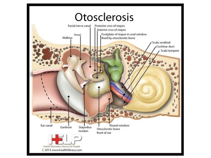
Otosclerosis
Definition • Localized fibrous dystrophy of the bony otic capsule characterized by alternating phases of bone resorption & formation • Most often, otosclerotic focus involves the stapes region leading to stapes fixation and conductive deafness. However, it may involve certain other areas of the bony labyrinth where it may cause sensory-neural hearing loss or no symptoms at all.
Epidemiology • Incidence of clinical otosclerosis 0. 5% to 1. 0% • 50% of patients have family history of otosclerosis • If a person has +ve family history of otosclerosis there is 60% chance of developing Otosclerosis • Bilateral DX: 80%
• Presenting age: 20 - 30 • Women: men 2: 1 • About 50% of otosclerotics have positive family history; rest are sporadic. • Hormonal influences during pregnancy and menopause may cause a more rapid progression in women, bringing them to clinical attention sooner.
• Symptoms 1. Hearing loss. This is the presenting symptom and usually starts in twenties. It is painless and progressive with insidious onset. Often it is bilateral conductive type. 2. Paracusis Willisii. An otosclerotic patient hears better in noisy than in quiet surroundings. This is because a normal person will raise his voice in noisy surroundings. 3. Tinnitus. It is more commonly seen in cochlear otosclerosis and in active lesions. 4. Vertigo. It is an uncommon symptom. 5. Speech. Patient has a monotonous, well-modulated soft speech.
Audiometric findings: Pure-tone audiometry: • pure CHL seen in a completely fixed stapes is limited to a 60 to 65 -d. B (More at the lower frequency = raising configuration) • Carhart notch: loss of 20 -30 d. B in bone conduction at 2 k. Hz (non-specific, and is an artifact not a true loss) • Definitive diagnosis is via exploratory tympanotomy
Management • Surgical: stapedotomy/ stapedectomy – Placement of prosthesis is the treatment of choice. The fixed otosclerotic stapes is removed and a prosthesis inserted between the incus and oval window Absolute Contra indications: » Only hearing ear. » Endolymph hydrops/ Meniere disease » Cochlear otosclerosis • Amplification: Hearing aids, in case the patient is unfit of refused surgery.