PANCREAS The pancreas is a glandular organ in


/sympathetic (NE) GLP‐ 1 = glucagon‐like peptide 1 ATP =")


 is a 37–amino acid peptide hormone that is")
 is a 36 amino acid peptide v PP secreted by F")

 is located behind the sternum and")
- Slides: 23
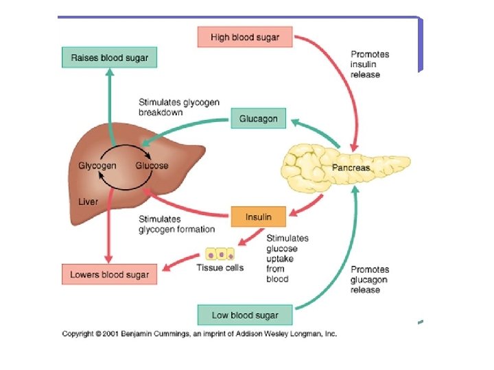
PANCREAS The pancreas is a glandular organ in the upper abdomen posterior to stomach. It serves as two glands in one: a digestive exocrine gland a hormone‐producing endocrine gland. Functioning as an exocrine gland, the pancreas excretes enzymes to break down the proteins, lipids, carbohydrates in food. Functioning as an endocrine gland, the pancreas secretes the hormones insulin and glucagon to control blood sugar levels throughout the day
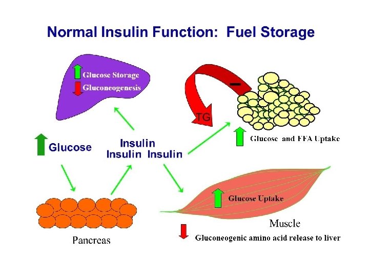
Unstable if taken orally, metabolised by the kidney Insulin maintains normal blood glucose levels by Øfacilitating cellular glucose uptake Øregulating carbohydrate, lipid and protein metabolism Øpromoting cell division and growth through its mitogenic effects.
INSULIN SECRETION IN BETA CELLS
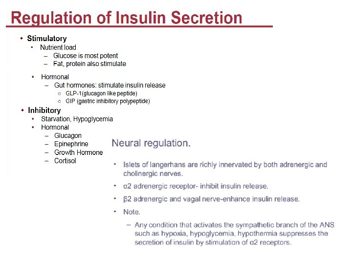
REGULATION OF INSULIN SECRETION Parasympathetic(AC)/sympathetic (NE) GLP‐ 1 = glucagon‐like peptide 1 ATP = adenosine triphosphate ADP = adenosine diphosphate c. AMP= cyclic adenosine monophosphate PACAP = pituitary adenylate cyclase‐activating polypeptide DAG = diacylglycerol GIP = gastric inhibitory peptide / glucose‐dependent insulinotropic polypeptide PKC = protein kinase C Glucose‐ 6‐ P = glucose 6 phosphate GLUT 2 = glucose transport protein 2 GLUT 4 = glucose transport protein 4 VIP= vasoactive intestinal peptide
RAS independent RAS dependent RTK Akt/PKB = protein kinase B PIPD 1 & 2 = phosphatidylinositol dependent protein kinases 1 & 2 IRS = insulin receptor substrate ADP = adenosine diphosphate MAP kinase = mitogen activated protein kinase c. AMP= cyclic adenosine monophosphate DAG = diacylglycerol PI 3 K = phosphatidylinositol 3 -kinase PKC = protein kinase C RAS = rat sarcoma protein GLUT 2 = glucose transport protein 2 SHC =adaptor protein with src-homology GLUT 4 = glucose transport protein 4 Increased protein synthesis- increased tissue growth
Functions of Insulin
Diabetes/ Diabetes mellitus, describes a group of metabolic diseases in which the person has high blood glucose (blood sugar), either because insulin production is inadequate, or because the body's cells do not respond properly to insulin, or both.
Increased breathing to compensate for increased plasma Pa. CO 2 and decreased p. H
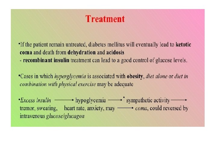
DIAGNOSIS • Urinary Glucose • Fasting Blood Glucose and Insulin Levels. • FBG levels in the early morning is normally 80‐ 90 mg/100 ml. • FBG above 110 mg/100 ml often indicates diabetes • In type I diabetes, plasma insulin levels are very low or undetectable during fasting and even after a meal. • Acetone Breath • Increased Acetoacetic acid in the blood is converted to acetone. This is volatile and vaporized into the expired air. • Consequently, one can frequently make a diagnosis of type I diabetes mellitus simply by smelling acetone on the breath of a patient.
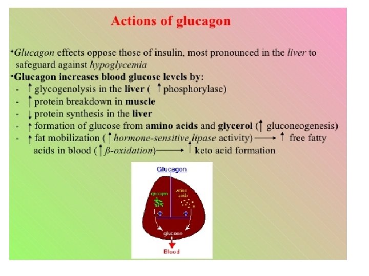
Glucagon is a 29 amino acid protein with a very short half-life in the blood. Glucagon serves as the counter-balancing hormone to insulin, having largely the opposite effects. Insulin is secreted and active during feeding and elevated blood glucose, ensuring storage of glucose in liver and other tissues (anabolic activity) glucagon ensures the release of glucose from liver when blood glucose is low during fasting and exercise (catabolic activity).
glucose-dependent regulation of glucagon secretion in the α-cell. ØGlucose is incorporated into the α‐cell by the transporter SLC 2 A 1. ØAt low‐glucose concentrations, the moderate activity of KATP channels situates the α‐cell membrane potential in a range that allows the opening of voltage‐dependent T‐ and N‐type Ca 2+ channels and voltage‐dependent Na+ channels. ØTheir activation triggers action potentials, Ca 2+ influx and exocytosis of glucagon granules.
GPCRs, β‐adrenergic receptor Glucagon receptors are mainly expressed in liver and in kidney with lesser amounts found in heart, adipose tissue, spleen, thymus, adrenal glands, pancreas, cerebral cortex, and gastrointestinal tract. The binding of glucagon to the receptors results in activation of adenylyl cyclase and generation of the second messenger cyclic AMP, which in turn activates protein kinase, leading to phosphorylation that results in the activation or deactivation of a number of enzymes.
The hormones glucagon, epinephrine, and insulin all bind reversible to receptors on the cell surface. The binding of glucagon and epinephrine ultimately lead to protein phosphorylation by the activation of protein kinases while the binding of insulin ultimately leads to activation of protein phosphatases which remove the phosphate groups from enzymes.
Amylin (islet amyloid polypeptide, or IAPP) is a 37–amino acid peptide hormone that is cosecreted with insulin from the pancreatic β‐cell and is thus deficient in diabetic people. It inhibits glucagon secretion, delays gastric emptying, and acts as a satiety agent. A stable analog, pramlintide, new approach for diabetes treatment Its analogous Gastric inhibitory polypeptide (GIP) and glucagon‐like peptide‐ 1 (GLP‐ 1), secreted from the intestine on ingestion of glucose or nutrients to stimulate insulin secretion from pancreatic β cells. Both GLP 1 and Amylin have anti‐obesity potential Gastric grinding, liquefication
Pancreatic Polypeptide (PP) is a 36 amino acid peptide v PP secreted by F cells & belongs to the family of PYY & NPY peptides. v PP has effects on GI motility, metabolism and food intake and is stimulated by vagus nerve. v Acts as satiety factor, PP secretion is absent in obese children with Prader‐Willi syndrome. v Its primary action on the exocrine pancreas is to inhibit secretion in vivo by acting on receptors in the brain leading to inhibition of vagal output to the pancreas.
Diseases of pancreas • Pancreatitis -inflammation of the pancreas that occurs when pancreatic enzyme secretions build up and begin to digest the organ itself. It can occur as acute painful attacks lasting a matter of days, or it may be a chronic condition that progresses over a period of years. • Precursors to Pancreatic Cancer-known risk factors that increase the risk of developing the disease are cigarette smoking, a family history of pancreatic cancer or hereditary cancer syndromes, and chronic pancreatitis are some of these factors. In addition, certain pancreatic lesions such as Intraductal Papillary Mucinous Neoplasms (IPMNs) and Pancreatic Intraepithelial Neoplasia (Pan. IN) are considered precursors to pancreatic cancer. • Pancreatic Cancer-The most common form of pancreatic cancer is pancreatic adenocarcinoma, an exocrine tumor arising from the cells lining the pancreatic duct. A far less common form, endocrine tumors, account for less than 5% of all pancreatic tumors and are sometimes referred to as neuroendocrine or islet cell tumors.
The thymus gland (endocrine as well as lymphatic) is located behind the sternum and between lungs, is only active until puberty. The thymus (a gland associated with the immune system), is enclosed in a capsule and divided internally by cross‐walls into many lobules (full of T‐lymphocytes). In relation to body size thymus is largest at birth. It doubles in size by puberty, after which it gradually shrinks, its functional tissue being replaced by fatty tissue In infancy the thymus controls the development of lymphoid tissue and the immune response to microbes and foreign proteins. T‐lymphocytes migrate from the bone marrow to the thymus, where they mature and differentiate until activated by antigen. Thymosin Activates the immune system by activating the T‐Cells (T‐Killer Cells; T‐Helper Cells; T‐Memory Cells). helps the body protect itself against autoimmunity, There are two main kinds of thymus cancer: thymoma and thymic carcinoma, and both are rare.