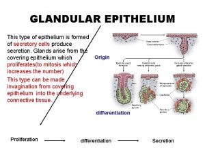
Glandular Epithelium Origin differentiation Types of glandular epithelium


- Slides: 24

Glandular Epithelium Origin differentiation

Types of glandular epithelium It is classified according to: 1 - Number of cells 2 - Presence or absence of a duct system 3 - Mode of secretion (mechanism) 4 - Nature of secretion 5 - Shape of the secretory portion 6 - Branching of duct

Number of cells Unicellular (goblet cell) Multicellular (Most of the glands e. g. Salivary glands)

Mechanism (Mode) of Glandular secretions • Merocrine glands • The secretion released through exocytosis e. g. Pancreas • Apocrine glands The secretion involves the loss of both product and apical cytoplasm e. g. Mammary glands . Holocrine gland The secretion destroys the cell Sebaceous glands

Presence of a duct system Exocrine Endocrine mixed

Nature of Glandular secretions - Serous glands: parotid gland -Mucous glands: sublingual gland -Mixed glands: submandibular gland -Glands with special secretion: • sebaceous gland (oily secretion) • lacrimal gland watery secretion • Mammary gland : Milk secretion • Glands in the ear : wax

shape of secretory portion o-

Classification of Tubular Glands Intestinal glands Sweat glands Fundic glands

Classification of Alveolar Glands Sebaceous glands Tarsal glands

Classification of Compound Glands Compound: branched duct, branched secretory portion Liver mammary glands salivary glands

Goblet cells • Unicellular • Exocrine • Shape of the cell : flask shape with basal nuclei • Mode of secretion: Merocrine • Nature of secretion : Mucus • Site : Respiratory system , GIT

Sebaceous gland • • Exocrine Mode : Holocrine Nature : (oily secretion) Shape of secretory units : Branched alveolar • Site : Related to hair follicles • Activity of the gland increase at the age of puberty • Obstruction of the duct by thick secretion & keratin Acne

Mammary gland • • Exocrine Mode : Apocrine Nature : (milk secretion) Shape of secretory units : Compound alveolar • Site : Related to skin

Special types of epithelium • 1 -Neuroepithelium • E. g. Taste buds • Site : dorsal surface of the tongue • Function : sensation

Special types of epithelium 2. Germinal epithelium Testis: sperm Ovary: ovum Function: Reproduction :

3 - Myoepithelium Shape : Irregular with many processes Contain actin & myosin in the cytoplasm Site : Acini & ducts of the gland Function : Contraction for squeezing the secretion

Epithelial polarity • Cells have a top , lateral side and a bottom • So different activities take place at different places • Apical modifications • Basal modifications • Lateral modifications

Apical modifications • Cilia • Microvilli • Stereocilia

Apical modifications

Intercellular junctions (cell to cell adhesion) • The intercellular junctions are more numerous between the epithelial cells. They are three types 1 - Occluding junctions: (Tight ) link cells to form an impermeable barrier. 2 - Anchoring junctions: (Adhering) • provide mechanical stability to the epithelial cells. • • Zonula adherens: Macula adherens = desmosomes: 3 - Communicating junctions: (Gap) allow movement of molecules between cells It permits the exchange of molecules e. g. ions, amino acids allowing integration, communication and coordination between cells It is found mainly in cardiac and smooth muscle cells

Intercellular junctions

Basal modifications • Basement membrane • Basal infolding • Hemidesmosome

Basement membrane • Thin extracellular layer having two parts: • Basal lamina : type IV collagen + laminin • Produced by epithelial cell • Reticular lamina : Type VII collagen + type III collagen (reticular F) • Secreted by C. T. cells Function : 1. Attach epithelium to C. T. 2. Separate epithelium from other tissue 3. Regulate (filter) substances passing from C. T. to epithelium 4. Guide during tissue regeneration

 Characteristics of glandular epithelium
 Tissue fixation
 Insufficient glandular tissue pictures
 Site:slidetodoc.com
 Testicul histologie
 Estomago de ave
 Epitelio glandular holocrino
 Glandular meats
 Glandular epithelia
 Pseudostratified columnar epithelium
 Labiaceous hair
 Somatostatin
 Holocrinas merocrinas y apocrinas
 Tejido conectivo fibroso
 Simple branched acinar gland
 Que es el sistema glandular
 Parakeratinised
 Palate epithelium
 Wida language objectives examples
 Difference between udl and differentiation
 High accuracy differentiation formulas
 Algebraic differentiation
 Porters generic competitive strategies
 Social differentiation definition
 T-tess goal setting template
