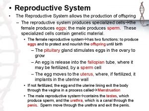
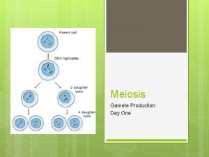
Reproductive System Reproductive system functions in gamete Production


- Slides: 44

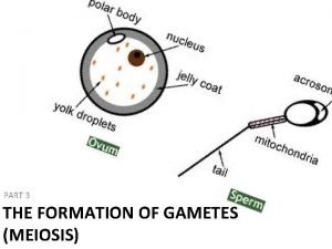
Reproductive System • Reproductive system functions in gamete • Production • Storage • Nourishment • Transport • Fertilization • Fusion of male and female gametes to form a zygote Copyright © 2004 Pearson Education, Inc. , publishing as Benjamin Cummings

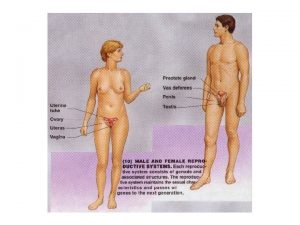
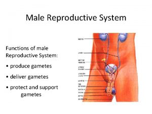
Male Reproductive System • Pathway of spermatozoa • Epididymis • Ductus deferens (Vas deferens) • Ejaculatory duct • Accessory organs • Seminal vesicles • Prostate gland • Bulbourethral glands • Scrotal sac encloses testes • Penis Copyright © 2004 Pearson Education, Inc. , publishing as Benjamin Cummings

Figure 28. 1 The Male Reproductive System Copyright © 2004 Pearson Education, Inc. , publishing as Benjamin Cummings Figure 28. 1

Figure 28. 3 The Male Reproductive System in Anterior View Copyright © 2004 Pearson Education, Inc. , publishing as Benjamin Cummings Figure 28. 3

Figure 28. 4 The Structure of the Testes Copyright © 2004 Pearson Education, Inc. , publishing as Benjamin Cummings Figure 28. 4

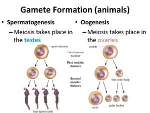
Spermatogenesis • Seminiferous tubules • Contain spermatogonia • Stem cells involved in spermatogenesis • Contain sustentacular cells • Sustain and promote development of sperm Copyright © 2004 Pearson Education, Inc. , publishing as Benjamin Cummings

Figure 28. 5 The Seminiferous Tubules Copyright © 2004 Pearson Education, Inc. , publishing as Benjamin Cummings Figure 28. 5 a, b

Figure 28. 5 The Seminiferous Tubules Copyright © 2004 Pearson Education, Inc. , publishing as Benjamin Cummings Figure 28. 5 c

Figure 28. 7 Spermatogenesis Copyright © 2004 Pearson Education, Inc. , publishing as Benjamin Cummings Figure 28. 7

Figure 28. 8 Spermiogenesis and Spermatozoon Structure Copyright © 2004 Pearson Education, Inc. , publishing as Benjamin Cummings Figure 28. 8

Male reproductive tract • Testes produce mature spermatozoa • Sperm enter epididymus • Elongated tubule with head, body and tail regions • Monitors and adjusts fluid in seminiferous tubules • Stores and protects spermatozoa • Facilitates functional maturation of spermatozoa Copyright © 2004 Pearson Education, Inc. , publishing as Benjamin Cummings

Figure 28. 9 The Epididymus Copyright © 2004 Pearson Education, Inc. , publishing as Benjamin Cummings Figure 28. 9

Accessory glands • Seminal vesicles • Active secretory gland • Contributes ~60% total volume of semen • Secretions contain fructose, prostaglandins, fibrinogen Copyright © 2004 Pearson Education, Inc. , publishing as Benjamin Cummings

Accessory glands • Prostate gland • Secretes slightly acidic prostate fluid • Bulbourethral glands • Secrete alkaline mucus with lubricating properties Copyright © 2004 Pearson Education, Inc. , publishing as Benjamin Cummings

Figure 28. 10 The Ductus Deferens and Accessory Glands Copyright © 2004 Pearson Education, Inc. , publishing as Benjamin Cummings Figure 28. 10 a-e

Contents of Semen • Typical ejaculate = 2 -5 ml fluid • Contains between 20 – 100 million spermatozoa per ml • Seminal fluid • A distinct ionic and nutritive glandular secretion Copyright © 2004 Pearson Education, Inc. , publishing as Benjamin Cummings

Figure 28. 11 The Penis Copyright © 2004 Pearson Education, Inc. , publishing as Benjamin Cummings Figure 28. 11

Hormones and male reproductive function • FSH (Follicle stimulating hormone) • Targets sustentacular cells to promote spermatogenesis • LH (leutinizing hormone) • Causes secretion of testosterone and other androgens • Gn. RH (Gonadotropin releasing hormone) • Testosterone • Most important androgen PLAY Animation: Male Reprroductive System Flythrough Copyright © 2004 Pearson Education, Inc. , publishing as Benjamin Cummings

Figure 28. 12 Hormonal Feedback and the Regulation of the Male Reproductive Function Copyright © 2004 Pearson Education, Inc. , publishing as Benjamin Cummings Figure 28. 12

SECTION 28 -3 The Reproductive System of the Female Copyright © 2004 Pearson Education, Inc. , publishing as Benjamin Cummings

Principle organs of the female reproductive system • Ovaries • Uterine tubes • Uterus • Vagina Copyright © 2004 Pearson Education, Inc. , publishing as Benjamin Cummings

Figure 28. 13 The Female Reproductive System Copyright © 2004 Pearson Education, Inc. , publishing as Benjamin Cummings Figure 28. 13

Figure 28. 14 The Ovaries and Their Relationships to the Uterine Tube and Uterus Copyright © 2004 Pearson Education, Inc. , publishing as Benjamin Cummings Figure 28. 14 a, b

Oogenesis • Ovum production • Occurs monthly in ovarian follicles • Part of ovarian cycle • Follicular phase (preovulatory) • Luteal phase (postovulatory) Copyright © 2004 Pearson Education, Inc. , publishing as Benjamin Cummings

Figure 28. 15 Oogenesis Copyright © 2004 Pearson Education, Inc. , publishing as Benjamin Cummings Figure 28. 15

Figure 28. 16 The Ovarian Cycle Copyright © 2004 Pearson Education, Inc. , publishing as Benjamin Cummings Figure 28. 16

Figure 28. 16 The Ovarian Cycle Copyright © 2004 Pearson Education, Inc. , publishing as Benjamin Cummings Figure 28. 16

Figure 28. 17 The Uterine Tubes Copyright © 2004 Pearson Education, Inc. , publishing as Benjamin Cummings Figure 28. 17 a-c

The uterus • Muscular organ • Mechanical protection • Nutritional support • Waste removal for the developing embryo and fetus • Supported by the broad ligament and 3 pairs of suspensory ligaments Copyright © 2004 Pearson Education, Inc. , publishing as Benjamin Cummings

Uterine wall consists of three layers: • Myometrium – outer muscular layer • Endometrium – a thin, inner, glandular mucosa • Perimetrium – an incomplete serosa continuous with the peritoneum Copyright © 2004 Pearson Education, Inc. , publishing as Benjamin Cummings

Figure 28. 18 The Uterus Copyright © 2004 Pearson Education, Inc. , publishing as Benjamin Cummings Figure 28. 18 a, b

Figure 28. 18 The Uterus Copyright © 2004 Pearson Education, Inc. , publishing as Benjamin Cummings Figure 28. 18 c

Figure 28. 19 The Uterine Wall Copyright © 2004 Pearson Education, Inc. , publishing as Benjamin Cummings Figure 28. 19 a

Figure 28. 19 The Uterine Wall Copyright © 2004 Pearson Education, Inc. , publishing as Benjamin Cummings Figure 28. 19 b

Uterine cycle • Repeating series of changes in the endometrium • Continues from menarche to menopause • Menses • Degeneration of the endometrium • Menstruation • Proliferative phase • Restoration of the endometrium • Secretory phase • Endometrial glands enlarge and accelerate their rates of secretion Copyright © 2004 Pearson Education, Inc. , publishing as Benjamin Cummings

Figure 28. 20 The Uterine Cycle Copyright © 2004 Pearson Education, Inc. , publishing as Benjamin Cummings Figure 28. 20

External genitalia • Vulva • Vestibule • Labia minora and majora • Paraurethral glands • Clitoris • Lesser and greater vestibular glands Copyright © 2004 Pearson Education, Inc. , publishing as Benjamin Cummings

Figure 28. 22 The Female External Genitalia Copyright © 2004 Pearson Education, Inc. , publishing as Benjamin Cummings Figure 28. 22

Figure 28. 23 The Mammary Glands Copyright © 2004 Pearson Education, Inc. , publishing as Benjamin Cummings Figure 28. 23 a-c

Hormones of the female reproductive cycle • Control the reproductive cycle • Coordinate the ovarian and uterine cycles Copyright © 2004 Pearson Education, Inc. , publishing as Benjamin Cummings

Hormones of the female reproductive cycle • Key hormones include: • FSH • Stimulates follicular development • LH • Maintains structure and secretory function of corpus luteum • Estrogens • Have multiple functions • Progesterones • Stimulate endometrial growth and secretion Copyright © 2004 Pearson Education, Inc. , publishing as Benjamin Cummings

Figure 28. 25 The Hormonal Regulation of Ovarian Activity Copyright © 2004 Pearson Education, Inc. , publishing as Benjamin Cummings Figure 28. 25

Figure 28. 26 The Hormonal Regulation of the Female Reproductive Cycle Copyright © 2004 Pearson Education, Inc. , publishing as Benjamin Cummings Figure 28. 26 a-c

Figure 28. 26 The Hormonal Regulation of the Female Reproductive Cycle PLAY Animation: Regulation of the Female Reproductive Cycle Copyright © 2004 Pearson Education, Inc. , publishing as Benjamin Cummings Figure 28. 26 d-f
 Gamete production
 Chromosomes form tetrads during
 Diagram alur produksi
 Gamete vs zygote
 Gametes vs somatic cells
 Gamete in meiosis
 Gamete intrafallopian transfer
 What is gamete
 Intra fallopian transfer
 Ch
Ch What is the difference between somatic and gamete cells
 Gamete
 Meiosis facts
 Gametophytes have gamete-producing organs called _____.
 Female gamete
 Gàmete
 Http://www.cellsalive.com/
 Lucy and maria twins
 What are gametes
 Mendel law
 Male and female gamete
 Somatic mutations
 Is it male or female
 Male reproductive system and its function
 Sperm duct
 Male reproductive system diagram
 Main function of female reproductive system
 Functions of testis
 Ram reproductive system
 Labeled female reproductive system
 Endocrine system and reproductive system
 Absolute value as a piecewise function
 Evaluating functions and operations on functions
 Evaluating functions and operations on functions
 Function of the vagina
 Development of female reproductive system
 Female reproductive system with baby
 Ovarian ligament.
 Anatomy of the reproductive system exercise 42
 Ovarian ligament.
 Male reproductive system lateral view
 Female reproductive system pregnancy
 Epidiymitis
 Dot quizlet
 Reproductive system jeopardy
