GI Pathology I Case 3 37 yearold man

 and labeled structures Describe the gross morphologic changes Diagnosis?")
 The surgical specimen (opened)")

 A close up view of the")


 Description:")

 Associated with a perforated")


- Slides: 17
GI Pathology I, Case 3 • 37 -year-old man presents with epigastric pain relieved by eating. • Stool is positive for occult blood
Identify the organ(s) and labeled structures Describe the gross morphologic changes Diagnosis?
A Pylorus B Pyloric Ring C Ulcer Peptic Ulcer (Duodenum) The surgical specimen (opened) of the distal stomach and proximal duodenum reveals an irregular ulcer of the duodenal mucosa. The ulcer is filled with clotted blood. The duodenal mucosa is stained with blood.
Identify the structures
A Artery B Ulcer Bed Peptic Ulcer (Duodenum) A close up view of the duodenal mucosa reveals a sharply demarcated peptic ulcer. The ulcer bed contains a ruptured small artery.
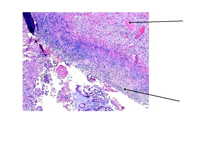
Identify the organ. Describe the histologic findings
Duodenal mucosa Brunner’s Glands Ulcer
Floor of ulcer with granulation tissue fibrosis Superficial surface of ulcer with debris, acute inflammatory cells
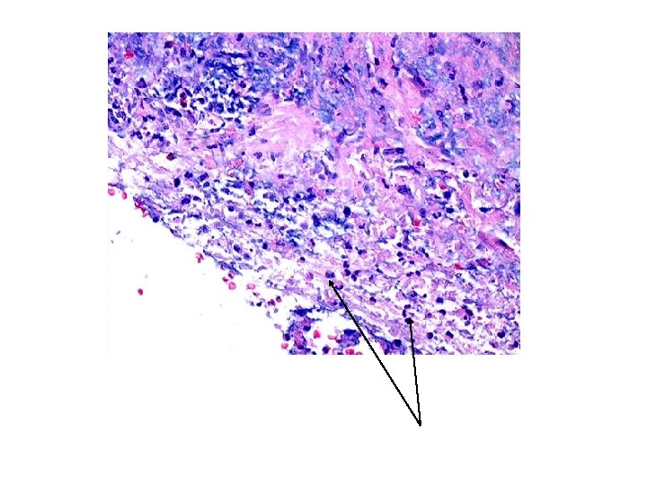
Ulcer surface neutrophils
Identify the organs and structures Diagnosis?
A B C D E Esophagus Liver Gallbladder Spleen Stomach Peptic Ulcer (Duodenum) Description: This autopsy specimen of the upper gastrointestinal tract, liver, and spleen, reveals a perforated (probe) peptic ulcer of the proximal duodenum. The probe passes from the gastric lumen to the exterior of the duodenum. The liver is folded back to reveal the gall bladder and ulcer.
Identify the abnormality
Free air in the abdomen (air under the right diaphragm) Associated with a perforated viscus (example: perforated peptic ulcer)
What is the most probable diagnosis?
Melena The specimen consists of a black tarry stool passed per anus. Note the mahogany color at the edge of the specimen (filter paper).