Xlinked Hypophosphatemic Rickets and its Treatment with AntiFGF


, Aluminum-containing antacids")


 is the most well characterized phosphatonin. Ø It")


")
 in a 48 -yearold woman with Xlinked hypophosphatemic")


 Ø")


")


 was cloned as")

 is a phosphaturic hormone produced by")


2 D by suppressing the expression of")
 is caused by impaired")


. Burosumab is")

 in the")


 was supported by a randomized, double-blind, placebo-controlled")


 blocking antibody")

 in pediatric XLH patients are: headache,")

 have been reported in")
 The recommended starting")


 serum phosphorus levels from")

 total RSS was 2. 9 (1. 37) in")
 serum total alkaline phosphatase activity")

 is a randomized, double-blind, placebocontrolled study in 134 adult XLH")
 is a 48 -week, open-label, single-arm study in 14 adult")
 serum phosphorus was 1. 9 (0. 32)")
 ratio of renal tubular maximum reabsorption rate of phosphate")


- Slides: 133
X-linked Hypophosphatemic Rickets and its Treatment with Anti-FGF 23 Bita Alimardani
Causes of Rickets Vitamin D disorders Nutritional vitamin D deficiency; Congenital vitamin D deficiency; Secondary vitamin D deficiency; Malabsorption ; Increased degradation; Decreased liver 25 -hydroxylase; Vitamin D-dependent rickets type 1; Vitamin D-dependent rickets type 2; Chronic renal failure Calcium deficiency: Low intake, Calcium deficient Diet, Premature infants (rickets of prematurity), Malabsorption, Dietary inhibitors of calcium absorption
Causes of Rickets Phosphorus deficiency inadequate intake, Premature infants (rickets of prematurity), Aluminum-containing antacids Renal Losses X-linked hypophosphatemic rickets; Autosomal dominant hypophosphatemic rickets; Hereditary hypophosphatemic rickets with hypercalciuria; Overproduction of phosphatonin (Tumor-induced rickets, Mc. Cune-Albright syndrome, Epidermal nevus syndrome, Neurofibromatosis), Fanconi syndrome, Dent disease Distal Renal Tubular Acidosis
Clinical features of Rickets GENERAL Failure to thrive; Listlessness; Protuding abdomen; Muscle weakness (especially proximal); Fractures. HEAD Craniotabes; Frontal bossing; Delayed fontanelle closure; Delayed dentition; caries; Craniosynostosis CHEST Rachitic rosary; Harrison groove; Respiratory infections and atelectasis BACK Scoliosis , Kyphosis , Lordosis EXTREMITIES Enlargement of wrists and ankles; Valgus or varus deformities Windswept deformity (combination of valgus deformity of 1 leg with varus deformity of the other leg); Anterior bowing of the tibia and femur; Coxa vara; Leg pain. HYPOCALCEMIC SYMPTOMS Tetany ; Seizures; Stridor due to laryngeal spasm
Extra skeletal findings in Rickets Extraskeletal manifestation of rickets vary depending upon the mineral deficiency. Hypoplasia of the dental enamel is typical for hypocalcemic rickets, whereas abscesses of the teeth occur more often in phosphopenic rickets. Hypocalcemic seizures, decreased muscle tone leading to delayed motor milestones, recurrent infections, increased sweating.
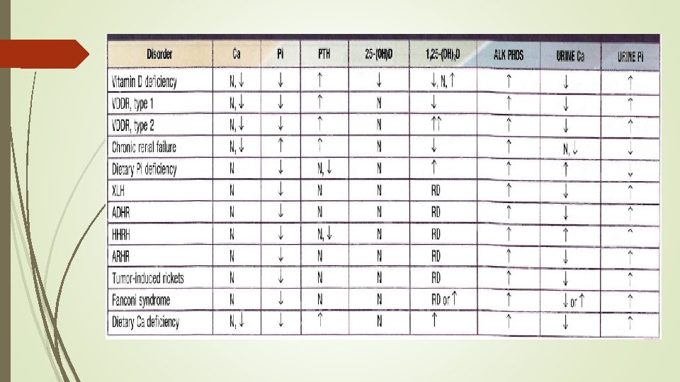
Biochemical findings in rickets Alkaline phosphatase usually is ↑in all forms of rickets. Serum phosphorus concentrations usually are↓ in both hypocalcemic and hypophosphatemic rickets. Serum Ca is ↓only in hypocalcemic rickets. Serum parathyroid hormone typically is ↑in hypocalcemic rickets, in contrast it is N in hypophosphatemic rickets. 25 -OH vitamin D reflect the amount of vitamin D stored in the body, and is ↓in vit D deficiency. 1, 25 -OH 2 vitamin D can be↓, N or ↑in hypocalcemic rickets and usually is N or slightly ↑in hypophosphatemic rickets.
Hypophosphatemic rickets
History Ø It was first described in 1937 by Albright etal who reported that patients afflicted by rickets did not respond to usual vitamin-D treatment Ø Most of these cases were caused by renal phosphate wasting, leading to the alternate name of "phosphate diabetes. " Ø This disorder is now called hereditary hypophosphatemic rickets because the primary problem is phosphate wasting rather than true vitamin D resistance.
ETIOLOGY Genetic hypophosphatemic rickets Ø X-linked hypophosphatemic rickets Ø Autosomal dominant hypophosphatemic rickets Ø Autosomal recessive hypophosphatemic rickets Ø Fibrous dysplasia Ø Epidermal nevus syndrome Ø Hereditary hypophosphatemic rickets with hypercalciuria Aquired hypophosphatemia Ø Ø Tumor induced osteomalacia Fanconi syndrome
In addition to hypophosphatemia, these disorders have: normal calcium Normal PTH Most have high levels of fibroblast growth factor 23 (FGF 23), a circulating phosphatonin
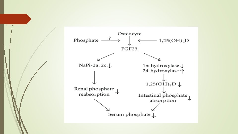
Phosphatonin Ø Fibroblast growth factor-23 (FGF-23) is the most well characterized phosphatonin. Ø It is a humoral mediator that decreases renal tubular reabsorption of phosphate and decreases the activity of renal 1α-hydroxylase
Hormonal Response to hypo. Pi
X-LINKED hypophosphatemic rickets
prevalence Ø approximately one case per 20, 000 live births Ø X-linked form is most common Ø other forms are rare
Pathogenesis Ø not fully understood Ø caused by one or more circulating factors rather than by a defect in the kidney Ø termed “ phosphatonins" or "minhibins"
Phosphatonin Ø is a humoral mediator that decreases renal tubular reabsorption of phosphate Ø decreases the activity of renal 1α-hydroxylase Ø FGF-23 is the most well characterized phosphatonin
Ø The principal phosphatonin involved in the pathogenesis of XLH is fibroblast growth factor 23 (FGF 23) Ø FGF 23 acts as a counter-regulatory hormone to inhibit phosphate reabsorption by the sodium/phosphate cotransporter in the kidney Ø Mutations in PHEX alter the degradation and production of FGF 23 causing increased circulating levels of the FGF 23, acting through specific FGF receptors with the important co-factor klotho protein
Ø The gene responsible is on chromosome Xp 22. 1 Ø named PHEX (Phosphate Regulating Endopeptidase on the X chromosome) Ø PHEX mutations were found in 87 percent of familial cases and 72 percent of sporadic cases Ø PHEX is expressed predominantly in bone and teeth
Diagnosis
The diagnosis is based on Ø clinical findings Ø radiographic findings Ø biochemical testing Ø family history Ø Determination of the PHEX mutation may be useful
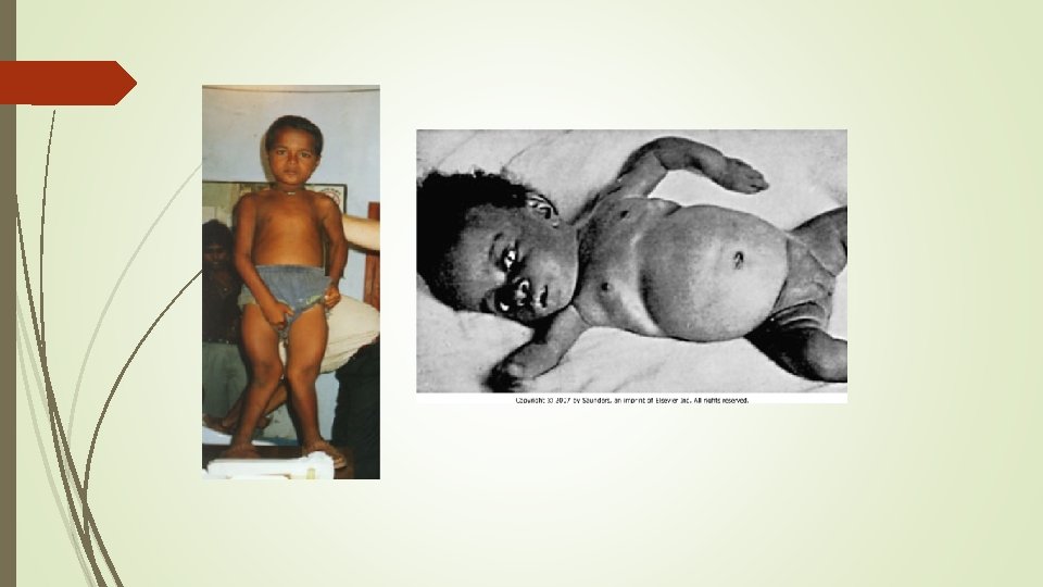
Clinical features in child Ø first finding frontal bossing appear in 6 months due to sagittal suture synostosis Ø slow growth Ø rickets and osteomalacia Ø leg deformities , bowing initiates at around 18 months of age Ø Delayed dentition and tooth abscesses in young Ø craniosynostosis Ø muscle weakness and waddle gait
Clinical features in adults Ø bone pain Ø Enthesopathy and calcification of tendons, ligaments Østress-fractures Ø short stature Ø Hypertension an association between hypertension and hyperparathyroidism Ø left ventricular hypertrophy Ø sensorineural hearing loss
Clinical features Ø abnormalities of the lower extremities and poor growth are the dominant features Ø Some have hypophosphatemia and short stature without clinically evident bone disease
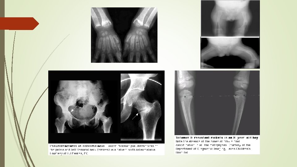
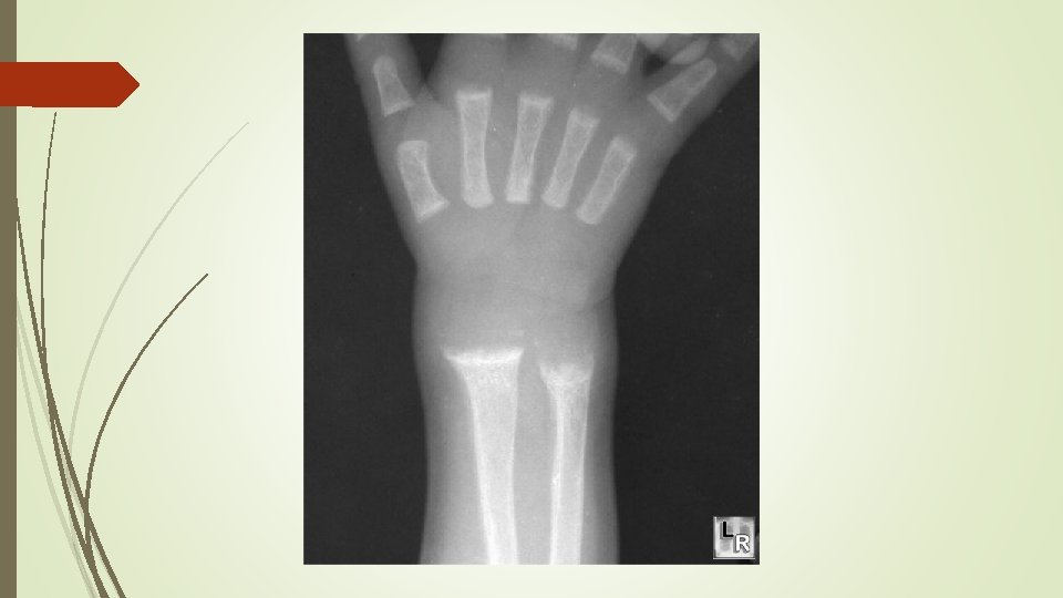
Radiographic findings Ø cupping Ø widening of the epiphyseal plate Ø fraying of the metaphysis
radiographs of a child with hypophosphatemic rickets, Metaphyseal widening in wrists And knees bone rarefaction
Anteroposterior radiograph of the knee in a 5 -year-old girl with XLHR partial fraying irregularity of the distal femoral proximal tibial growth plates (upper and lower arrows, respectively)
Looser zone (pseudofracture)
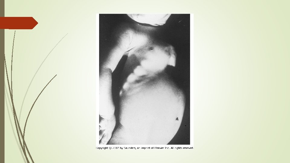
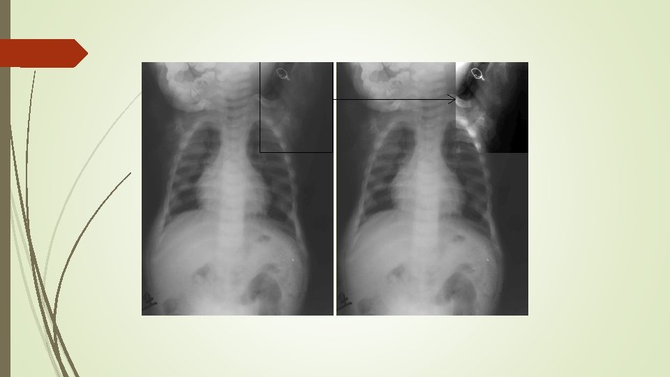
Anteroposterior pelvic show enthesopathy (arrows) in a 48 -yearold woman with Xlinked hypophosphatemic osteomalacia. Note sacroiliac joint sclerosis (arrowheads )
Radiographs in a 62 -year-old man with hypophosphatemic osteomalacia. Lateral lumbar spine image shows apophyseal joint fusion (arrows) and enthesopathy (arrowhead) of the anterior vertebral bodies
. Three-year-old boy with x-linked hypophosphatemic rickets abscess on tooth
Lab finding Ø The two main characteristic of XLH are: - Low phosphate - Reduced tubular resorption of phosphate for GFR(Tm. P/GFR) Ø calcium and 25 -(OH) vit. D are normal Ø PTH is normal to slightly elevated Ø Alk ph elevated Ø Absence of glycosuria, bicarbonaturia, proteinuria, or amino aciduria Ø Inappropriately normal serum calcitriol in the presence of hypophosphatemia
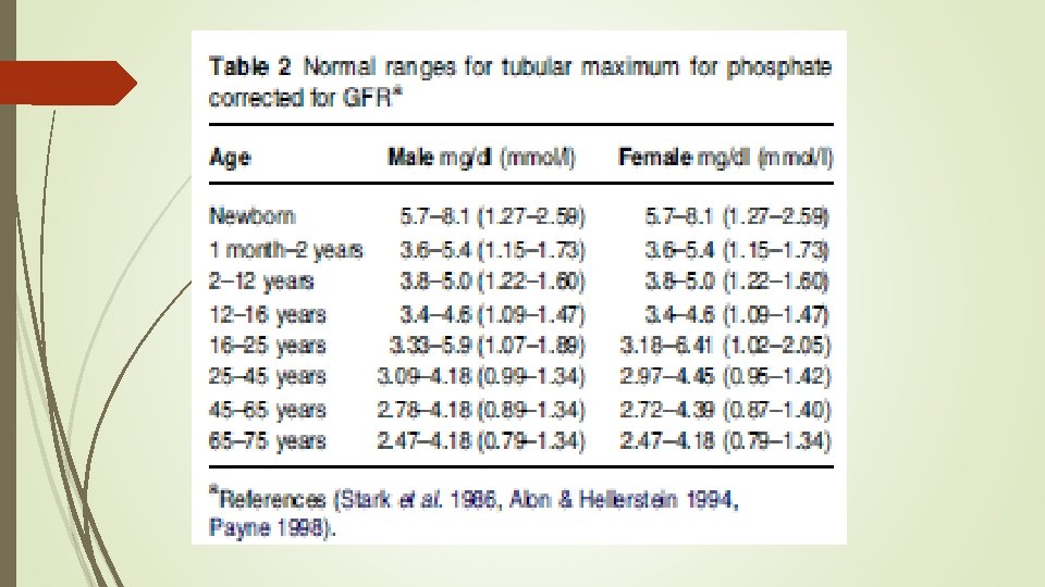
evaluate urinary phosphate wasting Ø tubular maximum for phosphate corrected for GFR(Tm. P/GFR) Ø tubular reabsorption of phosphate (%TRP) can be calculated at any time using the following formula:
TREATMENT
Children Ø Treatment more than 30 years consists oral phosphate and calcitriol Ø phosphate transiently increases the plasma phosphate concentration, lowers the plasma ionized calcium , reduces the plasma calcitriol concentration (by removing the hypophosphatemic stimulus to its synthesis). Ø This causes secondary hyperparathyroidism because of hypocalcemia and removal of the normal inhibitory effect of calcitriol on PTH synthesis
Ø The elevated PTH levels can aggravate the bone disease and increase urinary phosphate excretion, thereby defeating the aim of oral therapy. Ø The administration of calcitriol is necessary to increase the intestinal absorption of calcium, and to a lesser degree phosphate, to prevent secondary hyperparathyroidism; Ø calcitriol also suppresses PTH release directly
Ø Children Ø In children, treatment generally begins at the time of diagnosis Ø continues until long bone growth is complete(usually up to the age of 15 to 17 years) Calcitriol 10 -20 ng/kg/dose in 2 doses per day Phosphate four to five times. a nighttime dose is important to achieve satisfactory results. the starting dose is 40 mg of elemental phosphorus /kg per day. catch-up growth should be noticeable within the first year. If not, thdose should be increased in steps of 250 mg to 500 mg up to a maximum of 3500 mg/day.
Children Ø If diarrhea is a problem the dose of phosphorus should be decreased and then gradually re-increased in steps of 125 mg Ø Slow growth and persistently elevated alk-ph indicate: inadequate dose of phosphorus compliance with therapy
Adult Ø Contraversy for treatment in adults Ø Most have few if any skeletal complaint Ø employ empiric criteria for pharmacologic therapy: 1. Non union fractures 2. orthopaedic procedures treatment reduces recovery time Therapy should start 3 -6 months before surgery 3. Elevated alk ph 4. skeletal pain and muscle weakness
Adult Ø Calcitriol in two doses per day (10 to 20 ng/kg per dose) Ø Phosphate 1 to 4 g/day in three to four divided doses Ø goal of therapy is to manage generalized bone pain and enhance limited mobility, and to cure any non-union fractures
Complications There are two important complications of the treatment of XLH: v Nephrocalcinosis v Hyperparathyroidism
Nephrocalcinosis Ø on renal ultrasonography in up to 80 percent of patients Ø are composed calcium phosphate Ø long-term effect of nephrocalcinosis on renal function is not known Ø Isolated cases of renal insufficiency have been reported Ø The development of nephrocalcinosis linked to intermittent episodes of hypercalcemia and hypercalciuria Ø These can result from an excessive calcitriol dose or from noncompliance with oral phosphate
Nephrocalcinosis Ø serial measurements of renal function , serum and urine calcium is necessary Ø dose of calcitriol should be reduced Ø use of phosphorus supplementation emphasized Ø thiazide diuretics with or without amiloride may arrest the progression of nephrocalcinosis
Hyperparathyroidism q occurs after years of treatment, may be present early q increases phosphate lowers ionized calcium , Results in PTH release and secondary hyperparathyroidism q if secondary hyperparathyroidism is not adequately controlled, autonomous (tertiary) hyperparathyroidism can occur, necessitating surgical intervention q There is preliminary evidence that addition of a calcimimetic (cinacalcet) to the regimen may prevent secondary hyperparathyroidism
Adjuvant therapy q hydrochlorothiazide and amiloride, decreases urinary calcium excretion and prevents progression of nephrocalcinosis
Adjuvant therapy Growth Hormone Ø GH can improve short-term growth in children with XLH Ø it is possible that aggravates the preexistent disproportionate stature Ø a three-year study in rh. GH-treated children showed improvement in predicted adult height, but failure to normalize the body disproportion Ø Available data do not support the recommendation of growth hormone therapy
Surgical care Ø indicated for severe bowing or tibial torsion unlikely to improve with medical management alone Ø osteotomies are not usually performed in children under 6 years of age Ø Ø usually deferred until growth has nearly ceased
dental care Ø Some children are prone to dental abscesses Ø one abscess predicts future abscesses Ø Rigorous dental hygiene is recommended Ø brushing 2 -3 times daily Ø regular dental hygienist visits
Autosomal dominant hypophosphatemic rickets
Pathogenesis Ø ADHR results from missense mutations in FGF 23 that prevent its proteolytic cleavage and thereby increase circulating FGF 23 levels Ø serum FGF 23 concentrations are not consistently elevated Ø renal phosphate wasting may wax and wane;
Ø Hypophosphatemic flares often coincide with the onset of menses and following pregnancy, when iron deficiency is common. Ø In addition, high FGF 23 concentrations and low serum phosphate in patients with ADHR are associated with low levels of serum iron and ferritin. Ø So iron deficiency is an environmental trigger that stimulates expression of bone FGF 23 m. RNA and protein in both normal subjects and in patients with ADHR
Clinical findings Ø clinical, biochemical, radiographic and treatment similar to Xlinked Ø age of onset varies : early-onset disease Ø may lose the phosphate-wasting defect after puberty Ø Patients who present after growth plate closure have bone pain, weakness, and fractures, but no lower extremity deformities
Autosomal Recessive Hypophosphatemic Rickets
Ø extremely rare Ø Bone abnormalities generally include rickets and osteomalacia, some develop osteosclerosis and bone overgrowth Ø The features of ARHR may be variable and mutation-specific or age– dependent, Ø include nerve deafness, facial and dental abnormalities, learning disabilities, joint pain, contractures and immobilization of the spine, and short and deformed long bones
ØThe disorder has been divided into three subtypes: ØARHR 1 is caused by inactivating mutations in the DMP 1 gene, which encodes Dentin matrix protein 1; ØARHR 2 is caused by an inactivating mutation in the ENPP 1 gene, which encodes ectonucleotide pyrophosphatase/ phosphodiesterase 1 Ø AHRH 3 is associated with mutations in the FAM 20 C gene encoding a protein kinase. Ø mutations result in increased FGF 23 protein production and defective osteocyte maturation.
Hypophosphatemic rickets with hypercalciuria
Pathogenesis Ø mutations of the renal type 2 c sodium-phosphate cotransporter Clinical features Ø In most patients, disease onset in childhood, feature of rickets and/ or osteomalacia Ø hypophosphatemia, short stature, and secondary absorptive hypercalciuria.
Ø differs from XLH and ADHR in that the impairment is restricted to phosphate transport, and plasma calcitriol normal or appropriately elevated Ø Hypercalciuria because of high calcitriol levels and increased intestinal calcium absorption.
Treatment vphosphorus supplementation alone, the same dosing as XLH v. Endogenous calcitriol levels are elevated and the addition of exogenous calcitriol may be harmful v Monitor and follow up similar to XLH
TUMOR-INDUCED OSTEOMALACIA
TUMOR-INDUCED OSTEOMALACIA van acquired disorder in association with a tumor v The tumors, typically benign, often are small and of mesenchymal origin v primarily described in adults, but can occur in children and adolescents v. Severe hypophosphatemia with osteomalacia and, if growth plates are still open, rickets v. Longstanding bone pain and weakness
Pathogenesis v Extracts of the tumor inhibit phosphate transport in renal epithelial cells vproduce both hypophosphatemia and reduced calcitriol production v. The phosphaturic hormone implicated in tumor-induced osteomalacia is FGF 23
TUMOR-INDUCED OSTEOMALACIA Diagnosis Ø Associated with renal phosphate wasting (but no other proximal tubular defects) Ø inappropriately low calcitriol Ø family history is negative Finding the tumors • can be a major diagnostic challenge • total body MRI or scintigraphy using octreotide labeled with indium-111
TUMOR-INDUCED OSTEOMALACIA Treatment Ø Limited experience is available in the treatment Ø similar to those of X-linked disease regimens are used Ø Therapy is continued until the causative tumor can be removed Ø Removal of the tumor leads to healing of the bone disease Ø In some patients, hypophosphatemia were corrected with octreotide therapy
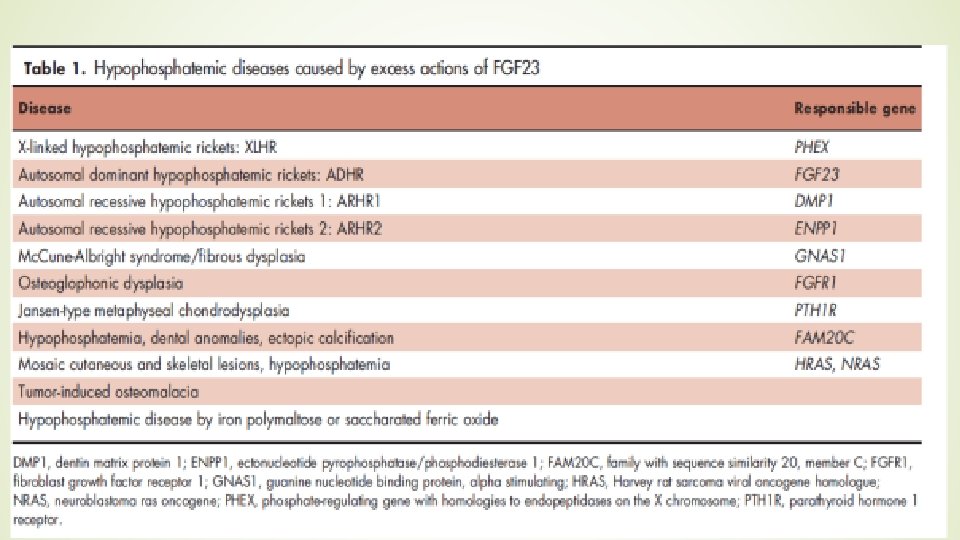
FGF 23 and Hypophosphatemic Diseases Fibroblast growth factor 23 (FGF 23) was cloned as a responsible gene for autosomal dominant hypophosphatemic rickets (ADHR) in 2000. Almost simultaneously, FGF 23 was also identified as a responsible humoral factor for tumor-induced osteomalacia (TIO). hypophosphatemia usually enhances serum 1, 25(OH)2 D level, but 1, 25(OH)2 D remains to be low to low normal in ADHR and TIO
FGF 23 and Hypophosphatemic Diseases Similar biochemical changes are seen in patients with X-linked hypophosphatemic rickets (XLH), the most prevalent cause of vitamin D-resistant rickets circulatory FGF 23 levels were shown to be high in patients with XLH, TIO, and ADHR FGF 23 -related hypophosphatemic diseases can be diagnosed by high FGF 23 levels in the presence of hypophosphatemia because FGF 23 levels are low in hypophosphatemic patients from other causes such as vitamin D deficiency, Fanconi syndrome, and so on
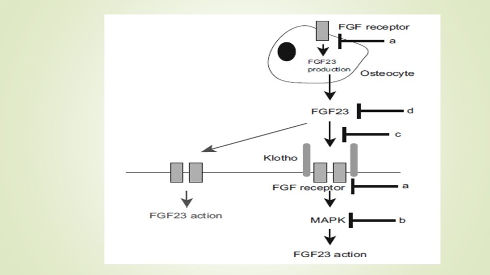
FGF 23 Fibroblast growth factor 23 (FGF 23) is a phosphaturic hormone produced by bone, mainly by osteocytes, works by binding to Klotho–FGF receptor complex. Klotho is a co-receptor for FGF 23
excessive and deficient actions of FGF 23 cause hypophosphatemic and hyperphosphatemic diseases, respectively FGF 23 is involved in the pathogenesis of chronic kidney disease–mineral and bone disorder (CKD–MBD)
These findings suggest that FGF 23 -FGF receptor/Klotho pathway can be a new drug target for disorders of mineral and bone metabolism several preclinical studies indicated that the inhibition of FGF 23 activities may be useful for hypophosphatemic diseases caused by excessive actions of FGF 23
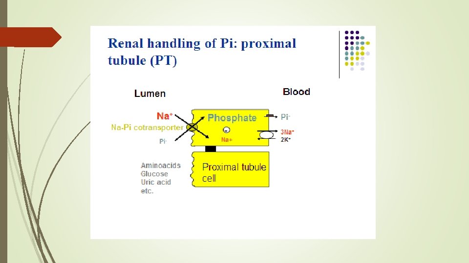
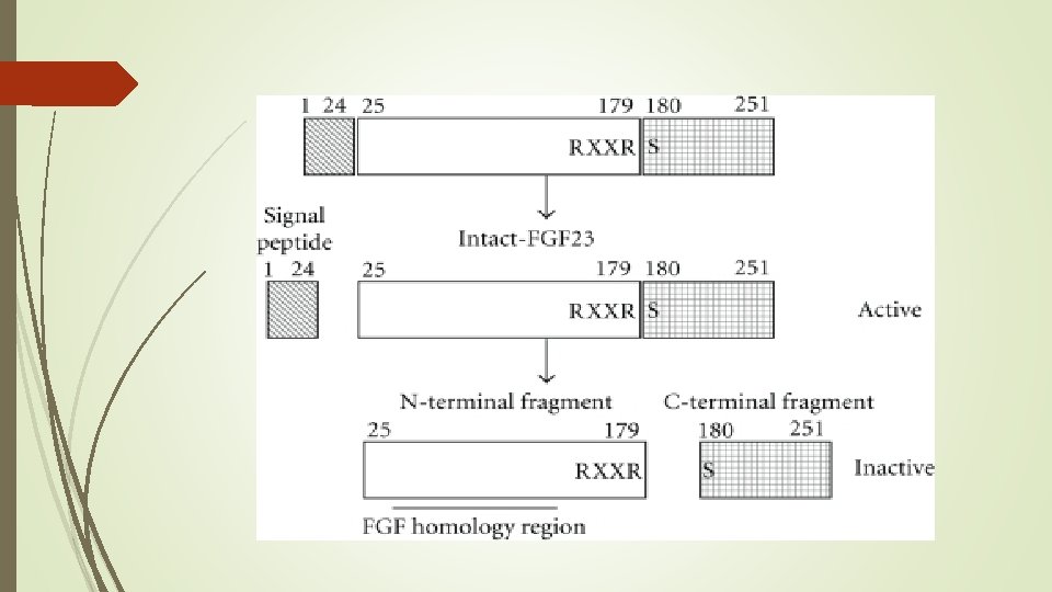
Actions of FGF 23 is a peptide with 251 amino acids. FGF 23 inhibits proximal tubular phosphate reabsorption by reducing the expression levels of type 2 a and 2 c sodiumphosphate cotransporters.
Actions of FGF 23 reduces serum 1, 25(OH)2 D by suppressing the expression of CYP 27 B 1 and enhancing the CYP 24 A 1 expression. CYP 27 B 1 encodes an enzyme that converts 25 -OH D to 1, 25(OH)2 D, and CYP 24 A 1 gene product inactivates 1, 25(OH)2 D into more hydrophilic metabolite. 1, 25(OH)2 D enhances intestinal phosphate absorption, so FGF 23 reduces serum phosphate by inhibiting both proximal tubular phosphate reabsorption and intestinal phosphate absorption.
FGF 23 and Hyperphosphatemic Diseases hyperphosphatemic familial tumoral calcinosis (HFTC) is caused by impaired actions of FGF 23 Patients with this disease show hyperphosphatemia associated with enhanced proximal tubular phosphate reabsorption and high 1, 25(OH)2 D, Three responsible genes for HFTC have been identified, FGF 23, GALNT 3, and Klotho
FGF 23 and CKD–MBD FGF 23 was shown to increase before the development of secondary Hyperparathyroidism in CKD indicated that this increased FGF 23 prevents the development of hyperphosphatemia by enhancing urinary phosphate excretion At the same time, high FGF 23 in early CKD decreases 1, 25(OH)2 D and contributes to the development of secondary hyperparathyroidism
FGF 23 and CKD–MBD It is also reported that FGF 23 inhibits the production and secretion of PTH. Therefore, the precise role of FGF 23 in the development of secondary hyperparathyroidism is complex. unknown what triggers the increase of FGF 23 in early CKD with severely reduced nephron mass, hyperphosphatemia eventually develops even with extremely high FGF 23 levels in patients with end-stage renal disease
FGF 23 and CKD–MBD FGF 23 levels have been shown to be associated with various adverse events such as cardiovascular events, fractures, progression of CKD, and higher mortality especially in patients with CKD. FGF 23 was also shown to induce left ventricular hypertrophy in a Klotho independent way
Ø FGF 23 is a humoral mediator that decreases renal tubular reabsorption of phosphate and decreases the activity of renal 1α-hydroxylase
FGF 23 -FGF Receptor/Klotho Pathway as a Drug Target for Hypophosphatemic Diseases excessive actions of FGF 23 cause several kinds of hypophosphatemic diseases, Most of these diseases are treated with phosphate supplementation and calcitriol These medications are associated with several adverse events such as gastrointestinal symptoms, secondary– tertiary hyperparathyroidism, hypercalcemia, hypercalciuria and nephrocalcinosis
it is presumed that the inhibition of FGF 23 actions corrects hypophosphatemia in these diseases FGF 23 works by binding to FGF receptor/Klotho complex.
How FGF 23 exerts its bioactivities?
FGF 23 -FGF Receptor/Klotho Pathway as a Drug Target for Hypophosphatemic Diseases Isolated C-terminal fragment of FGF 23 competes with fulllength FGF 23 for binding to FGF receptor/Klotho complex. This C-terminal fragment inhibited signaling by FGF 23 in in vitro experiments. In addition, injection of isolated C-terminal fragment of FGF 23 inhibited urinary phosphate excretion and increased serum phosphate levels both in wild-type and Hyp mice.
several reports indicate that FGF 23 production is stimulated by signaling from FGF receptor. Therefore, the suppression of FGF receptor was expected to inhibit both the production and actions of FGF 23 Another way to inhibit FGF 23 actions is to suppress the downstream signals from FGF receptor/Klotho complex
These antibodies were shown to inhibit FGF 23 actions in in vitro experiments The antibodies also synergistically enhanced serum phosphate and 1, 25(OH)2 D levels In addition, single injection of these antibodies increased serum phosphate, 1, 25(OH)2 D, and renal tubular phosphate reabsorption in Hyp mice. Once weekly injections of the antibodies into young Hyp mice for five times resulted in enhanced bone growth and mineralization, increased bone mineral density, and correction of disorganized growth plates
These results suggested that the inhibition of FGF 23 activities by anti- FGF 23 antibodies can correct biochemical, morphological, histological, and clinical features of Hyp mice. Based on these backgrounds, the clinical trial of humanized anti- FGF 23 was conducted.
In the phase I trial, single intravenous or subcutaneous administration of various amounts of anti-FGF 23 antibody was tested in 38 adult patients with XLH. The antibody increased tubular maximum transport of phosphate per glomerular filtration rate (Tm. P/GFR) and serum phosphate in a dose-dependent manner both by intravenous and subcutaneous administration.
The peak serum phosphate was observed on days 4– 5 after intravenous administration. peak phosphate occurred between days 8 and 15 after subcutaneous injection. Serum phosphate returned toward baseline within 29 and 50 days in intravenous and subcutaneous groups, respectively. The antibody also transiently increased serum 1, 25(OH)2 D. There were no serious adverse events.
From these results, repeated subcutaneous injections of FGF 23 were conducted Twenty-two adult patients with XLH were treated with up to 16 doses of subcutaneous injections of anti-FGF 23 antibody every 28 days.
Maximum serum phosphate levels were observed 7– 14 days after each dose. While serum phosphate was2. 5 mg/dl at baseline, serum phosphate was between 2. 5 and 3. 5 mg/dl in more than 70 % of subjects on day 7. This antibody also increased Tm. P/GFR and 1, 25(OH)2 D by each injection. there were no serious adverse events.
In April 2018, the FDA approved burosumab, for X-linked hypophosphatemia (XLH). Burosumab is a monoclonal Ig. G 1 antibody that binds excess fibroblast growth factor 23 (FGF 23). This action normalizes phosphorus levels, improves bone mineralization, improves rickets in children, and helps to heal fractures in adults.
In a placebo-controlled trial, 94% of adults receiving burosumab once a month achieved normal phosphorus levels compared with 8% of those receiving placebo. In children, 94% to 100% of patients treated with burosumab every 2 weeks achieved normal phosphorus levels. In both children and adults, radiograph findings associated with XLH improved with burosumab treatment
Pharmacokinetics and pharmacodynamics of a human monoclonal anti‐FGF 23 antibody (KRN 23) in the first multiple ascending‐dose trial treating adults with X‐linked hypophosphatemia
a phase 1 randomized, placebo‐controlled, double‐blind trial of KRN 23 in adults with XLH demonstrated that a single intravenous or subcutaneous dose of KRN 23 resulted in prolonged inhibition of FGF 23 activity. Serum Pi, Tm. P per glomerular filtrate (Tm. P/GFR), and serum 1, 25(OH)2 D increased in a dose‐dependent manner, and the half‐life of KRN 23 given subcutaneously was 13 to 19 days.
The greater than 4‐week duration of the Pi effect suggested that subcutaneous injection of KRN 23 every 4 weeks might be an effective treatment to increase serum Pi in XLH patients. Subsequently a phase 1/2 dose‐escalation study of repeated subcutaneous doses of KRN 23 at 28‐day intervals in adults with XLH was undertaken to assess the efficacy and safety of KRN 23
Approval in adults with XLH (n=134) was supported by a randomized, double-blind, placebo-controlled study. Compared with patients on placebo, a higher proportion of burosumab-treated adults attained serum phosphorus levels above the lower limit of normal. In addition, the rate of complete healing for active fractures and pseudofractures for burosumab treatment outpaced that of placebo therapy.
A 48 -week, open-label, single-arm bone biopsy study in 14 adults also bolstered the adult indication, with osteomalacia healing revealed by decreases in osteoid volume/bone volume, osteoid thickness, and mineralization lag time
Approval for children followed a 64 -week, randomized, openlabel study in 52 patients aged 5 to 12 years, burosumab therapy was associated with an improvement in rickets, a rise in serum phosphorus levels, a reduction in serum alkaline phosphatase activity, and an increase in growth. Forty-week data from an open-label study in 13 patients aged 1 to 4 years also back up the indication, with burosomab showing similar benefits to the 64 -week study
Effects of KRN 23, an Anti-FGF 23 Antibody in Patients With Tumor-Induced Osteomalacia and Epidermal Nevus Syndrome: Results from an Ongoing Phase 2 Study Summary and Conclusions. In: patients with TIO or ENS, KRN 23 improved mean serum phosphorus, 1, 25(OH) 2 D, and Tm. P/GFR over 24 weeks of treatment KRN 23 improved bone mineralization as demonstrated by the first bone biopsy results The increases in BMD and bone turnover markers over 24 weeks provide additional clinical evidence of an improvement in skeletal health Treatment-related adverse events were mild in severity These data suggest KRN 23 has the potential to reverse hypophosphatemia and provide clinical benefit to patients with TIO and ENS
Protein convertase
However, several questions need to be answered before the clinical use of anti-FGF 23 antibody. First, it has not been established in human whether the antibody improves growth and prevents the development of bone deformities in child patients with FGF 23 -related hypophosphatemic rickets. Second, it is not known either whether the inhibition of FGF 23 activity ameliorates the symptoms of adult patients with FGF 23 -related hypophosphatemic osteomalacia. Third, long-term safety clearly needs to be evaluated in more studies.
FGF 23 Antibody BUROSOMAB https: //www. accessdata. fda. gov/drugsatfda_docs/label/2018/761068 s 000 lbl. pdf
INDICATIONS AND USAGE CRYSVITA is a fibroblast growth factor 23 (FGF 23) blocking antibody indicated for the treatment of X-linked hypophosphatemia (XLH) in adult and pediatric patients 1 year of age and older.
CONTRAINDICATIONS Do not use CRYSVITA with oral phosphate and active vitamin D analogs. Do not initiate CRYSVITA if serum phosphorus is within or above the normal range for age. CRYSVITA is contraindicated in patients with severe renal impairment or end stage renal disease, because these conditions are associated with abnormal mineral metabolism
ADVERSE REACTIONS Most common adverse reactions (≥ 25%) in pediatric XLH patients are: headache, injection site reaction, vomiting, pyrexia, pain in extremity, vitamin D decreased. Other adverse reactions: Rash, Toothache, Myalgia, Tooth abscess, Dizziness Most common adverse reactions (≥ 5% and in at least 2 patients more than placebo) in adult XLH patients are: back pain, headache, tooth infection, restless leg syndrome, vitamin D decreased, dizziness, constipation, blood phosphorus increased.
ADVERSE REACTIONS Spinal Stenosis: Spinal stenosis is prevalent in adults with XLH and spinal cord compression has been reported. In the CRYSVITA phase 2 and phase 3 studies of adults with XLH (total N=176), a total of 6 patients underwent spinal surgery. Most of these cases appeared to involve progression of a pre -existing spinal stenosis. It is unknown if CRYSVITA therapy exacerbates spinal stenosis or spinal cord compression.
WARNINGS AND PRECAUTIONS Hypersensitivity: Hypersensitivity reactions (e. g. rash, urticaria) have been reported in patients with CRYSVITA. Hyperphosphatemia and Risk of Nephrocalcinosis: Increases in serum phosphorus to above the upper limit of normal may be associated with an increased risk of nephrocalcinosis. For patients already taking CRYSVITA, dose interruption and/or dose reduction may be required based on a patient’s serum phosphorus levels. Injection Site Reactions: Administration of CRYSVITA may result in local injection site reactions. Discontinue CRYSVITA if severe injection site reactions occur and administer appropriate medical treatment.
Pediatric Patients with XLH(1 to less than 18 years of age) The recommended starting dose regimen is 0. 8 mg/kg of body weight, rounded to the nearest 10 mg, administered every two weeks. The minimum starting dose is 10 mg up to a maximum dose of 90 mg. After initiation of treatment with CRYSVITA, measure fasting serum phosphorus every 4 weeks for the first 3 months of treatment, and thereafter as appropriate. If serum phosphorus is above the lower limit of the reference range for age and below 5 mg/d. L, continue treatment with the same dose. Follow dose adjustment schedule to maintain serum phosphorus within the reference range for age.
CLINICAL STUDIES Pediatric X-linked Hypophosphatemia
CRYSVITA has been evaluated in 65 pediatric patients with XLH. § Study 1 is a randomized, open-label study in 52 prepubescent XLH patients, 5 to 12 years old, which compared treatment with CRYSVITA administered every 2 weeks versus every 4 weeks. § Following an initial 16 -week dose titration phase, patients completed 48 -weeks of treatment with CRYSVITA every 2 weeks. § All 52 patients completed at least 64 weeks on study; no patient discontinued. § Burosumab-twza dose was adjusted to target a fasting serum phosphorus concentration of 3. 5 to 5. 0 mg/d. L based on the fasting phosphorus level the day of dosing.
§ Twenty-six of 52 patients received CRYSVITA every two weeks up to a maximum dose of 2 mg/kg. § The average dose was 0. 73 mg/kg (range: 0. 3, 1. 5) at week 16, 0. 98 mg/kg (range: 0. 4, 2. 0) at week 40 and 1. 04 mg/kg (range: 0. 4, 2. 0) at week 60. § The remaining 26 patients received CRYSVITA every four weeks. § At study entry, the mean age of patients was 8. 5 years and 46% were male. Ninety-six percent had received oral phosphate and active vitamin D analogs for a mean (SD) duration of 7 (2. 4) years. § Oral phosphate and active vitamin D analogs were discontinued prior to study enrollment. § Ninety-four percent of patients had radiographic evidence of rickets at baseline.
Study 2 is a 64 -week open-label study in 13 pediatric XLH patients, 1 to 4 years old. § Patients received CRYSVITA at a dose of 0. 8 mg/kg every two weeks with titration up to 1. 2 mg/kg based on serum phosphorus measurements. § All patients completed at least 40 weeks on study; no patients discontinued. § At study entry, the mean age of patients was 2. 9 years and 69% were male. § All patients had radiographic evidence of rickets at baseline and had received oral phosphate and active vitamin D analogs for a mean (SD) duration of 16. 9 (13. 9) months. § Oral phosphate and active vitamin D analogs were discontinued prior to study enrollment.
Serum Phosphorus § In Study 1, CRYSVITA increased mean (SD) serum phosphorus levels from 2. 4 (0. 40) at baseline to 3. 3 (0. 40) and 3. 4 (0. 45) mg/d. L at week 40 and week 64 in the patients who received CRYSVITA every 2 weeks. § The ratio of renal tubular maximum reabsorption rate of phosphate to glomerular filtration rate (Tm. P/GFR) increased in these patients from mean (SD) of 2. 2 (0. 49) at baseline to 3. 3 (0. 60) and 3. 4 (0. 53) mg/d. L at week 40 and week 64. § In Study 2, CRYSVITA increased mean (SD) serum phosphorus levels from 2. 5 (0. 28) mg/d. L at baseline to 3. 5 (0. 49) mg/d. L at week 40.
Radiographic Evaluation of Rickets Radiographs from 52 CRYSVITA-treated XLH patients in Study 1 and 13 patients in Study 2 were examined to assess XLH-related rickets using the 10 -point Thacher Rickets Severity Score (RSS) and the 7 -point Radiographic Global Impression of Change (RGI-C). The RSS score is assigned based on images of the wrist and knee from a single timepoint, with higher scores indicating greater rickets severity. The RGIC score is assigned based on side-by-side comparisons of wrist and knee radiographs from two timepoints, with higher scores indicating greater improvement in radiographic evidence of rickets. A RGI-C score of +2. 0 was defined as radiographic evidence of substantial healing. In Study 1, baseline mean (SD) RSS total score was 1. 9 (1. 17) in patients receiving CRYSVITA every two weeks. After 40 weeks of treatment with CRYSVITA, mean total RSS decreased from 1. 9 to 0. 8 (see Table 6). After 40 weeks of treatment with CRYSVITA, the mean RGI-C Global score was +1. 7 in patients receiving CRYSVITA every two weeks. Eighteen out of 26 patients achieved an RGI-C score of ≥ +2. 0. These findings were maintained at week 64 as shown in Table 6.
In Study 2, baseline mean (SD) total RSS was 2. 9 (1. 37) in 13 patients. After 40 weeks of treatment with CRYSVITA, mean total RSS decreased from 2. 9 to 1. 2 and the mean (SE) RGI-C Global score was +2. 3 (0. 08). All 13 patients achieved a RGI-C global score ≥ +2. 0. The mean (SE) lower limb deformity as assessed by RGI-C, using standing long leg radiographs, was +1. 3 (0. 14)
Serum Alkaline Phosphatase Activity For Study 1, mean (SD) serum total alkaline phosphatase activity was 462 (110) U/L at baseline and decreased to 354 (73) U/L at Week 64 (-23%, p < 0. 0001) in the patients who received CRYSVITA every 2 weeks. For Study 2, mean (SD) serum total alkaline phosphatase activity was 549 (194) U/L at baseline and decreased to 335 (88) U/L at Week 40 (mean change: -36%). Growth In Study 1, CRYSVITA treatment for 64 weeks increased standing mean (SD) height Z score from -1. 72 (1. 03) at baseline to -1. 54 (1. 13) in the patients who received CRYSVITA every two weeks (LS mean change of +0. 19 (95% CI: 0. 09 to 0. 29).
Adult X-linked Hypophosphatemia
Study 3 (NCT 02526160) is a randomized, double-blind, placebocontrolled study in 134 adult XLH patients. The study comprises a 24 -week placebo-controlled treatment phase. CRYSVITA was administered at a dose of 1 mg/kg every 4 weeks. At study entry, the mean age of patients was 40 years (range 19 to 66 years) and 35% were male. All patients had skeletal pain associated with XLH/osteomalacia at baseline. The baseline mean (SD) serum phosphorus concentration was below the lower limit of normal at 1. 98 (0. 31) mg/d. L. Oral phosphate and active vitamin D analogs were not allowed during the study. One patient in the CRYSVITA group discontinued treatment.
Study 4 (NCT 02537431) is a 48 -week, open-label, single-arm study in 14 adult XLH patients to assess the effects of CRYSVITA on improvement of osteomalacia as determined by histologic and histomorphometric evaluation of iliac crest bone biopsies. Patients received 1 mg/kg CRYSVITA every four weeks. At study entry, the mean age of patients was 40 years (range 25 to 52 years) and 43% were male. Oral phosphate and active vitamin D analogs were not allowed during the study.
In Study 3 at baseline, mean (SD) serum phosphorus was 1. 9 (0. 32) and 2. 0 (0. 30) mg/d. L in the placebo and CRYSVITA groups respectively. During the initial 24 weeks of treatment, mean (SD) serum phosphorus across the midpoints of dose intervals (2 weeks post dose) was 2. 1 (0. 30) and 3. 2 (0. 53) mg/d. L in the placebo and CRYSVITA groups, and mean (SD) serum phosphorus across the ends of dose intervals was 2. 0 (0. 30) and 2. 7 (0. 45) mg/d. L in the placebo and CRYSVITA groups. A total of 94% of patients treated with CRYSVITA achieved a serum phosphorus level above the lower limit of normal (LLN) compared to 8% in the placebo group through week 24 (Table 7).
At baseline, the mean (SD) ratio of renal tubular maximum reabsorption rate of phosphate to glomerular filtration rate (Tm. P/GFR) was 1. 60 (0. 37) and 1. 68 (0. 40) mg/d. L in the placebo and CRYSVITA groups respectively. At week 22 (midpoint of a dose interval), mean (SD) Tm. P/GFR was 1. 69 (0. 37) and 2. 73 (0. 75) mg/d. L in the placebo and CRYSVITA groups. At week 24 (end of a dose interval), mean (SD) Tm. P/GFR was 1. 73 (0. 42) and 2. 21 (0. 48) mg/d. L in the placebo and CRYSVITA groups.
Radiographic Evaluation of Osteomalacia In Study 3, a skeletal survey was conducted at baseline to identify osteomalaciarelated fractures and pseudofractures. Osteomalacia-related fractures are defined as atraumatic lucencies extending across both bone cortices and pseudofractures are defined as atraumatic lucencies extending across one cortex. There were 52% of patients who had either active (unhealed) fractures (12%) or active pseudofractures (47%) at baseline. The active fractures and pseudofractures were predominantly located in the femurs, tibia/fibula, and metatarsals of the feet. Assessment of these active fracture/pseudofracture sites at week 24 demonstrated a higher rate of complete healing in the CRYSVITA group compared to placebo as shown in Table 8. During treatment through week 24, a total of 6 new fractures or pseudofractures appeared in 68 patients receiving CRYSVITA, compared to 8 new abnormalities in 66 patients receiving placebo
Bone Histomorphometry In Study 4, after 48 weeks of treatment, healing of osteomalacia was observed in ten patients as demonstrated by decreases in Osteoid volume/Bone volume (OV/BV) from a mean (SD) score of 26% (12. 4) at baseline to 11% (6. 5), a change of -57%. Osteoid thickness (O. Th) declined in eleven patients from a mean (SD) of 17 (4. 1) micrometers to 12 (3. 1) micrometers, a change of -33%. Mineralization lag time (MLt) declined in 6 patients from a mean (SD) of 594 (675) days to 156 (77) days, a change of -74%.
THANKS