
Rickets Done by Areej ALHadidi Reference baby Nelson

Rickets Done by : Areej AL-Hadidi Reference: baby Nelson
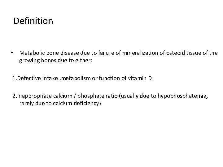
Definition • Metabolic bone disease due to failure of mineralization of osteoid tissue of the growing bones due to either: 1. Defective intake , metabolism or function of vitamin D. 2. Inappropriate calcium / phosphate ratio (usually due to hypophosphatemia, rarely due to calcium deficiency)
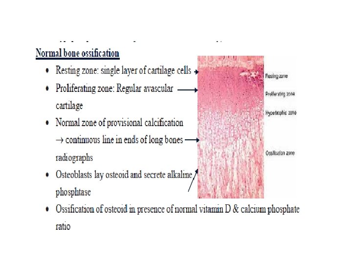
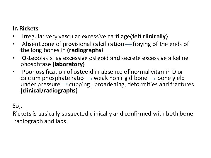
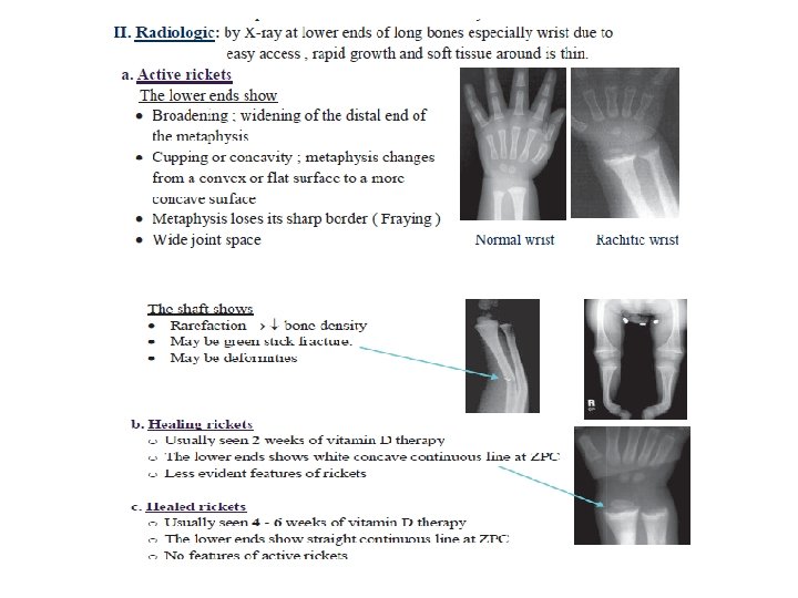
In Rickets • Irregular very vascular excessive cartilage(felt clinically) • Absent zone of provisional calcification fraying of the ends of the long bones in (radiographs) • Osteoblasts lay excessive osteoid and secrete excessive alkaline phosphtase (laboratory) • Poor ossification of osteoid in absence of normal vitamin D or calcium phosphate ratio weak non rigid bone yield under pressure cupping , broadening, deformities and fractures (clinical/radiographs) So, , Rickets is basically suspected clinically and confirmed with bone radiograph and labs

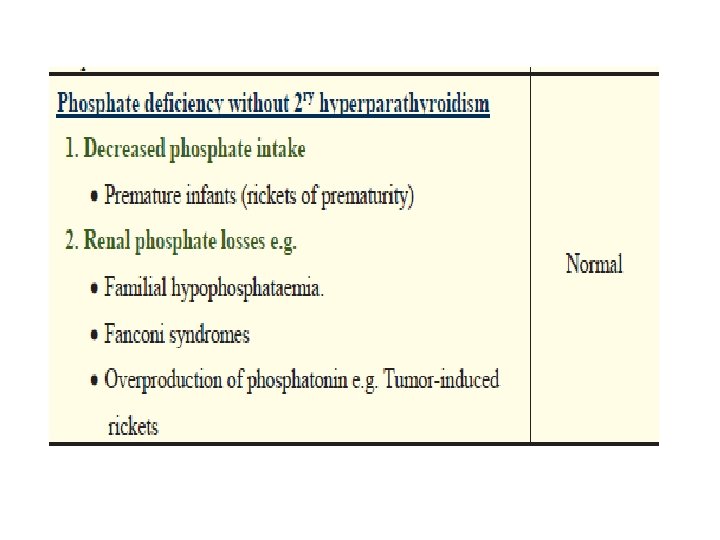
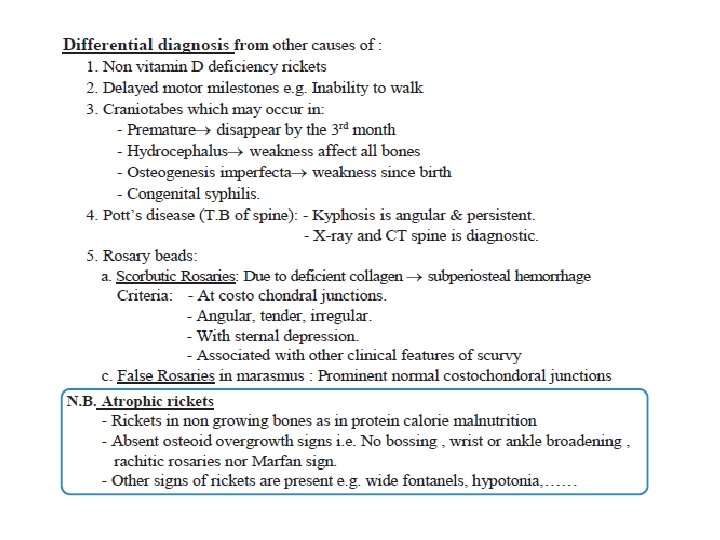
Cont, , Causes of rickets other than nutritional rickets are referred to as: • Non vitamin D deficiency rickets • Vitamin D refractory or resistant rickets as they are not cured with the same dose or form of vitamin D that cures nutritional rickets
Vitamin D deficiency rickets Predisposing factors • Season : - Commoner in winter • Age : - Commonest age : 6 months - 24 month. • Growth : - More in rapidly growing infant e. g. twins & preterm. - Less in infants with arrested growth e. g. PCM & cretinism. Etiology A. Decreased vitamin D intake due to: 1. Lack of rich sources of vitamin D e. g. egg yolk, meat, fortified milks, fish liver oil. 2. Use of rachitogenic diet with: - Poor sources of vitamin D as fresh animal milk , cereals and carbohydrates. - Poor sources of calcium as cereals , and excess leafy vegetables - Inappropriate calcium /phosphate ratio as in fresh animal milk B. Lack of access of ultra violet rays to the skin due to: 1. Lack of sun exposure 2. Poor sun exposure through glass windows, clouds & dust. 3. Excessive wrappings of the infants. 4. Poor penetration in dark skinned infants
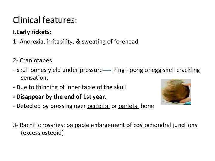
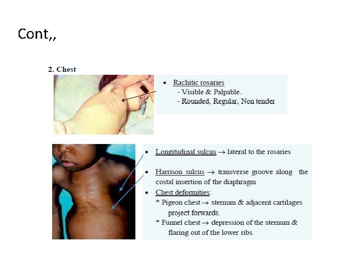
Clinical features: I. Early rickets: 1 - Anorexia, irritability, & sweating of forehead 2 - Craniotabes - Skull bones yield under pressure Ping - pong or egg shell crackling sensation. - Due to thinning of inner table of the skull - Disappear by the end of 1 st year. - Detected by pressing over occipital or parietal bone 3 - Rachitic rosaries: palpable enlargement of costochondral junctions (excess osteoid)
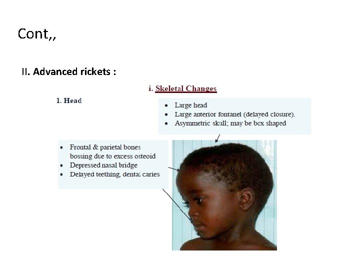
Cont, , II. Advanced rickets :
Cont, ,
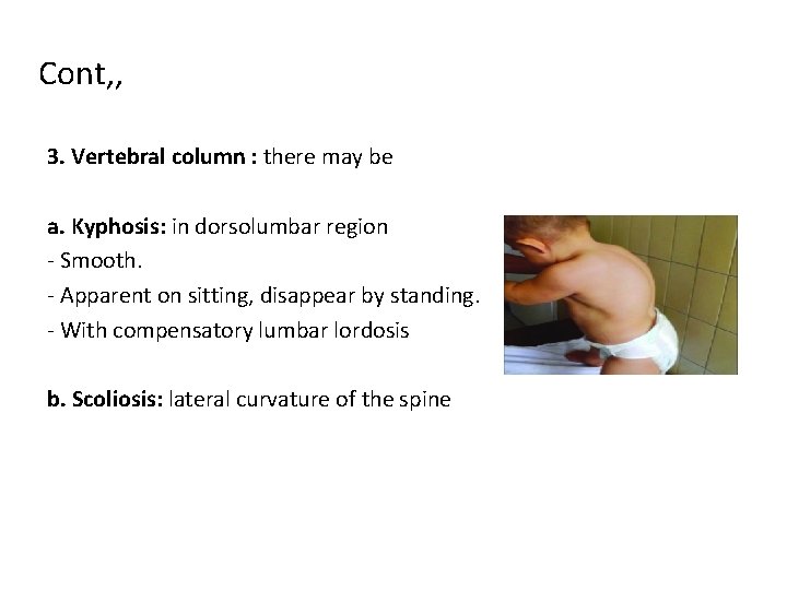
Cont, , 3. Vertebral column : there may be a. Kyphosis: in dorsolumbar region - Smooth. - Apparent on sitting, disappear by standing. - With compensatory lumbar lordosis b. Scoliosis: lateral curvature of the spine

Cont, ,
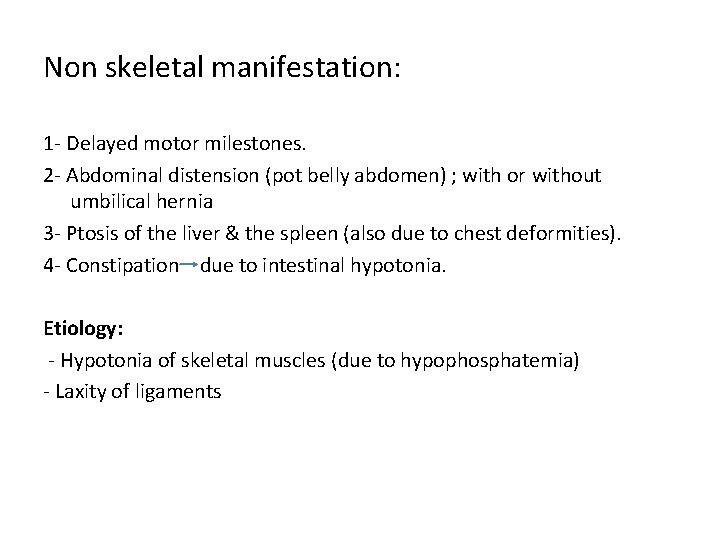
Non skeletal manifestation: 1 - Delayed motor milestones. 2 - Abdominal distension (pot belly abdomen) ; with or without umbilical hernia 3 - Ptosis of the liver & the spleen (also due to chest deformities). 4 - Constipation due to intestinal hypotonia. Etiology: - Hypotonia of skeletal muscles (due to hypophosphatemia) - Laxity of ligaments
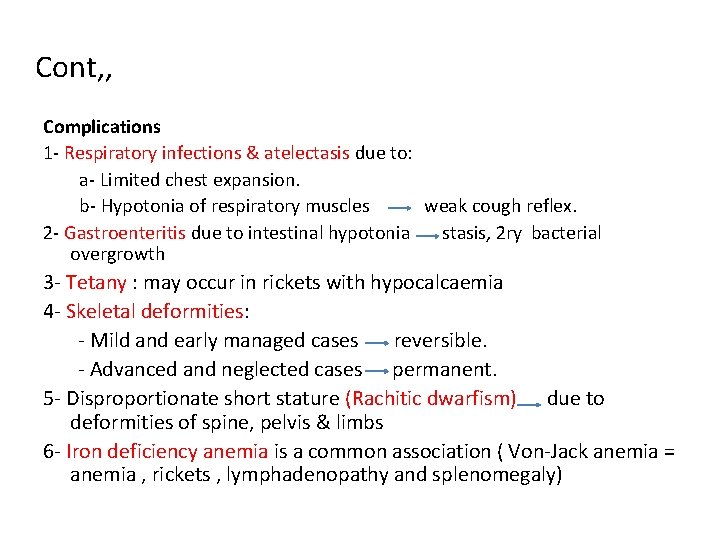
Cont, , Complications 1 - Respiratory infections & atelectasis due to: a- Limited chest expansion. b- Hypotonia of respiratory muscles weak cough reflex. 2 - Gastroenteritis due to intestinal hypotonia stasis, 2 ry bacterial overgrowth 3 - Tetany : may occur in rickets with hypocalcaemia 4 - Skeletal deformities: - Mild and early managed cases reversible. - Advanced and neglected cases permanent. 5 - Disproportionate short stature (Rachitic dwarfism) due to deformities of spine, pelvis & limbs 6 - Iron deficiency anemia is a common association ( Von-Jack anemia = anemia , rickets , lymphadenopathy and splenomegaly)
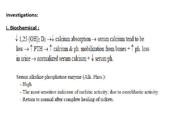
Investigations: I. Biochemical :
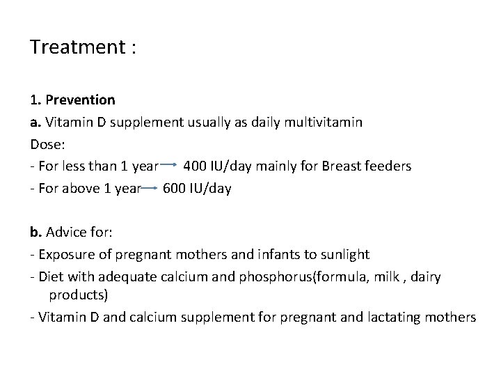
Treatment : 1. Prevention a. Vitamin D supplement usually as daily multivitamin Dose: - For less than 1 year 400 IU/day mainly for Breast feeders - For above 1 year 600 IU/day b. Advice for: - Exposure of pregnant mothers and infants to sunlight - Diet with adequate calcium and phosphorus(formula, milk , dairy products) - Vitamin D and calcium supplement for pregnant and lactating mothers
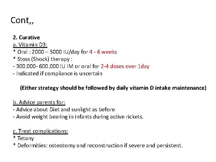
Cont, , 2. Curative a. Vitamin D 3: * Oral : 2000 – 5000 IU/day for 4 - 6 weeks * Stoss (Shock) therapy : - 300. 000 - 600. 000 IU IM or oral for 2 -4 doses over 1 day - Indicated if compliance is uncertain (Either strategy should be followed by daily vitamin D intake maintenance) b. Advice parents for: - Advice about Diet and sunlight as before - Avoid weight bearing in infants during active rickets. c. Treat complications: * Tetany * Deformities: osteotomy and reconstruction if severe and persistent.
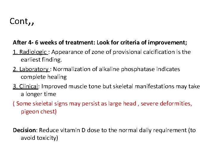
Cont, , After 4 - 6 weeks of treatment: Look for criteria of improvement; 1. Radiologic : Appearance of zone of provisional calcification is the earliest finding. 2. Laboratory : Normalization of alkaline phosphatase indicates complete healing 3. Clinical: Improved muscle tone but skeletal manifestations may take a longer time ( Some skeletal signs may persist as large head , severe deformities, pigeon chest) Decision: Reduce vitamin D dose to the normal daily requirement (to avoid toxicity)

Thank you
- Slides: 33