
Department of Pediatrics with a course of Pediatric

- Slides: 35
Department of Pediatrics with a course of Pediatric Infectious Disease RICKETS Etiology, pathogenesis, clinical features, diagnostics, treatment and prophilaxis
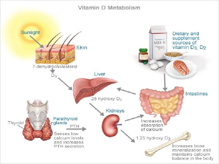
RICKETS is a condition that affects bone development in children due to deficiency or impaired metabolism of vitamin D, phosphorus or calcium
ETILOGY • Deficiency of sunlight exposure • Nutrition disbalance (lack of Vit D, Ca, P in food) • Perinatal factors (prematurity, placental insufficiency, etc. ) • Low motor activity • Chronic liver and kidney diseases, malabsorption • Medicaments • Hereditary abnormalities of Ca and P, Vit D metabolism • Environmental factors (excess of strontium, zinc and other microelements in soil, water, food) • Dark skin color
Classification 1. According to the level of severity -Mild (1 st degree) -Moderate (2 nd degree) -Severe (3 rd degree)
Classification 2. According to the course of disease -Acute -Subacute -Recurrent
Classification 3. According to the clinical variants -Calcipaenic Rickets -Phosphopaenic Rickets -with no significant changes in Ca and P level
PERIODS OF DISEASE • • INITIAL MANIFEST REPARATION RESIDUAL SYMPTOMS
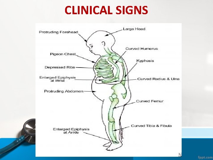
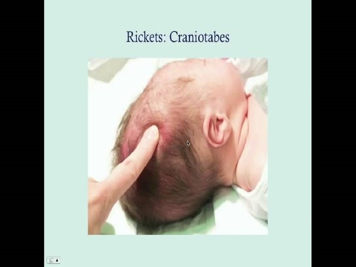
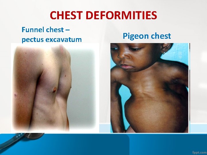
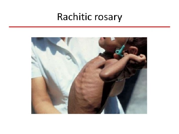
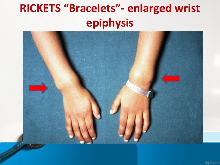
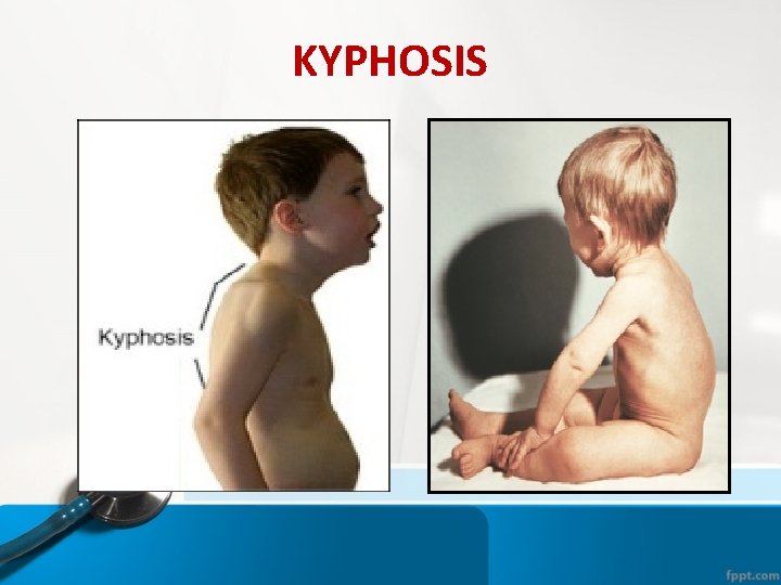
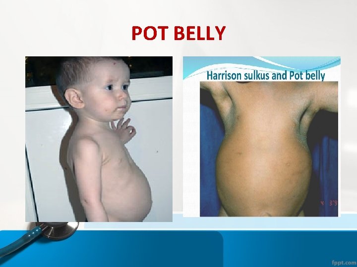
CLINICAL FEATURES • GENERAL: • a) Dysfunction of Nervous System (Irritability, Listlessness, Sleep Disorders, Sweating, Loss of Appetite, Dyspepsia, Lack of Motor Development, Nervous-Psychical Retardation) • b) Syndrome of Osseous System Lesions: • - Osteomalacia in form of softening of the Fontanel's Edges, Craniotabes, Softening of Ribs (Harrison's groove), Kyphosis, bandy and baker's legs; • - Hyperplasia of osteoid tissue - increase of frontal and occipital Tubers, Costohondral prominence (“Rachitic Rosary"), “Bracelets” on the upper and lower extremities, threads -of pearls on finger's phalanges;
CLINICAL FEATURES • c) muscular hypotonia • d) Other systems involvement: Dysfunction of Cardio-Vascular System, Respiratory Tract, Digestive Tract etc.
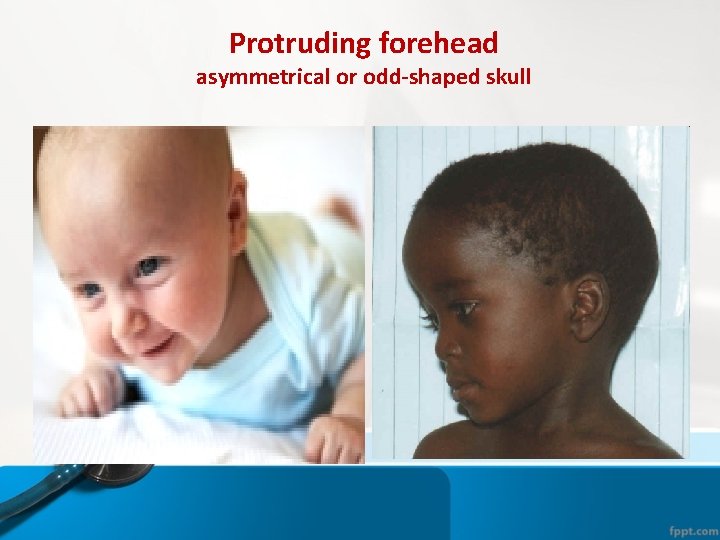
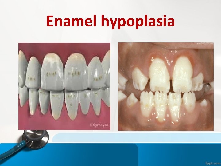
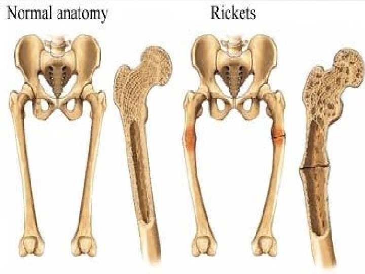
CLINICAL FEATURES • HEAD: Craniotabes; Frontal bossing; Delayed fontanelle closure; Delayed dentate; Enamel hypoplasia; Craniosynostosis; • CHEST: Rachitic Rosary; Harrison groove; Respiratory infections and atelectasis; • BACK: Scoliosis , Kyphosis , Lordosis; • EXTREMITIES: Enlargement of wrists and ankles; Valgus or varus deformities; Windswept deformity (combination of valgus deformity of 1 leg with varus deformity of the other leg); Anterior bowing of the tibia and femur; Coxa vara; pain; • HYPOCALCEMIC SYMPTOMS: Tetany ; Seizures; Stridor due to laryngeal spasm
• symptoms of Osteomalacia are predominant in case of acute course of rickets • symptoms of Hyperplasia of osteoid tissue – in case of subacute course
CLINICAL SIGNS
Protruding forehead asymmetrical or odd-shaped skull
Enamel hypoplasia
CHEST DEFORMITIES Funnel chest – pectus excavatum Pigeon chest
RICKETS “Bracelets”- enlarged wrist epiphysis
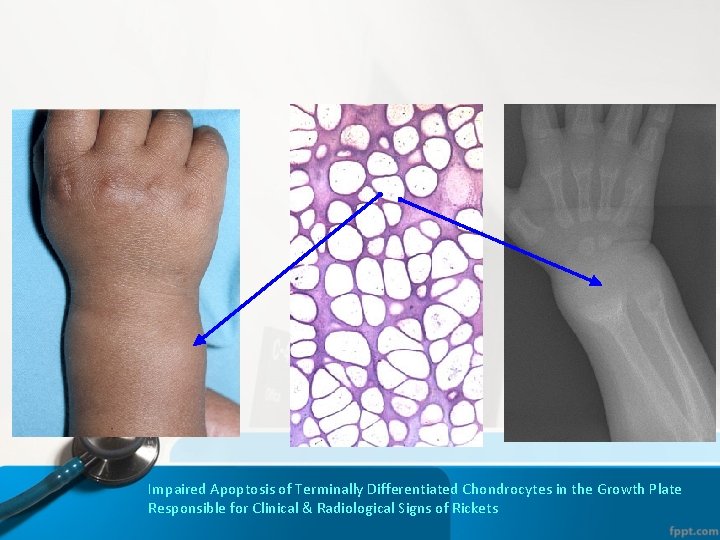
Impaired Apoptosis of Terminally Differentiated Chondrocytes in the Growth Plate Responsible for Clinical & Radiological Signs of Rickets
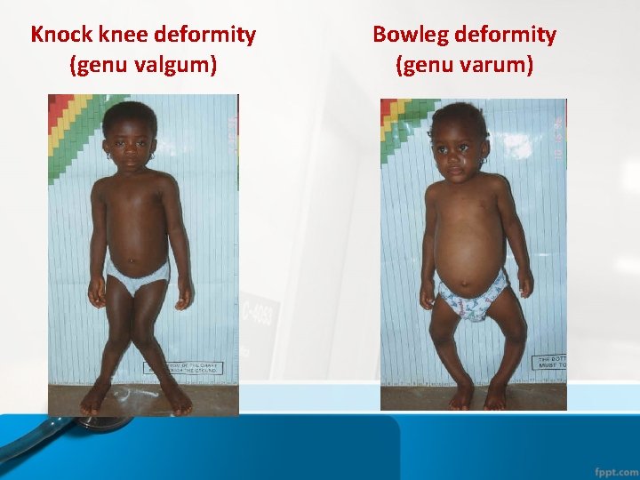
Knock knee deformity (genu valgum) Bowleg deformity (genu varum)
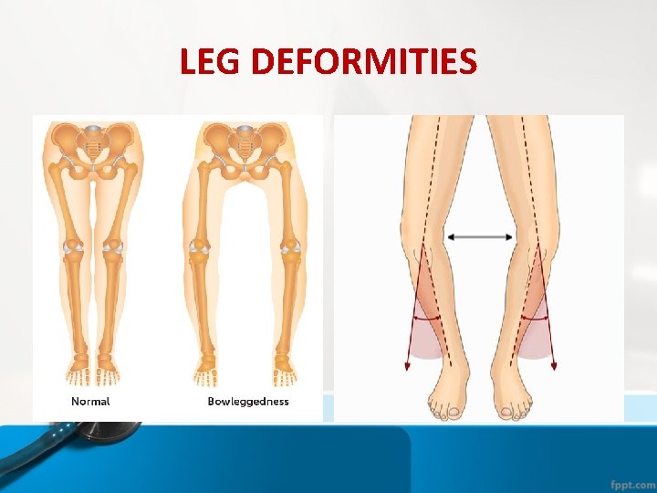
LEG DEFORMITIES
KYPHOSIS
POT BELLY
DIAGNOSTICS • • • Assessment according to the followings: 1. History of disease 2. Physical examination 3. Laboratory findings 4. X-ray changes evaluation
PHYSICAL EXAMINATION Hvostek sign — simple tapping the cheek over the facial nerve causes involuntary contraction of the muscles about the eye or mouth (spasm of facial muscles occurs when the facial nerve is tapped) Trousseau's Sign — is the carpal or pedal spasm, induced by compression of the arm or thigh during 3 -5 minutes (by the cuff for measurement of arterial pressure) when hands assume the socalled “obstetrical position”, while the feet are held in a position of equinas; Maslow Sign — stop of breathing at a mild skin pricks
DIAGNOSTICS • Laboratory findings: • CBC - hypochromic anemia; • Biochemical analyses of blood: – Alkaline phosphatase increasing the initial period of disease; – Hypophosphatemia; – Hypocalcemia:
DIAGNOSTICS • • X-ray: Reaction of the periosteum (may occur) Indistinct cortex Coarse trabeculation Knees, wrists, and ankles affected predominantly Epiphyseal plates, widened and irregular Tremendous metaphysis (cupping, fraying, splaying) Spur (metaphyseal)
Differential diagnosis – Down's syndrome, – Hypophosphatasia, – Chondrodystrophy, – Hypothyroidism, – Pseudohypoparathyreoidism, – Phosphat-Diabetes, – De Toni-Debre-Fanconi Syndrome, – Renal Tubular Acidosis, – Renal Osteodystrophy
TREATMENT • • Vit. D-3 (water solution): – 1 -degree (mild) -2000 IU daily – 2 -degree (moderate) - 4000 IU daily – 3 -degree (severe) - 5000 IU daily 30 -45 days Prophylactic of recurrence rickets 2000 IU daily, 30 days, 2 -3 times a year, interval between courses 3 month – Calcium supplements
PROPHYLAXIS • - Pregnant women: – physiological pregnancy - starting from 28 -32 week of pregnancy 500 IU of Vit. D-3 daily; 6 -8 weeks – with risks – starting from 28 -32 weeks of pregnancy 1000 -2000 IU daily, 6 -8 weeks;
POSTNATAL PROPHYLAXIS: • 1. Proper infant care, • 2. Full-term baby - within 2 month of life 500 IU daily 3 years, except for the summer months (June, July, August), • 3. Full-term baby with risk of Rickets development – within 2 -3 weeks of life - 500 -1000 IU daily, 3 years • 4. Sickly children – 400 IU
POSTNATAL PROPHYLAXIS: • 5. Preterm (1 st degree) infants -within 10 -14 day of life - 500 -1000 IU daily, 6 months. Then 2000 IU 30 days 2 -3 times in year, interval 3 -4 months • 6. Preterm (2 - 3 rd degree) infants –within 10 -20 day of life, 1000 -2000 IU, 6 months. Then 2000 IU 30 days 2 -3 times in year, interval 3 -4 months
Thank you for attention!