EKA AGUSTIA RINI RACHITIS VITAMIN D RICKETSIA OSTEOMALACIA
- Slides: 43
EKA AGUSTIA RINI RACHITIS, VITAMIN D RICKETSIA, OSTEOMALACIA
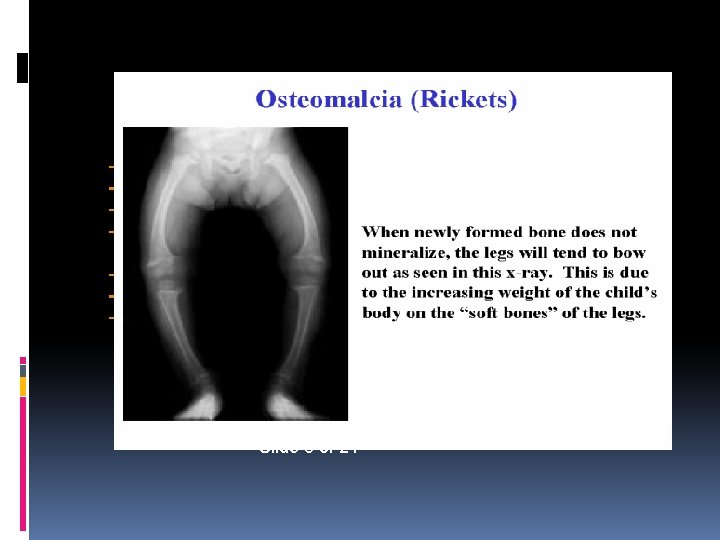
RICKETS Disorder of mineralization of the bone matrix / osteoid in growing bone Involved : growth plate Newly trabecular formed Cortical bone Osteomalacia After cessation of growth Involves only a bone, not the growth plate
Risk factors Living in northern latitudes (>30 o); Dark skinned children; Decreased exposure to sunlight Maternal vitamin D deficiency; Diets low in calcium, phosphorus and vit. D Prolonged parenteral nutrition in infancy with an inadequate supply of intravenous calcium and phosphate; Intestinal malabsorption
Defective production of 1, 25(OH)2 D 3 Hereditary type I vitamin D-resistant (or dependent) rickets (mutation which abolishes activity of renal hydroxylase); Familial (X-linked ) hypophosphataemic rickets – renal tubular defect in phosphate transport; Chronic renal disease; Fanconi syndrome (renal loss of phosphate) Target organ resistance to 1, 25(OH)2 D 3 - hereditary vitamin D-dependent rickets type II (due to mutations in vitamin D receptor gene).
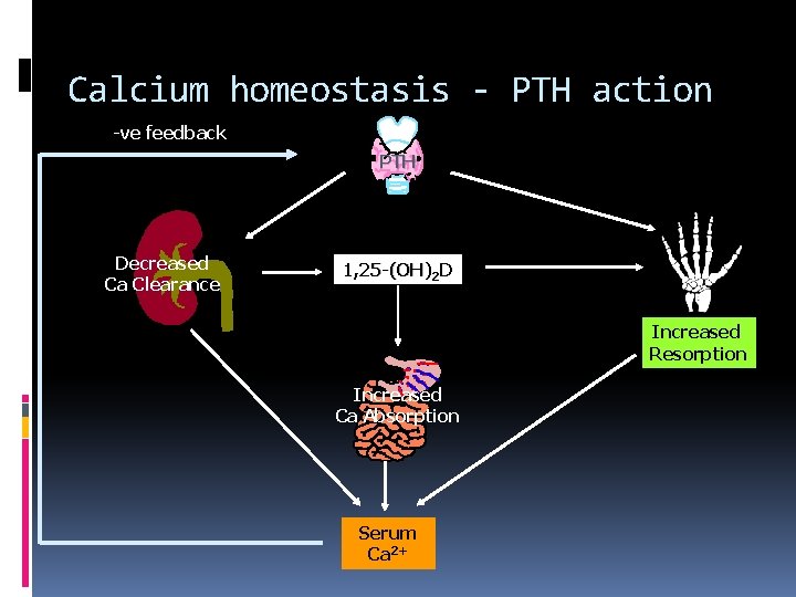
Calcium homeostasis - PTH action -ve feedback PTH Decreased Ca Clearance 1, 25 -(OH)2 D Increased Resorption Increased Ca Absorption Serum Ca 2+
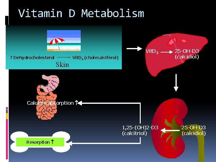
Vitamin D Metabolism Vit. D 3 7 Dehydrocholesterol Skin Vit. D 3 (cholecalciferol) 25 -OH-D 3 (calcidiol) Calcium absorption 1, 25 -(OH)2 -D 3 (calcitriol) Resorption 25 -OH-D 3 (calcidiol)
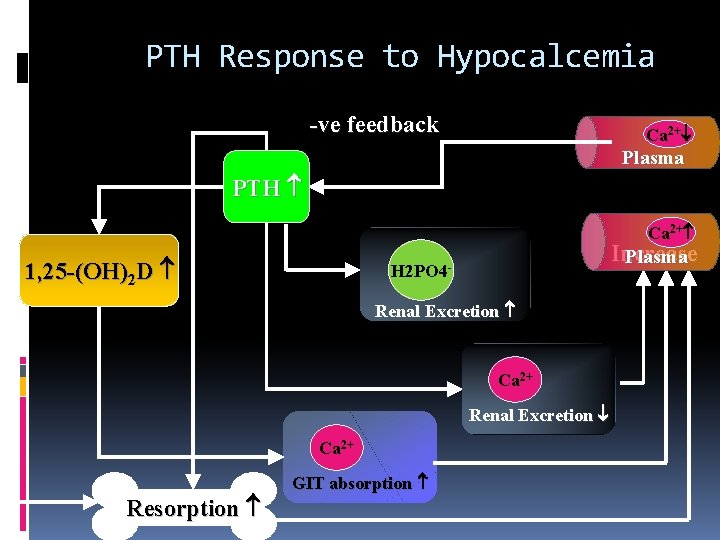
PTH Response to Hypocalcemia -ve feedback Ca 2+ Plasma PTH Ca 2+ 1, 25 -(OH)2 D Increase Plasma H 2 PO 4 Renal Excretion Ca 2+ Resorption GIT absorption
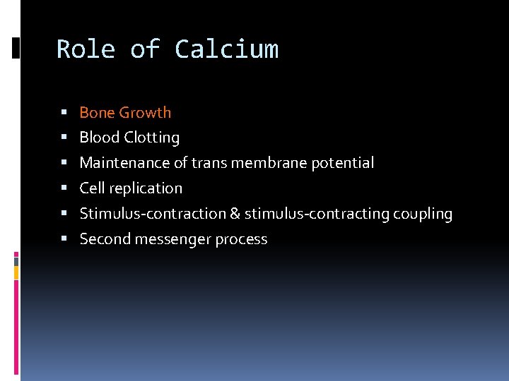
Role of Calcium Bone Growth Blood Clotting Maintenance of trans membrane potential Cell replication Stimulus-contraction & stimulus-contracting coupling Second messenger process
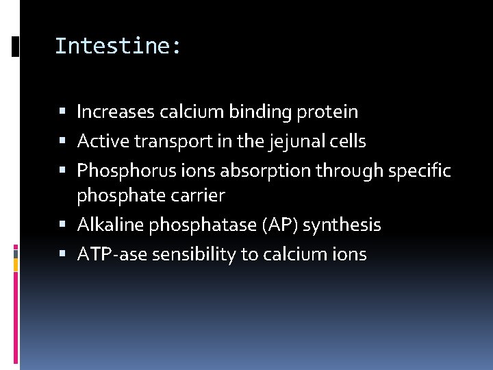
Intestine: Increases calcium binding protein Active transport in the jejunal cells Phosphorus ions absorption through specific phosphate carrier Alkaline phosphatase (AP) synthesis ATP-ase sensibility to calcium ions
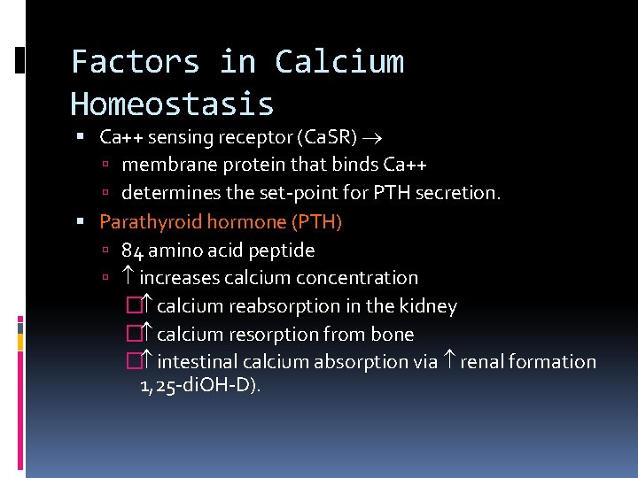
Factors in Calcium Homeostasis Ca++ sensing receptor (Ca. SR) membrane protein that binds Ca++ determines the set-point for PTH secretion. Parathyroid hormone (PTH) 84 amino acid peptide increases calcium concentration � calcium reabsorption in the kidney � calcium resorption from bone � intestinal calcium absorption via renal formation 1, 25 -di. OH-D).
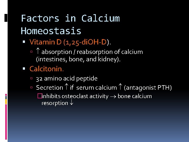
Factors in Calcium Homeostasis Vitamin D (1, 25 -di. OH-D). absorption / reabsorption of calcium (intestines, bone, and kidney). Calcitonin. 32 amino acid peptide Secretion if serum calcium (antagonist PTH) �inhibits osteoclast activity bone calcium resorption
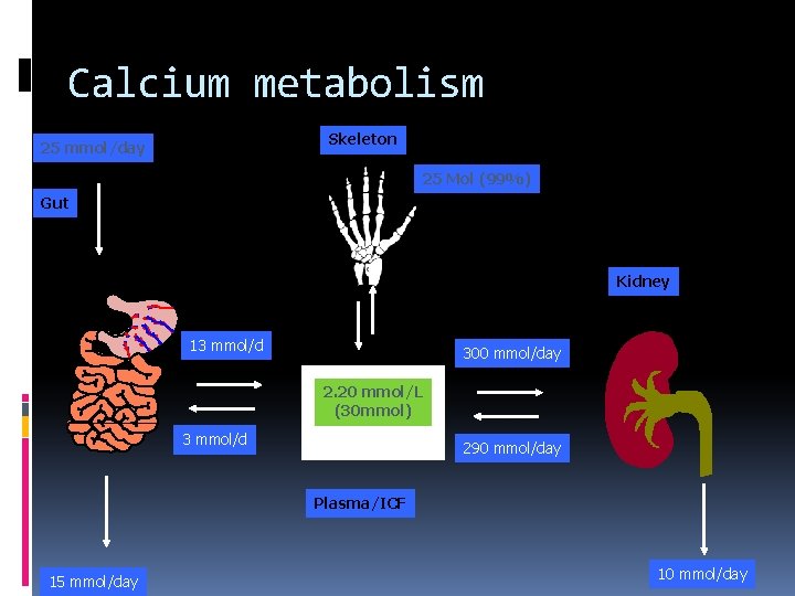
Calcium metabolism Skeleton 25 mmol/day 25 Mol (99%) Gut Kidney 13 mmol/d 300 mmol/day 2. 20 mmol/L (30 mmol) 3 mmol/d 290 mmol/day Plasma/ICF 15 mmol/day 10 mmol/day
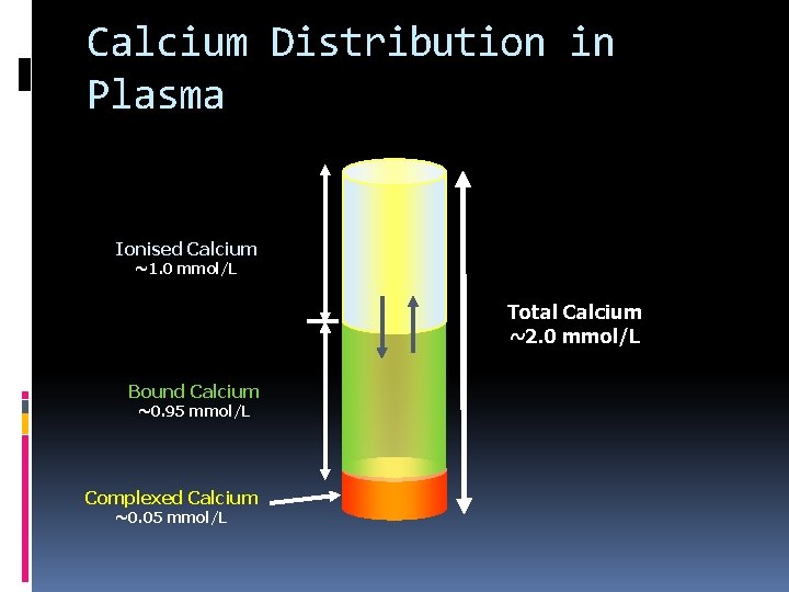
Calcium Distribution in Plasma Ionised Calcium ~1. 0 mmol/L Total Calcium ~2. 0 mmol/L Bound Calcium ~0. 95 mmol/L Complexed Calcium ~0. 05 mmol/L
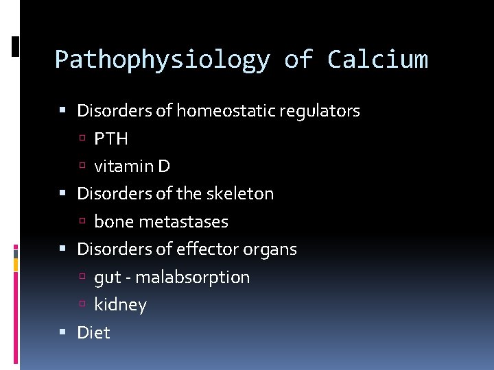
Pathophysiology of Calcium Disorders of homeostatic regulators PTH vitamin D Disorders of the skeleton bone metastases Disorders of effector organs gut - malabsorption kidney Diet
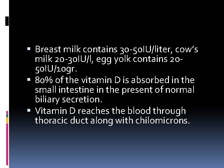
Breast milk contains 30 -50 IU/liter, cow’s milk 20 -30 IU/l, egg yolk contains 2050 IU/10 gr. 80% of the vitamin D is absorbed in the small intestine in the present of normal biliary secretion. Vitamin D reaches the blood through thoracic duct along with chilomicrons.
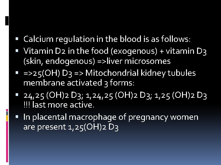
Calcium regulation in the blood is as follows: Vitamin D 2 in the food (exogenous) + vitamin D 3 (skin, endogenous) =>liver microsomes =>25(OH) D 3 => Mitochondrial kidney tubules membrane activated 3 forms: 24, 25 (OH)2 D 3; 1, 25 (OH)2 D 3 !!! last more active. In placental macrophage of pregnancy women are present 1, 25(OH)2 D 3
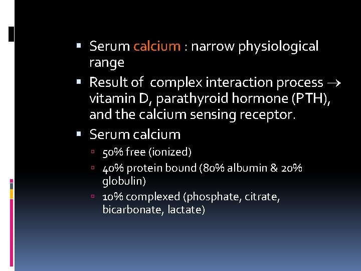
Serum calcium : narrow physiological range Result of complex interaction process vitamin D, parathyroid hormone (PTH), and the calcium sensing receptor. Serum calcium 50% free (ionized) 40% protein bound (80% albumin & 20% globulin) 10% complexed (phosphate, citrate, bicarbonate, lactate)
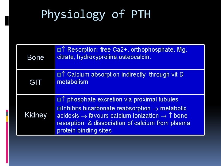
Physiology of PTH o Resorption: free Ca 2+, orthophosphate, Mg, Bone citrate, hydroxyproline, osteocalcin. o Calcium absorption indirectly through vit D GIT metabolism o phosphate excretion via proximal tubules Kidney o. Inhibits bicarbonate reabsorption metabolic acidosis favours calcium ionization bone resorption & dissociation of calcium from plasma protein binding sites
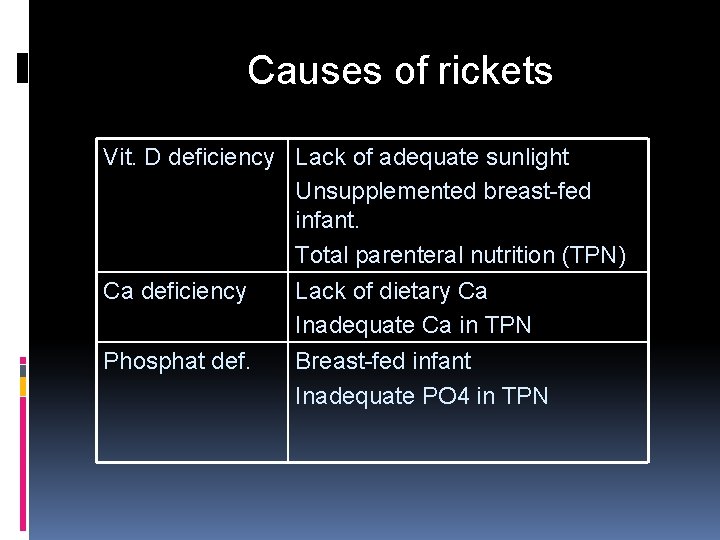
Causes of rickets Vit. D deficiency Lack of adequate sunlight Unsupplemented breast-fed infant. Total parenteral nutrition (TPN) Ca deficiency Phosphat def. Lack of dietary Ca Inadequate Ca in TPN Breast-fed infant Inadequate PO 4 in TPN
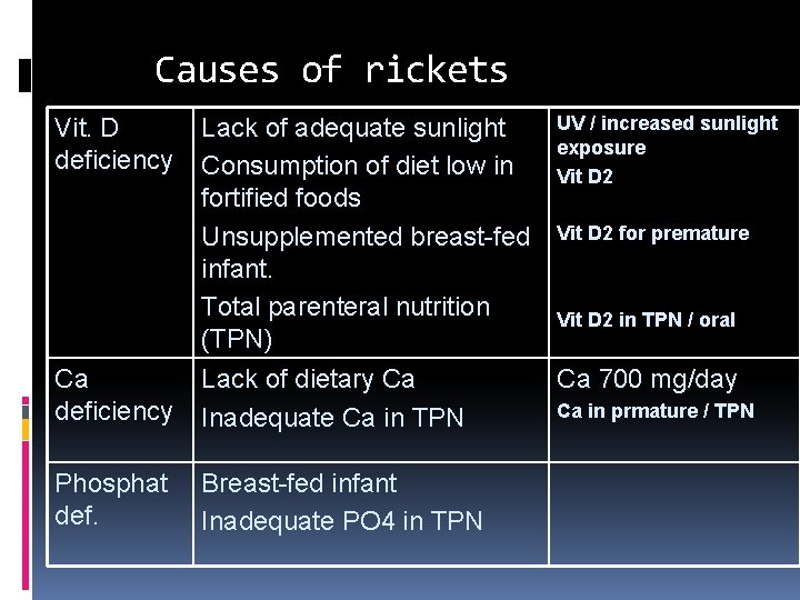
Causes of rickets Ca deficiency UV / increased sunlight Lack of adequate sunlight exposure Consumption of diet low in Vit D 2 fortified foods Unsupplemented breast-fed Vit D 2 for premature infant. Total parenteral nutrition Vit D 2 in TPN / oral (TPN) Lack of dietary Ca Ca 700 mg/day Ca in prmature / TPN Inadequate Ca in TPN Phosphat def. Breast-fed infant Inadequate PO 4 in TPN Vit. D deficiency
CLINICAL MANIFESTATIONS Rickets may develop in any age of an infant, more frequent at 3 -6 mo, early in prematures. The first signs of hypocalcaemia are CNS changes- excitation, restlessness, excessive sweated during sleep and feeding, tremors of the chin and extremities.
Skin and muscle changes- pallor, occipital alopecia, fragile nails and hair, muscular hypotony, motor retardation. Complications- apnoea, stridor, low calcium level with neuromuscular irritability (tetany). CNS changes are sometimes interpreted as CNS trauma and the administration of the
ACUTE SIGNS Have acute and subacute clinical signs Craniotabes – acute sign of rickets, osteolyses detected by pressing firmly over the occipital or posterior parietal bones, pingpong ball sensation will be felt. Large anterior fontanella, with hyperflexible borders, cranial deformation with asymmetric occipital flattening.
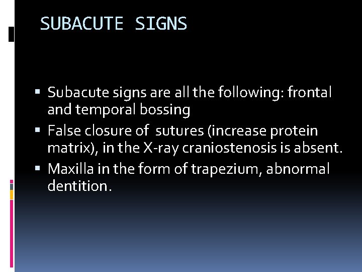
SUBACUTE SIGNS Subacute signs are all the following: frontal and temporal bossing False closure of sutures (increase protein matrix), in the X-ray craniostenosis is absent. Maxilla in the form of trapezium, abnormal dentition.
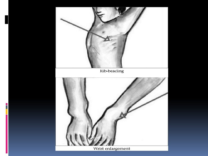
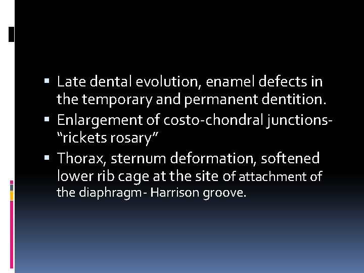
Late dental evolution, enamel defects in the temporary and permanent dentition. Enlargement of costo-chondral junctions“rickets rosary” Thorax, sternum deformation, softened lower rib cage at the site of attachment of the diaphragm- Harrison groove.
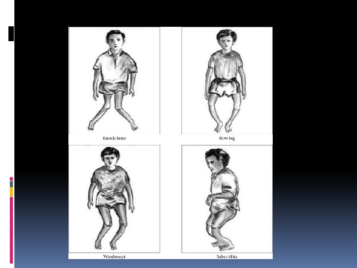
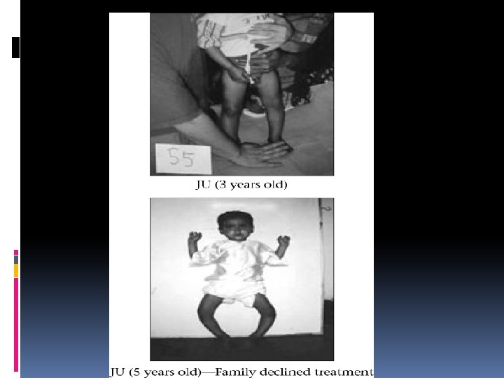
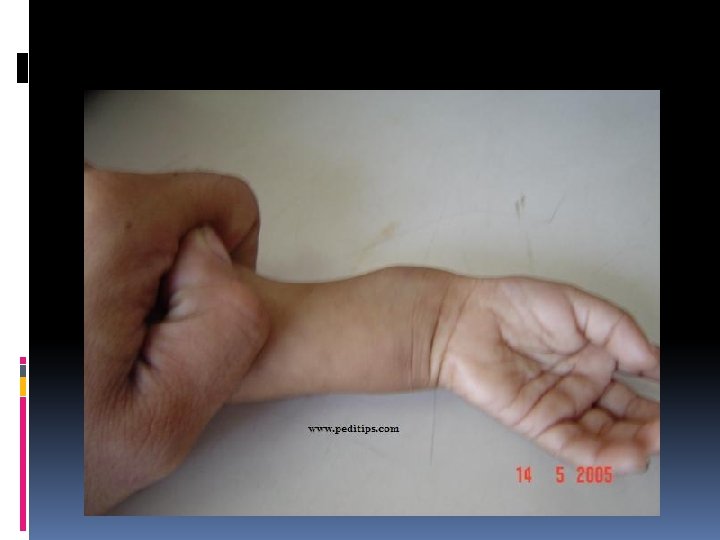
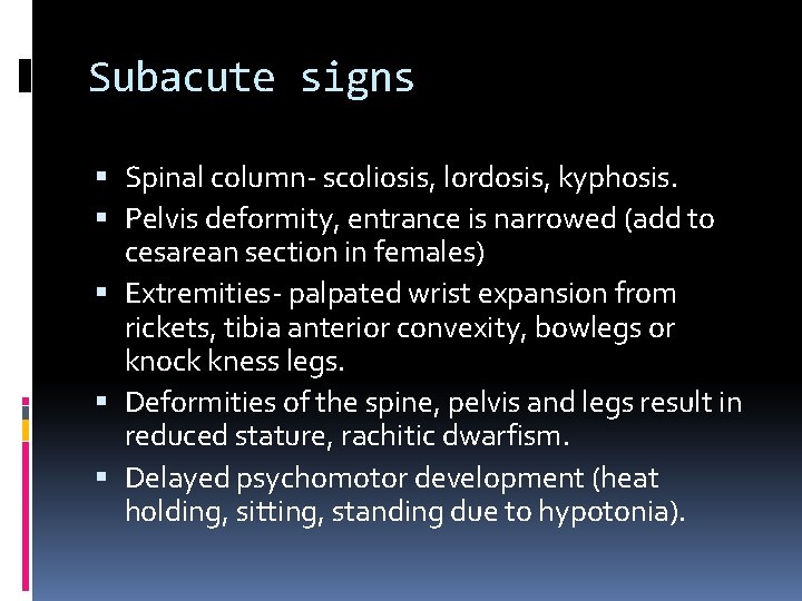
Subacute signs Spinal column- scoliosis, lordosis, kyphosis. Pelvis deformity, entrance is narrowed (add to cesarean section in females) Extremities- palpated wrist expansion from rickets, tibia anterior convexity, bowlegs or knock kness legs. Deformities of the spine, pelvis and legs result in reduced stature, rachitic dwarfism. Delayed psychomotor development (heat holding, sitting, standing due to hypotonia).
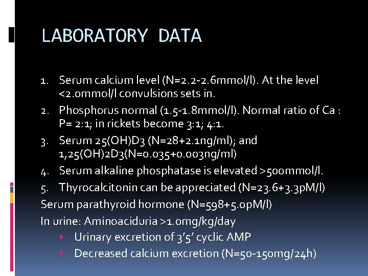
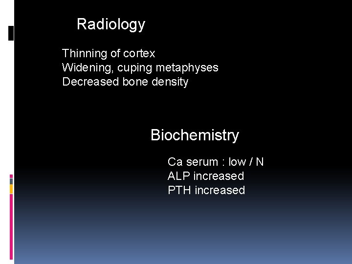
LABORATORY DATA 1. Serum calcium level (N=2. 2 -2. 6 mmol/l). At the level <2. 0 mmol/l convulsions sets in. 2. Phosphorus normal (1. 5 -1. 8 mmol/l). Normal ratio of Ca : P= 2: 1; in rickets become 3: 1; 4: 1. 3. Serum 25(OH)D 3 (N=28+2. 1 ng/ml); and 1, 25(OH)2 D 3(N=0. 035+0. 003 ng/ml) 4. Serum alkaline phosphatase is elevated >500 mmol/l. 5. Thyrocalcitonin can be appreciated (N=23. 6+3. 3 p. M/l) Serum parathyroid hormone (N=598+5. 0 p. M/l) In urine: Aminoaciduria >1. 0 mg/kg/day • Urinary excretion of 3’ 5’ cyclic AMP • Decreased calcium excretion (N=50 -150 mg/24 h)
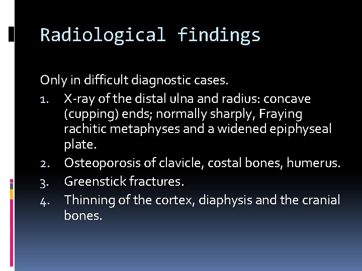
Radiological findings Only in difficult diagnostic cases. 1. X-ray of the distal ulna and radius: concave (cupping) ends; normally sharply, Fraying rachitic metaphyses and a widened epiphyseal plate. 2. Osteoporosis of clavicle, costal bones, humerus. 3. Greenstick fractures. 4. Thinning of the cortex, diaphysis and the cranial bones.
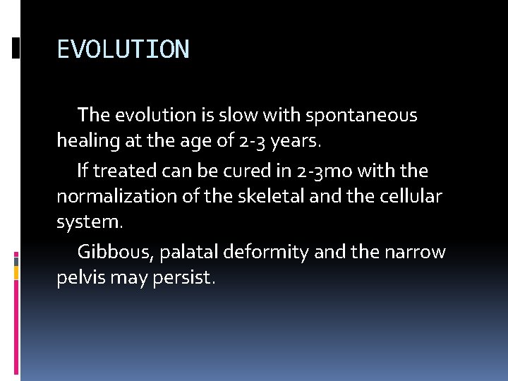
EVOLUTION The evolution is slow with spontaneous healing at the age of 2 -3 years. If treated can be cured in 2 -3 mo with the normalization of the skeletal and the cellular system. Gibbous, palatal deformity and the narrow pelvis may persist.
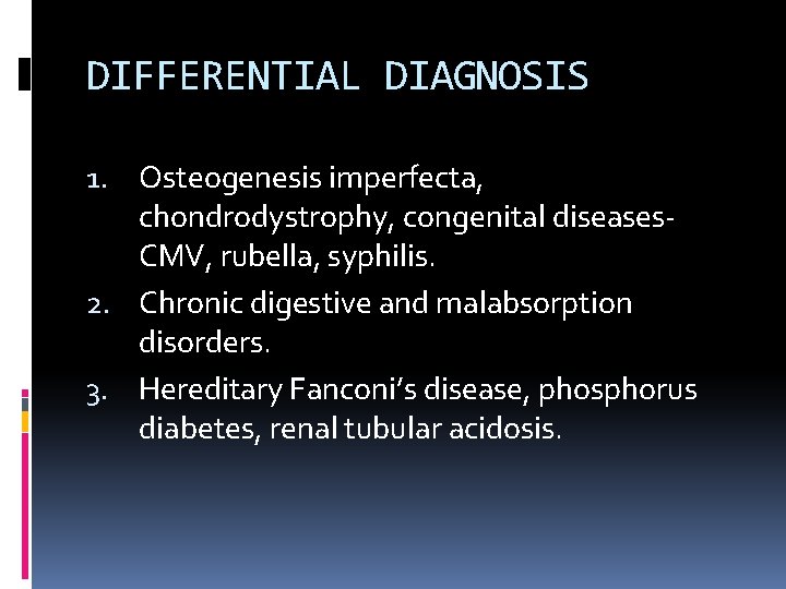
DIFFERENTIAL DIAGNOSIS 1. Osteogenesis imperfecta, chondrodystrophy, congenital diseases- CMV, rubella, syphilis. 2. Chronic digestive and malabsorption disorders. 3. Hereditary Fanconi’s disease, phosphorus diabetes, renal tubular acidosis.
Slide 3 of 21
Radiology Thinning of cortex Widening, cuping metaphyses Decreased bone density Biochemistry Ca serum : low / N ALP increased PTH increased
PROPHILAXIS IN RICKETS Specific antenatal prophylactic dose administration : 500 -1000 IU/day of vitamin D 3 solution at the 28 -th week of pregnancy. The total dose administered is 135000180000 IU. In term infants prophylactic intake of vitamin D 2 700 IU/d started at 10 days of age during the first 2 years of life; in premature the dose may increase to 1000 IU/day.
PROPHILAXIS IN RICKETS WHO recommendation for rickets prophilaxis in a children coming from unfavorable conditions and who have difficult access to hospitals is 200000 IU vitamin D 2 i/muscular, On the 7 day, 2, 4, 6 month- total dose 800000 IU. In case of the necessary prolongation 700 IU/day till 24 mo are given.
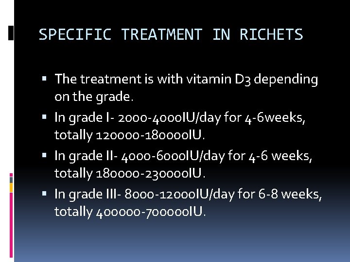
SPECIFIC TREATMENT IN RICHETS The treatment is with vitamin D 3 depending on the grade. In grade I- 2000 -4000 IU/day for 4 -6 weeks, totally 120000 -180000 IU. In grade II- 4000 -6000 IU/day for 4 -6 weeks, totally 180000 -230000 IU. In grade III- 8000 -12000 IU/day for 6 -8 weeks, totally 400000 -700000 IU.
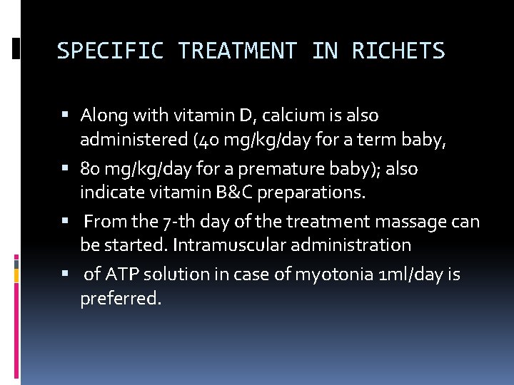
SPECIFIC TREATMENT IN RICHETS Along with vitamin D, calcium is also administered (40 mg/kg/day for a term baby, 80 mg/kg/day for a premature baby); also indicate vitamin B&C preparations. From the 7 -th day of the treatment massage can be started. Intramuscular administration of ATP solution in case of myotonia 1 ml/day is preferred.
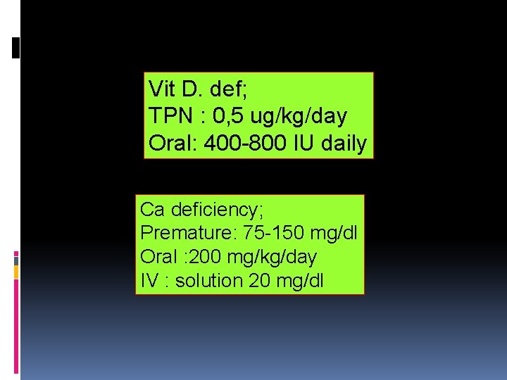
Vit D. def; TPN : 0, 5 ug/kg/day Oral: 400 -800 IU daily Ca deficiency; Premature: 75 -150 mg/dl Oral : 200 mg/kg/day IV : solution 20 mg/dl
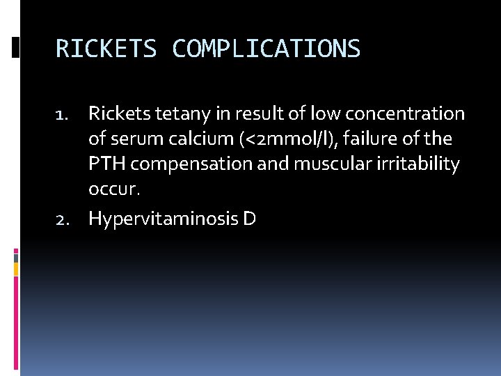
RICKETS COMPLICATIONS 1. Rickets tetany in result of low concentration of serum calcium (<2 mmol/l), failure of the PTH compensation and muscular irritability occur. 2. Hypervitaminosis D
HYPERVITAMINOSIS D Symptoms develop in hypersensitivity to vitamin D children or after 1 -3 mo of high doses intakes of vitamin D; they include hypotonia, anorexia, vomiting, irritability, constipation, polydipsia, polyuria, sleep disorder, dehydration. High serum level of acetone, nitrogen and Ca>2. 9 mmol/l are found. Increase calcium concentration in urine may provoke incontinence, renal damage and calcification.