Pathophysiology of ascites in cirrhosis Khalid Alswat MD


 NASH Other causes Autoimmune (AIH, PBC,")


 is the first step toward fluid")

 Portal hypertension leads to VD Mechanisms of vasodilation Increase production of nitric oxide")

 –")


 at the")

 (extravasate)")


 High SAAG ≥ 1. 1 g/d. L • Cirrhosis Low")


- Slides: 27
Pathophysiology of ascites in cirrhosis Khalid Alswat, MD, MRCP, FACP Associate Professor, Consultant Gastroenterologist/hepatologist College of Medicine, King Saud University
Objectives To understand basic pathophysiologic steps in the development of ascites secondary to cirrhosis. To correlate the anatomic and pathophysiologic changes with clinical manifestations. To understand the basic steps in evaluation of patients with ascites
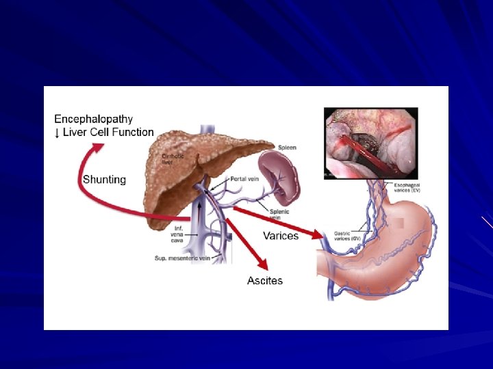
Cirrhosis: Late stage of chronic liver inflammation and fibrosis, in which liver parenchyma is distorted and replaced by fibrous tissue and regenerating nodules. NORMAL LIVER CIRRHOTIC LIVER
Causes of Cirrhosis Commonest Viral (HBV+- HDV, HCV) NASH Other causes Autoimmune (AIH, PBC, PSC) Metabolic & Hereditary WD, A 1 AT, HH Vascular BCS, HF Biliary SC Drugs
Definition The pathologic accumulation of fluid in the peritoneal cavity § It is the most common complication of cirrhosis Ascites: § Cirrhosis 85% § Other causes 15 %
The development of ascites is the final consequence of a series of anatomic, pathophysiologic, and biochemical abnormalities occurring in patients with cirrhosis. The formation of ascites is governed by the same principles as edema formation at other sites: net capillary permeability and the hydraulic and oncotic pressure gradients.
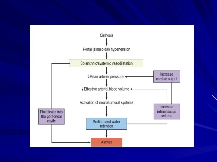
PORTAL HYPERTENSION The development of portal hypertension (PHT) is the first step toward fluid retention in the setting of cirrhosis. Patients with cirrhosis but without PHT do not develop ascites or edema A portal pressure >12 mm. Hg appears to be required for fluid retention
PORTAL HYPERTENSION 2 main mechanisms involved in portal hypertension: 1 -Mechanical (due to structural changes in the liver with fibrosis and regenerative nodules) 2 -Hemdynamic (circulatory, vascular, functional, and biochemical abnormalities)
Vasodilation(VD) Portal hypertension leads to VD Mechanisms of vasodilation Increase production of nitric oxide (NO), which is the primary mediator of VD in cirrhosis (for splanchnic and peripheral vasodilation). Increased levels of other circulating VDs. (Glucagon , vasoactive intestinal peptide(VIP), prostacyclin. production of these VDs may be stimulated by endotoxins or other bacterial products
VD VD initially develops in the arterial splanchnic circulation (i. e. , the mesenteric arteries). Subsequently, vasodilation develops in the arterial systemic circulation.
Hyperdynamic circulation Systemic consequences to VD – reduction in systemic vascular resistance (SVR) – reduction in mean arterial pressure (MAP) – increase in cardiac output hyperdynamic circulation Usually patients with cirrhosis and ascites have hyperdynamic circulation.
CONSEQUENCES OF VASODILATION Activation of endogenous vasoconstrictors Sodium and water retention Increase renal vasoconstriction.
Pathogenic mechanisms responsible for the activation of vasoactive systems and hyperdynamic circulation in cirrhosis
Activation of endogenous vasoconstrictor agents VD The reduction in pressure (or stretch) at the carotid and renal baroreceptors activation of the sodium-retaining neurohumoral mechanisms (in an attempt to restore perfusion pressure to normal – renin-angiotensin-aldosterone system – sympathetic nervous system – antidiuretic hormone (vasopressin). The net effect is avid sodium and water retention
Sodium retention impaired sodium excretion Water retention Initially water excretion is normal in patients with cirrhosis before the development of ascites and then becomes increasingly impaired as the liver disease progresses. (Increase ADH) Thus, patients with cirrhosis and ascites usually demonstrate urinary sodium retention, increased total body sodium, and dilutional hyponatremia. Renal vasoconstriction — VC renal hypoperfusion decrease GFR (in some patient hepatorenal syndrome)
So ultimately This excess retained blood volume is thought to leak-out (filtered ) (extravasate) directly from both the liver surface, and the mesenteric vessels. due to: – increased hydrostatics pressure – Increase vascular wall permeability – concurrently decreased oncotic (osmotic) pressure (hypoalbuminemia)
Abnormalities in patients with cirrhosis and ascites Circulatory Vascular Functional Reduced systemic vascular resistance Splanchnic vasodilation Activation of systemic Sodium retention vasodilator factors Reduced arterial pressure Renal artery vasoconstriction Activation of systemic Water retention vasoconstrictor factors Increased heart rate Pulmonary vasodilation Activation of renal vasodilator factors Increased systemic nitric oxide Reduced glomerular filtration rate Increased systemic prostaglandins Increased cardiac index Increased plasma volume Reduced renal blood flow Increased portal blood flow Biochemical Increased renal nitric oxide and prostaglandins
Any new ascites should be tapped analyzed!
Ascitic Fluid Analysis Measure SAAG
Serum-to-Ascites Albumin Gradient (SAAG) High SAAG ≥ 1. 1 g/d. L • Cirrhosis Low SAAG <1. 1 g/d. L • Peritoneal • Heart • • • failure/constrictive pericarditis Alcoholic hepatitis Budd chiari Massive hepatic metastases • • • carcinomatosis Peritoneal tuberculosis Secondary peritonitis Pancreatitis Serositis Nephrotic syndrome
Initial treatment of ascites 1 -Dietary sodium restriction – Limiting sodium intake to 88 meq (2000 mg) per day 2 -Diuretics (most successful therapeutic regimen is the combination of Spironolactone and Furosemide) – Monitor electrolytes and kidney function Discontinue non-steroidal anti-inflammatory drugs Rx of underlying cause Evaluation for liver transplantation
Take home message-1 Ascites is the most common liver cirrhosis complication. Development of ascites indicates advanced stage of liver disease and poorer prognosis. Development of ascites is complex process. Portal hypertension is first step in ascites development in patient with cirrhosis. Portal hypertension and possible bacterial toxin trigger VDs.
Take home message-2 VD with activation of secondary mechanisms; – renin-angiotensin-aldosterone system – sympathetic nervous system – antidiuretic hormone (vasopressin). LEADS TO SALT AND WATER RESTENTION and Increase plasma volume All these with hypoalbuminemia and increase vascular permeability lead to fluid extravasation.
Thank you