
Heart disease with pregnancy Dr Manal Shafik Swelem





- Slides: 67
Heart disease with pregnancy Dr. Manal Shafik Swelem Assistant Professor Obstetrics & Gynecology
Incidence n n n Heart disease is the third leading cause of death in 25 - 44 year old women. Because it is relatively common in women of childbearing age, heart disease of varying severity complicates about 1 % of pregnancies. Although maternal mortality related to cardiovascular disease has decreased remarkably over the past 50 years, heart disease still contributes significantly to maternal mortality.
Anatomical & physiological considerations During pregnancy, the circulation is embarrassed an extra strain is thrown on the heart due to the following factors: 1. Increase in body weight 2. Increase in blood volume, which is progressive; it rises steeply in the 2 nd half of pregnancy. 3. Increased cardiac output (most important) is increased 3050%. Almost half of the total increase occurs by 8 weeks, and it is maximized by midpregnancy. 4. The early increase in cardiac output results from an augmented stroke volume associated with decreased vascular resistance and corresponding diminished blood pressure.
Anatomical & physiological considerations During pregnancy, the circulation is embarrassed an extra strain is thrown on the heart due to the following factors: 5. The maternal placental circulation acts as an intravenous shunt. 6. The upward displacement of the diaphragm by the growing uterus leads to: compression of the lung bases, impeding ventillation, displacement of the heart causing cardiac embarrassment. 7. Later in pregnancy there is also an increased resting pulse, and stroke volume increases even more, presumably related to increased diastolic filling from augmented blood volume. 8. Each uterine contraction is accompanied by a 15 -20% increase in COP, while compression of the IVC by the gravid uterus reduces it by 25 -30%.
Types of cardiac lesions n n n Over 90% are cases of rheumatic disease; mitral stenosis is the commonest. Other types of cardiac disease are less common. Cases of congenital heart disease usually suffer from chronic ill-health & rarely marry or become pregnant. ? ? ? Hypertensive & ischemic heart disease (IHD) belong to a later period of life.
Diagnosis of heart disease: n Many of the physiologic changes of normal pregnancy tend to make the diagnosis of heart disease more difficult. For example, systolic heart murmurs, accentuated respiratory effort, and edema may occur during normal pregnancy as well as in association with cardiac disease. Clinical indicators of heart disease during pregnancy: Symptoms: n Progressive dyspnea or orthopnea, nocturnal cough, hemoptysis, syncope, chest pain. Clinical findings: n Cyanosis, clubbing of fingers, persistent neck vein distension, systolic murmur grade 3/6 or greater, diastolic murmur, cardiomegaly, persistent arrhythmia, persistent split-second sound.
Diagnostic studies n n n Most diagnostic studies are non-invasive and can be conducted safely in pregnant women. Conventional testing typically includes electrocardiography, echocardiography, and chest radiography. If indicated, heart catheterization can be performed with limited X-ray fluoroscopy.
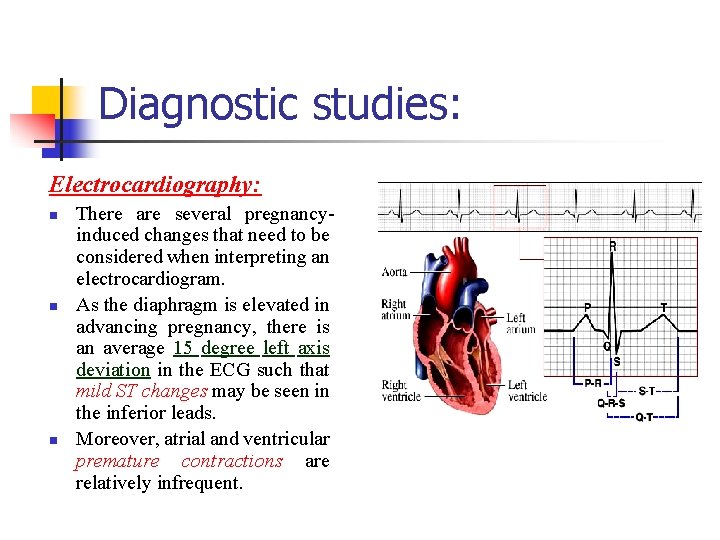
Diagnostic studies: Electrocardiography: n n n There are several pregnancyinduced changes that need to be considered when interpreting an electrocardiogram. As the diaphragm is elevated in advancing pregnancy, there is an average 15 degree left axis deviation in the ECG such that mild ST changes may be seen in the inferior leads. Moreover, atrial and ventricular premature contractions are relatively infrequent.
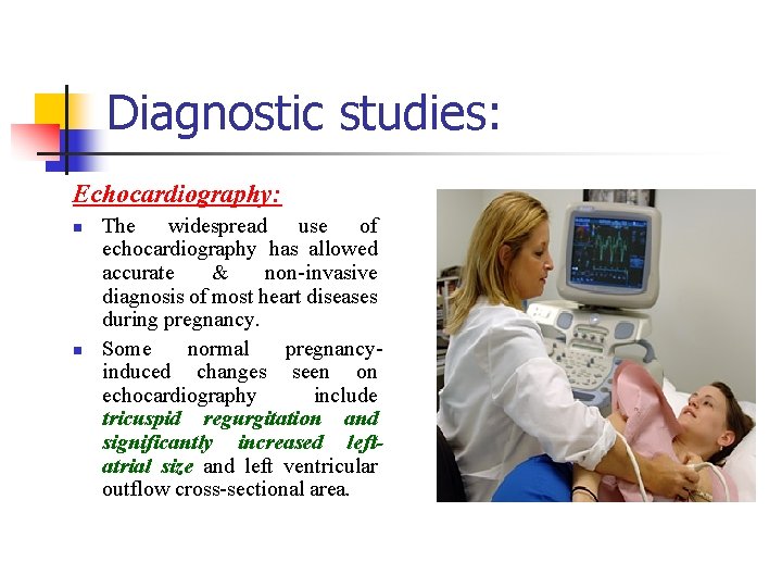
Diagnostic studies: Echocardiography: n n The widespread use of echocardiography has allowed accurate & non-invasive diagnosis of most heart diseases during pregnancy. Some normal pregnancyinduced changes seen on echocardiography include tricuspid regurgitation and significantly increased leftatrial size and left ventricular outflow cross-sectional area.
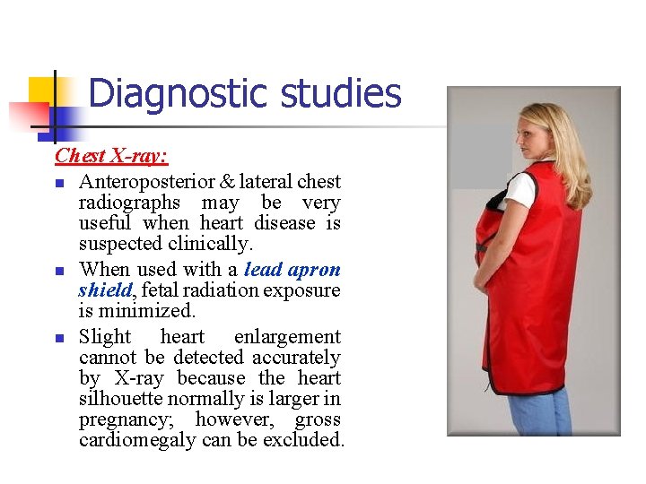
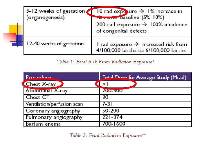
Diagnostic studies Chest X-ray: n Anteroposterior & lateral chest radiographs may be very useful when heart disease is suspected clinically. n When used with a lead apron shield, fetal radiation exposure is minimized. n Slight heart enlargement cannot be detected accurately by X-ray because the heart silhouette normally is larger in pregnancy; however, gross cardiomegaly can be excluded.
How do you grade the functional capacity of heart?
The New York Heart Association (NYHA) Grading of functional capacity of the heart: CLASS II No functional limitation of activity Mild limitation of physical activity. Symptoms with extra ordinary physical work. Symptoms with ordinary physical work CLASS III CLASS IV Marked limitation of physical activity Severe limitation of physical activity Symptoms with less than ordinary physical work Symptoms at rest
Classes III and IV n n n The important question in these women is whether pregnancy should be undertaken. Those who choose to become pregnant must understand the risks and cooperate fully. If seen early enough, women with some types of severe cardiac disease should consider pregnancy termination. If the pregnancy is continued, prolonged hospitalization or bed rest will often be necessary.
What is the mortality associated with the various cardiac lesions ?
Mortality associated with specific cardiac lesions; 1. Low risk of maternal mortality (less than 1%). (a) Septal defects (ASD, VSD). (b) New York Heart Association classes I and II. (c) Patent ductus arteriosus. (d) Pulmonary / tricuspid lesions. 2. Moderate risk of maternal mortality (5 -15%). (a) NYHA classes III and IV mitral stenosis. (b) Aortic stenosis. (c) Marfan’s syndrome with normal aorta. (d) Uncomplicated coarctation of aorta. (e) Past history of myocardial infarction. 3. High risk of maternal mortality (25 -50%). (a) Eissenmenger’s syndrome. (b) Pulmonary hypertension. (c) Marfan’s syndrome with abnormal aortic root. (d) Peripartum cardiomyopathy.
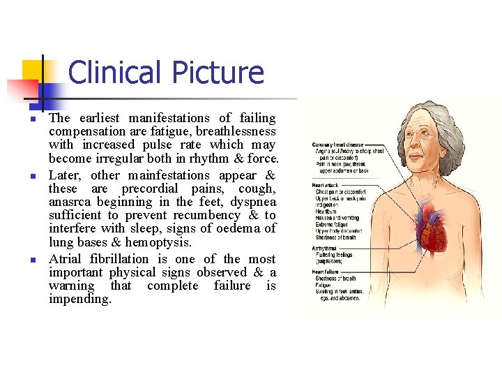
Clinical Picture n n n The earliest manifestations of failing compensation are fatigue, breathlessness with increased pulse rate which may become irregular both in rhythm & force. Later, other mainfestations appear & these are precordial pains, cough, anasrca beginning in the feet, dyspnea sufficient to prevent recumbency & to interfere with sleep, signs of oedema of lung bases & hemoptysis. Atrial fibrillation is one of the most important physical signs observed & a warning that complete failure is impending.
Critical Periods during pregnancy
Critical periods Compensation is least likely to break down in the early months of pregnancy. The patient however is exposed to serious risks at the following times: 1. Around the 30 th week of pregnancy when the blood volume & the cardiac output are at their maximum. 2. In the last month of pregnancy due to the maximal compression of the lung bases & displacement of the heart. 3. During the second stage of labour when the expulsive efforts of the mother throw a big strain on the right side of the heart.
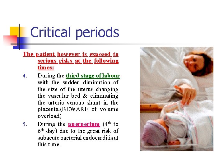
Critical periods The patient however is exposed to serious risks at the following times: 4. During the third stage of labour with the sudden diminution of the size of the uterus changing the vascular bed & eliminating the arterio-venous shunt in the placenta. (BEWARE of volume overload) 5. During the puerperium (4 th to 6 th day) due to the great risk of subacute bacterial endocarditis at this time.
Managing Cardiac Disease & Pregnancy
Preconception Counselling Discuss: n n n n n Characteristics of heart condition, functional & risk classifications Effects of cardiac condition on pregnancy Effects of pregnancy on cardiac condition Fetal & neonatal complications associated with her specific heart condition Need for multidisciplinary care Frequency of prenatal visits Need for anticoagulation & monitoring Timing of birth, type of hospital facility for childbirth Pain control, anesthesia, and potential need for CS
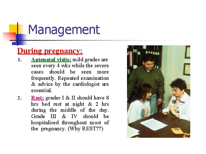
Management During pregnancy: 1. 2. Antenatal visits: mild grades are seen every 4 wks while the severe cases should be seen more frequently. Repeated examination & advice by the cardiologist are essential. Rest: grades I & II should have 8 hrs bed rest at night & 2 hrs during the middle of the day. Grade III & IV should be hospitalised throughout most of the pregnancy. (Why REST? ? )
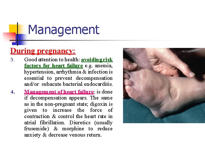
Management During pregnancy: 3. 4. Good attention to health: avoiding risk factors for heart failure e. g. anemia, hypertension, arrhythmia & infection is essential to prevent decompensation and/or subacute bacterial endocarditis. Management of heart failure: is done if decompensation appears. The same as in the non-pregnant state; digoxin is given to increase the force of contraction & control the heart rate in atrial fibrillation. Diuretics (usually frusemide) & morphine to reduce anxiety & decrease venous return.
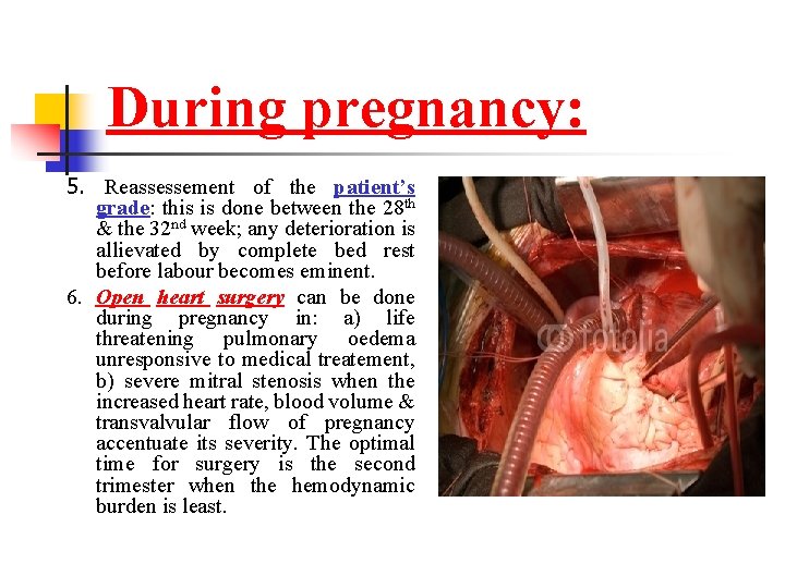
During pregnancy: 5. Reassessement of the patient’s grade: this is done between the 28 th & the 32 nd week; any deterioration is allievated by complete bed rest before labour becomes eminent. 6. Open heart surgery can be done during pregnancy in: a) life threatening pulmonary oedema unresponsive to medical treatement, b) severe mitral stenosis when the increased heart rate, blood volume & transvalvular flow of pregnancy accentuate its severity. The optimal time for surgery is the second trimester when the hemodynamic burden is least.
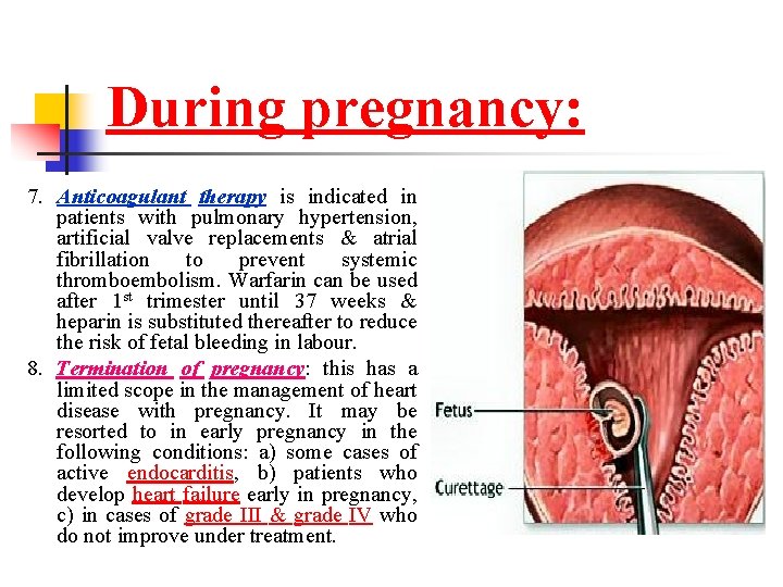
During pregnancy: 7. Anticoagulant therapy is indicated in patients with pulmonary hypertension, artificial valve replacements & atrial fibrillation to prevent systemic thromboembolism. Warfarin can be used after 1 st trimester until 37 weeks & heparin is substituted thereafter to reduce the risk of fetal bleeding in labour. 8. Termination of pregnancy: this has a limited scope in the management of heart disease with pregnancy. It may be resorted to in early pregnancy in the following conditions: a) some cases of active endocarditis, b) patients who develop heart failure early in pregnancy, c) in cases of grade III & grade IV who do not improve under treatment.
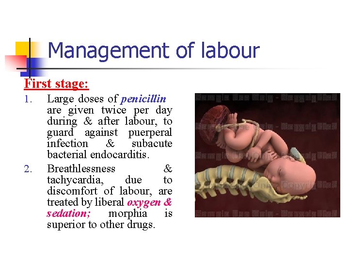
Management of labour First stage: 1. 2. Large doses of penicillin are given twice per day during & after labour, to guard against puerperal infection & subacute bacterial endocarditis. Breathlessness & tachycardia, due to discomfort of labour, are treated by liberal oxygen & sedation; morphia is superior to other drugs.
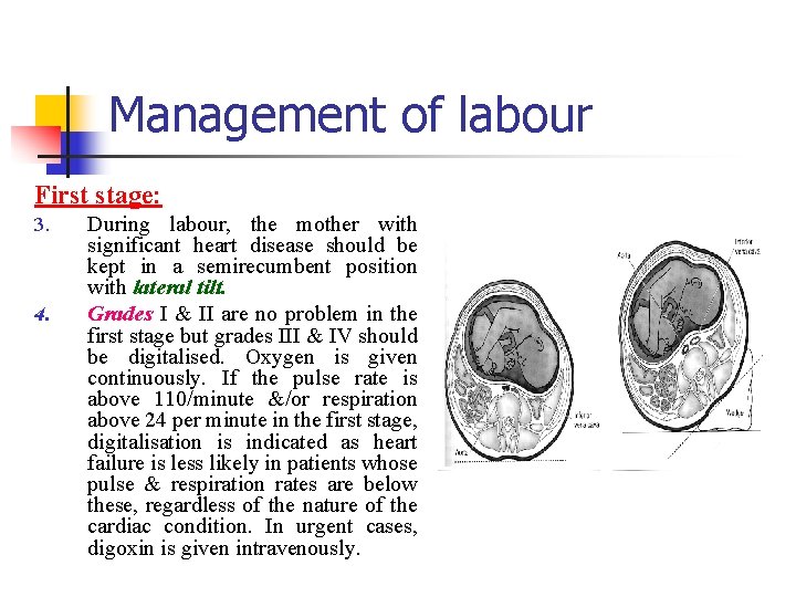
Management of labour First stage: 3. 4. During labour, the mother with significant heart disease should be kept in a semirecumbent position with lateral tilt. Grades I & II are no problem in the first stage but grades III & IV should be digitalised. Oxygen is given continuously. If the pulse rate is above 110/minute &/or respiration above 24 per minute in the first stage, digitalisation is indicated as heart failure is less likely in patients whose pulse & respiration rates are below these, regardless of the nature of the cardiac condition. In urgent cases, digoxin is given intravenously.
Is it better for cardiac pregnant females to have a vaginal delivery or a cesarean section? ? ?
Anesthesia n n n Relief from pain and apprehension is especially important. Although intravenous analgesic provide satisfactory pain relief for some women, continous epidural analgesia is recommended for most situations. The major danger of regional analgesia is maternal hypotension. For vaginal delivery in women with only mild cardiovascular compromise, epidural analgesia given along with intravenous sedation often suffices. This has been shown to minimize intrapartum cardiac output fluctuation and allows forceps or vacuum-assisted delivery. For cesarean delivery, epidural anesthesia is preferred by most clinicians. Spinal anesthesia is contraindicated with some lesions. Finally, general endotracheal anesthesia with thiopental, succinylcholine, nitrous oxide, and at least 30% oxygen has also proved satisfactory.
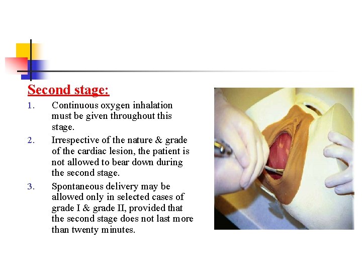
Second stage: 1. 2. 3. Continuous oxygen inhalation must be given throughout this stage. Irrespective of the nature & grade of the cardiac lesion, the patient is not allowed to bear down during the second stage. Spontaneous delivery may be allowed only in selected cases of grade I & grade II, provided that the second stage does not last more than twenty minutes.
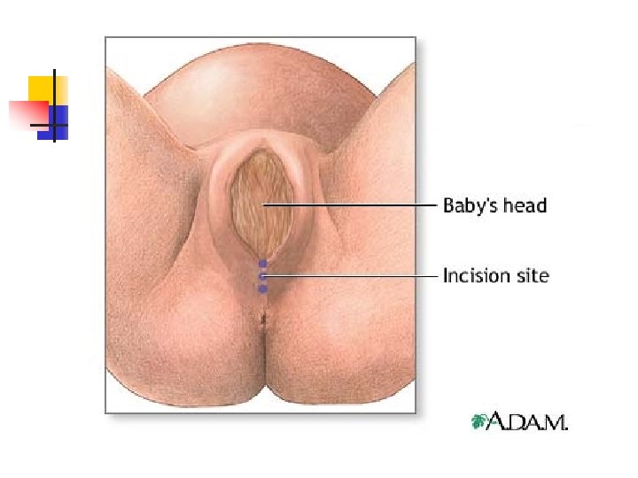
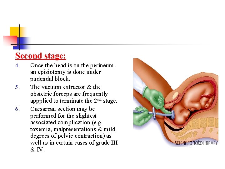
Second stage: 4. 5. 6. Once the head is on the perineum, an episiotomy is done under pudendal block. The vacuum extractor & the obstetric forceps are frequently appplied to terminate the 2 nd stage. Caesarean section may be performed for the slightest associated complication (e. g. toxemia, malpresentations & mild degrees of pelvic contraction) as well as in certain cases of grade III & IV.
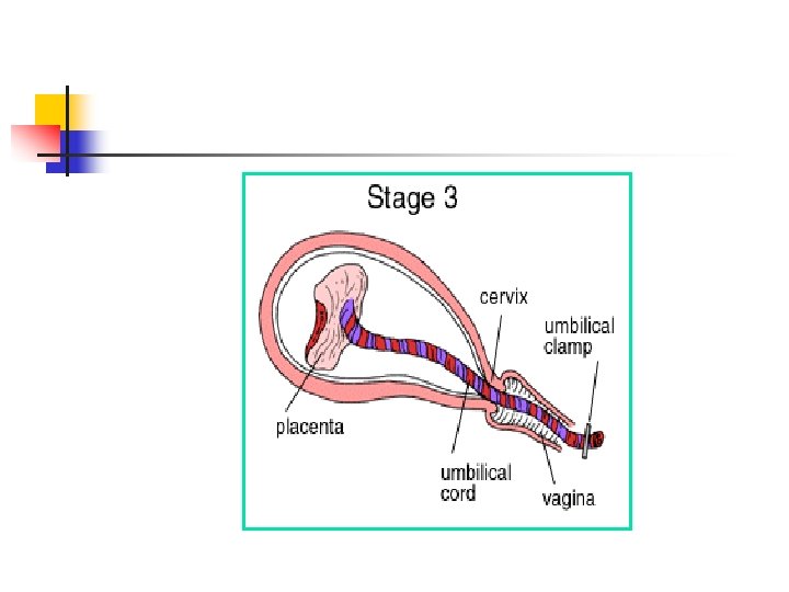
Third stage 1. Expulsion of the placenta should never be hurried. 2. Methyl ergometrin (methergin) should not be given, but intravenous oxytocin may be combined with frusemide to reduce the risk of postpartum hemorrhage. 3. Suturing of the episiotomy is done under local anesthesia. ? ? ? 4. After delivery, the patient is propped up in bed 45 degrees, given morphia & oxygen should be at hand.
Puerperium n n n Women, who have shown little or no evidence of cardiac distress during pregnancy, labour, or delivery may still decompensate after delivery. Therefore, it is important that meticulous care be continued into the puerperium. Postpartum hemorrhage, anemia, infection, and thromboembolism are much more serious complications with heart disease. Indeed, these factors frequently act in concert to precipitate postpartum heart failure in women with underlying heart disease. Breast feeding contraindicated in grade IV and in most of the grade III cases.
Which is the ideal contraceptive for women with heart disease ?
Contraception 1. OC pills are not ideal as they can cause thrombo embolism. 2. IUCD can cause infection- endocarditis. 3. Barrier contraceptives – Have high failure rates. 4. Progestin only pills or Long acting injectable progesterone are better 5. Sterilization.
Anaemias of pregnancy
Physiological considerations n n n During pregnancy, there is lowering of the concentration of Hb & RBC; but these return to normal values within a short time after delivery. This is due to a physiologic hemodilution which accompanies the increased blood volume. Anemia during pregnancy must be differentiated from the physiological hemodilution of pregnancy. The increased blood volume helps to compensate for the blood loss during labour.
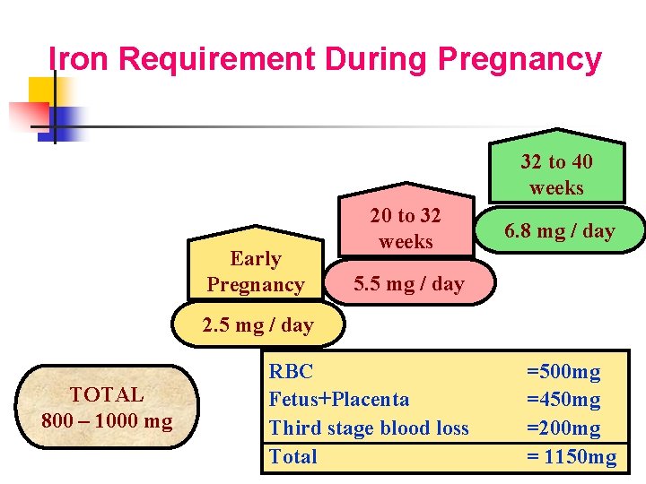
Iron Requirement During Pregnancy 32 to 40 weeks Early Pregnancy 20 to 32 weeks 6. 8 mg / day 5. 5 mg / day 2. 5 mg / day TOTAL 800 – 1000 mg RBC Fetus+Placenta Third stage blood loss Total =500 mg =450 mg =200 mg = 1150 mg
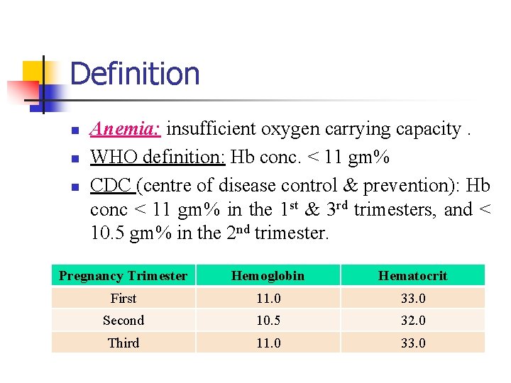
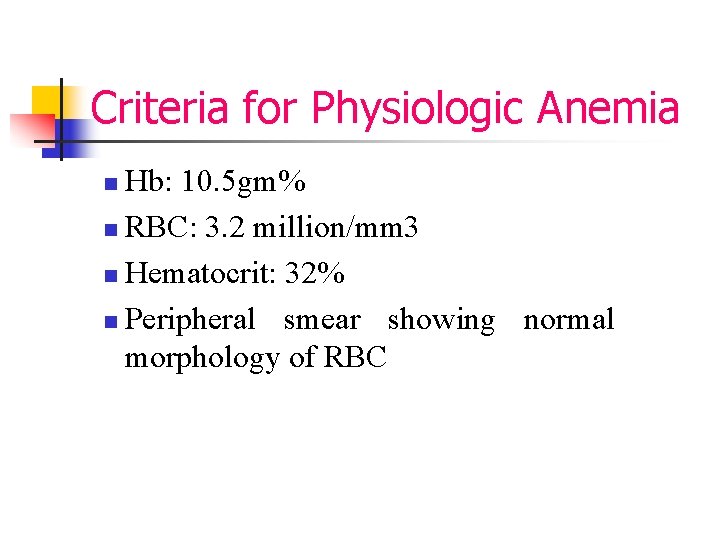
Definition n Anemia: insufficient oxygen carrying capacity. WHO definition: Hb conc. < 11 gm% CDC (centre of disease control & prevention): Hb conc < 11 gm% in the 1 st & 3 rd trimesters, and < 10. 5 gm% in the 2 nd trimester. Pregnancy Trimester Hemoglobin Hematocrit First 11. 0 33. 0 Second 10. 5 32. 0 Third 11. 0 33. 0
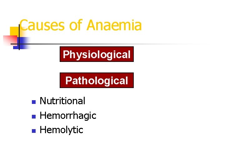
Causes of Anaemia Physiological Pathological n n n Nutritional Hemorrhagic Hemolytic
Criteria for Physiologic Anemia Hb: 10. 5 gm% n RBC: 3. 2 million/mm 3 n Hematocrit: 32% n Peripheral smear showing normal morphology of RBC n
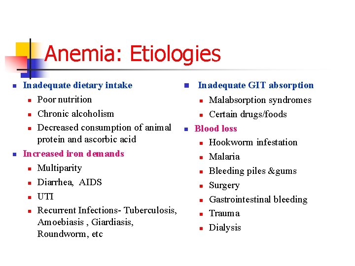
Anemia: Etiologies n n Inadequate dietary intake n Poor nutrition n Chronic alcoholism n Decreased consumption of animal protein and ascorbic acid Increased iron demands n Multiparity n Diarrhea, AIDS n UTI n Recurrent Infections- Tuberculosis, Amoebiasis , Giardiasis, Roundworm, etc n Inadequate GIT absorption Malabsorption syndromes n Certain drugs/foods Blood loss n Hookworm infestation n Malaria n Bleeding piles &gums n Surgery n Gastrointestinal bleeding n Trauma n Dialysis n n
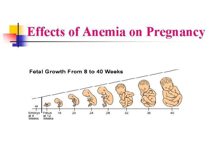
Effects of Anemia on Pregnancy
Effects of Anemia on Fetus n The fetus tolerates quite well even advanced degrees of maternal anemia, this is the result of the high oxygen affinity of fetal hemoglobin, and the efficiency of the maternal oxygen transport system which is capable of delivering adequate amounts of oxygen to the tissues despite low levels of hemoglobin.
Facts about fetal affection Mild and moderate anemia may not show significant effects. n Iron is actively transported across the placenta. n Fetal iron and ferritin levels > maternal levels 3 times n
Effects of Anemia on Fetus • • • PROM IUGR, IUFD Prematurity, Abnormal trophoblast invasion Fetal programming & disease of newborn: behavioral abnormalities, poor performance. • (Barker DJP, Bull AR, et all BMJ 1990; 301: 259 -262)
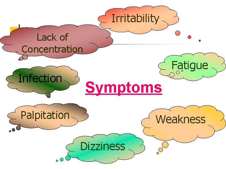
Irritability Lack of Concentration Infection Fatigue Symptoms Palpitation Weakness Dizziness
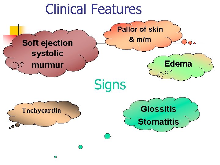
Clinical Features Soft ejection systolic murmur Pallor of skin & m/m Edema Signs Tachycardia Glossitis Stomatitis
Maternal Effects of Anemia § § Behavioral changes, irritability. Loss of appetite, indigestion, etc. due low performance of each organ. Increased morbidity and mortality due to PIH, APH, PPH, if associated. Reduced immune function - infection, ante-partum and puerperal sepsis. § § § Negative thermoregulation Increased risk of blood transfusion Preterm Labor Sub involution Failing lactation Pulmonary Venous thrombosis & embolism, due to thrombophlebitis.
Maternal Effects of Anemia n Many anemic women who were asymptomatic before delivery are at risk of developing severe symptoms including syncopal episodes in the first 24 -48 hours following delivery.
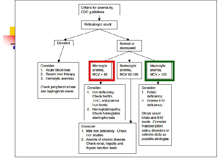
Hypochromic microcytic anaemia This may have been present before the pregnancy or may develop during its course. Aetiology: n Women who cannot afford iron-containing foods & those with rapid successive pregnancies are unable to replace the depleted iron stores, and are liable to this form of anemia. n
Hypochromic microcytic anaemia The causes that favour its development or aggravation in the pregnant woman are: a) The demands of the fetus for iron. b) A possible deficient iron intake. c) A possible deficient intake of proteins and vitamins d) Deficient iron- assimilation due to deficient gastric secretion Clinical picture The patient is weak, dyspneic on exertion with easy fatiguability. On examination, there is pallor, the pulse is rapid, the hemoglobin is less than 70%, the RBCs are less than 3. 5 millions per mm 3, and the colour index is less than one.
Stages of iron deficiency anemia Depletion of iron stores: n n n Ferritin < 20 ng/ml Normal Hb/Hct Normal RBC indicies Iron-deficient hematopoiesis: n n n Ferritin < 20 ng/ml MCV & MCH low Transferrin saturation & serum iron decreased Frank iron deficiency anemia: n n Ferritin < 20 ng/ml MCV & MCH decreased Transferrin saturation & serum iron decreased. Hb < 10 g/dl, Hct < 28%
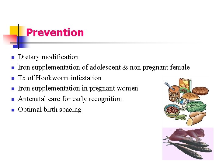
Prevention n n n Dietary modification Iron supplementation of adolescent & non pregnant female Tx of Hookworm infestation Iron supplementation in pregnant women Antenatal care for early recognition Optimal birth spacing
Hypochromic microcytic anaemia Treatment 1) Properly balanced diet including intake of iron containing vegetables and meat (liver) 2) An iron preparation is given either orally or in the form of injectables. 3) Ascorbic acid in doses of 500 mg is given, it plays an important role in the absorption and metabolism of iron. 4) If anemia is discovered late and is severe, repeated blood transfusions are effective (Rh & ABO matched blood).
How long? 3 months postpartum to replenish the iron stores
Megaloblastic anemia (hyperchromic macrocytic) This is much less common than the hypochromic anemia, but has severe effects on pregnant women. Its onset is usually sudden and occasionally it is precipitated by a mild infection. n Aetiology Deficiency of folic acid is the basic cause of the disease, or of vitamin B 12; this is extremely rare in egypt. n
NOTE n Sometimes hyperchromic anemia is only suspected when there is no improvement in the treatment of iron deficiency anemia.
Hyperchromic macrocytic anemia Clinical picture n 1. 2. 3. 4. 5. 6. 7. In addition to the classical picture of anemia i. e. lassitude, dyspnea, palpitation, pallor, etc, the following characteristics are found: The presence of macrocytic (megaloblast) cells & hypersegmented neutrophis in peripheral blood. Elevated MCV (D. D. hypothyroidism) The bone marrow is hyperplastic and megaloblasts are prominent. Deficient or absent hydrocholic acid in the gastric secretions (gastric bypass) Serum folate < 2 ng/ml, B 12 < 100 pg/ml Red cell folate < 150 ng/ml The RBCs < 3. 5 millions per mm 3, and the Hb < 70%
Hyperchromic macrocytic anemia Treatment n Intake of folic acid 1 mg/day and ascorbic acid by mouth n B 12 deficiency: 1000µg parenteral cyanocobalamin weekly for 6 wks followed by 1000µg IM monthly. (liver extract) n Iron by oral or intravenous route. N. B. : hypersegmentation of neutrophils disappears after 2 wks, reticulocyte count show appropriate response in 4 -6 wwks.
THANK YOU
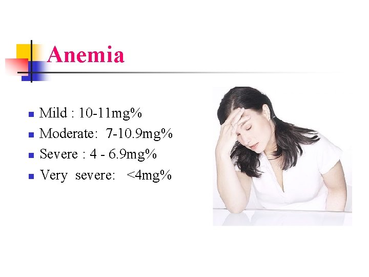
Anemia n n Mild : 10 -11 mg% Moderate: 7 -10. 9 mg% Severe : 4 - 6. 9 mg% Very severe: <4 mg%
Eat foods that are: n n Rich in iron - liver, beef, whole-grain breads , cereals, eggs, dark green vegetables and dried fruit. High in folic acid, such as wheat germ, beans, peanut butter, oatmeal, mushrooms, collards, broccoli, beef liver and asparagus. High in vitamin C, such as citrus fruits and fresh, raw vegetables. Vitamin C makes iron absorption more efficient. Take prenatal vitamin and mineral supplements, especially folic acid.