Skeletal Muscular and Integumentary Systems The explosive speed


 below: – Enables the skin to")


 radiation is one type of energy")


- Slides: 130
Skeletal, Muscular, and Integumentary Systems • The explosive speed needed for speed skating comes from a well-developed muscular
Skeletal, Muscular, and Integumentary Systems
The Skeletal System • To retain their shapes, all organisms need some type of structural support • Single-celled organisms have a cytoskeleton that provides structural support • In multicellular animals, support is provided by some form of skeleton, including the external exoskeletons of arthropods and the internal endoskeletons of vertebrates • The human skeleton is composed of a type of connective tissue called bone – Bones and other connective tissues, such as cartilage and ligaments, form the skeletal system
The Skeletal System • Scientists can infer a lot about the behavior of extinct species by studying fossil bones and reconstructing skeletons • The human skeleton also contains important clues – The shape of your hip bones shows that you walk upright on two legs – The structure of the bones in your hands, especially your opposable thumbs, indicates that you have the ability to grasp objects – The size and shape of your skull is a clue that you have a well-developed brain
The Skeleton • The skeletal system has many important functions • The skeleton supports the body, protects internal organs, provides for movement, stores mineral reserves, and provides a site for blood cell formation • The bones that make up the skeletal system support and shape the body much like an internal wooden frame supports a house – Just as a house could not stand without its wooden frame, the human body would collapse without its bony skeleton • Bones protect the delicate internal organs of the body – For example, the skull forms a protective shell around the brain, and the ribs form a basketlike cage that protects the heart and lungs
The Skeleton • Bones provide a system of levers on which muscles act to produce movement – Levers are rigid rods that can be moved about a fixed point • In addition, bones contain reserves of minerals, mainly calcium salts, that are important to many body processes • Finally, bones are the site of blood cell formation – Blood cells are produced in the soft marrow tissue that fills the internal cavities in some bones
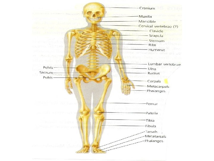
SKELETON • Human body contains 206 bones • Human skeleton is an internal structure referred to as a Endoskeleton • Bones: – Support the muscles and organs – Give shape and structure to the body – Store calcium and phosphorous which are important minerals used by the body in certain metabolic processes – Internal portions of certain bones manufacture blood cells – Protect delicate internal organs • Cranium (skull): protects the brain • Ribs: curved bones that form a cage to protect the heart and lungs
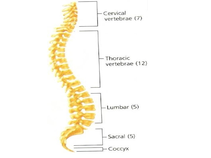
The Skeleton • There are 206 bones in the adult human 206 skeleton • These bones can be divided into two parts: – Axial skeleton: supports the central axis of Axial skeleton the body • Consists of the skull, the vertebral column, and the rib cage – Appendicular skeleton: skeleton • The bones of the arms and legs, along with the bones of the pelvis and shoulder area
SKELETON STRUCTURE • Composed of two parts: – Axial skeleton: consisting of about 80 bones, including the spine, ribs, sacrum, sternum, and cranium – Appendicular skeleton: contains 126 bones, including the bones of the arms, legs, pelvis, and shoulders
The Human Skeleton • The skeleton supports the body • The human skeleton is divided into two parts: – Axial skeleton – Appendicular skeleton
The Human Skeleton
Structure of Bones • It is easy to think of bones as nonliving – After all, most of the mass of bone is mineral salts—mainly calcium and phosphorus • However, bones are living tissue – Bones are a solid network of living cells and protein fibers that are surrounded by deposits of calcium salts
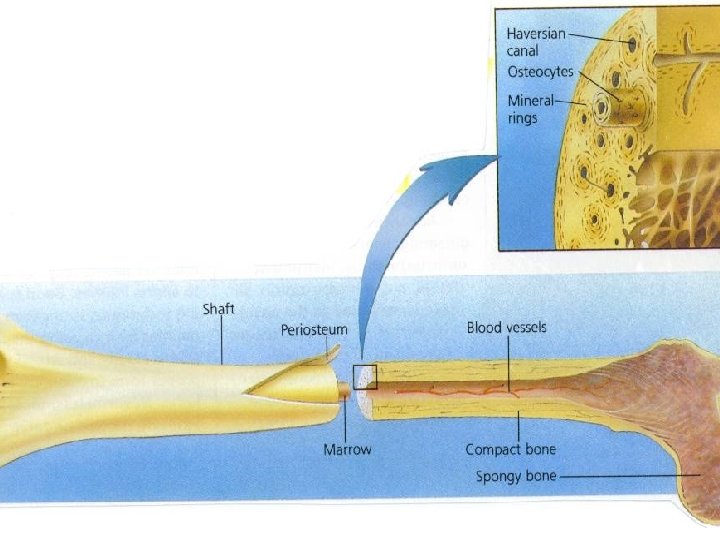
Structure of Bones • • The figure at right shows the structure of a typical bone The bone is surrounded by a tough layer of connective tissue called the periosteum Blood vessels that pass through the periosteum carry oxygen and nutrients to the bone Beneath the periosteum is a thick layer of compact bone – Although compact bone is dense, it is far from being solid • Running through compact bone is a network of tubes called Haversian canals that contain canals blood vessels and nerves
Structure of Bones • Bones are a solid network of living cells and protein fibers that are supported by deposits of calcium salts • A typical long bone such as the femur contains spongy bone and compact bone • Within compact bone are Haversian canals, which contain blood vessels
BONE STRUCTURE • • Bones are made up of both organic and inorganic material Internal structure of long bones (arms/legs) – – Periosteum: outer membrane of the bone which contains a network of blood vessels that supplies living bone cells with oxygen and nutrients and carries away carbon dioxide Compact bone: • • • Under the periosteum Hard material Composed of rings of mineral crystals and protein fibers – Osteocytes: – Haversian Canal: • • – Channel in the center of each mineral ring in the Compact Bone containing nerves and blood vessels Spongy bone: • • – Living bone cells interspersed throughout the mineral rings of the Compact Bone Connective tissue interior to the Compact Bone and at end of long bones Lacy structure adds strength to the bone without adding weight Bone marrow: • Red: – – – • Consist of blood vessels, fibers, cells Manufactures erythrocytes and white blood cells Found in spongy bone /ends of long bones/ribs/vertebrate/sternum/pelvis Yellow – – – Consist mostly of fat cells Serves as energy reserve Fill shafts of the long bones
Structure of Bones
Structure of Bones • A less dense tissue known as spongy bone is found inside the outer layer of compact bone – It is found in the ends of long bones and in the middle part of short, flat bones • Despite its name, spongy bone is not soft and spongy; it is actually quite strong – Near the ends of bones where force is applied, spongy bone is organized into structures that resemble the supporting girders in a bridge • This latticework structure of spongy bone helps to add strength to bone without adding mass
Structure of Bones • Osteocytes, which are mature bone cells, are Osteocytes embedded in the bone matrix • Two other kinds of bone cells—osteoclasts and osteoblasts line the Haversian canals and the osteoblasts surfaces of compact and spongy bone – Osteoclasts break down bone – Osteoblasts produce bone • Although we stop growing in our late teens, our bones are continuously remodeled through the activity of osteoclasts and osteoblasts
Structure of Bones • Within bones are cavities that contain a soft tissue called bone marrow • There are two types of bone marrow: – Yellow: • Made up primarily of fat cells – Red: • Produces red blood cells, some kinds of white blood cells, and cell fragments called platelets
Development of Bones • The skeleton of an embryo is composed almost entirely of a type of connective tissue called cartilage • The cells that make up cartilage are scattered in a network of protein fibers including both tough collagen and flexible elastin • Unlike bone, cartilage does not contain blood vessels – Cartilage cells must rely on the diffusion of nutrients from the tiny blood vessels in surrounding tissues • Because cartilage is dense and fibrous, it can support weight, despite its extreme flexibility
Development of Bones • Cartilage is replaced by bone during the process of bone formation called ossification – Ossification begins to take place up to seven months before birth • Bone tissue forms as osteoblasts secrete mineral deposits that replace the cartilage in developing bones • When the osteoblasts become surrounded by bone tissue, they mature into osteocytes
Development of Bones • Many long bones, including those of the arms and legs, have growth plates at either end – The growth of cartilage at these plates causes the bones to lengthen – Gradually, this new growth of cartilage is replaced by bone tissue, and the bones become larger and stronger – During late adolescence or early adulthood, the cartilage in the growth plates is replaced by bone, the bones become completely ossified, and the person “stops growing”
Development of Bones • In adults, cartilage is found in those parts of the body that are flexible, such as the tip of the nose and the external ears • Cartilage also is found where the ribs are attached to the sternum, which allows the rib cage to move during breathing
BONE DEVELOPMENT • Ossification: process by which bones develop – Two types: • Long bones first develop as cartilage that is later replaced by bone – Cartilage: tough, flexible connective tissue Cartilage: » Once osteocytes develop in the cartilage, they release minerals that lodge in the spaces between cartilage cells eventually replacing the cartilage » Some cartilage is never replaced: disc between vertebrae, joints, end of nose, external ear, trachea » Makes area flexible • Other bones develop directly from embryonic connective tissue without forming cartilage first – Clavicle, some parts of the skull
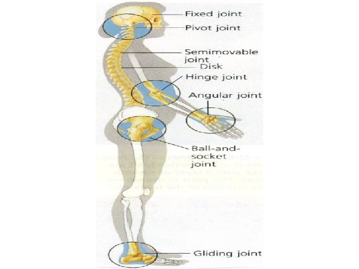
Types of Joints • A place where one bone attaches to another bone is called a joint – Joints permit bones to move without damaging each other – Some joints, such as those of the shoulder, allow extensive movement – Others, like the joints of the fully developed skull, allow no movement at all • Depending on its type of movement, a joint is classified as immovable, slightly movable, or freely movable
JOINTS • Location where two bones meet • Ligaments: tough bands of connective tissue that holds the bones of a joint in place – Stretch as the bones move • Three kinds: – Fixed: skull – Semimovable(bend and twist): vertebral column, ribs – Movable: • • • Hinge (back and forth movement): elbow Hinge Ball and socket (circular motion): hip, shoulder Ball and socket Pivot (side to side / up and down): cervical vertebrae Pivot Angular (twisting): wrists, ankles Angular Gliding (slide against each other): small bones of the hand feet Gliding (
Immovable Joints • Immovable joints, often called fixed Immovable joints, allow no movement – The bones at an immovable joint are interlocked and held together by connective tissue, or they are fused – The places where the bones in the skull meet are examples of immovable joints
Slightly Movable Joints • Slightly movable joints permit a small Slightly movable joints amount of restricted movement – Unlike the bones of immovable joints, the bones of slightly movable joints are separated from each other – The joints between the two bones of the lower leg and the joints between adjacent vertebrae are examples of slightly movable joints
Freely Movable Joints • Freely movable joints permit movement Freely movable joints in one or more directions – Freely movable joints are grouped according to the shapes of the surfaces of the adjacent bones
Freely Movable Joints • Ball-and-socket joints permit movement in Ball-and-socket joints many directions – They allow the widest range of movement of any joint • Hinge joints permit back-and-forth motion, Hinge joints like the opening and closing of a door • Pivot joints allow one bone to rotate around Pivot joints another • Saddle joints permit one bone to slide in two Saddle joints directions
Types of Freely Movable Joints • Freely movable joints are classified by the type of movement they permit • The joints illustrated are in the shoulder, knee, elbow, and hand
Types of Freely Movable Joints
FUNCTIONING OF JOINTS • Joints are subject to a great deal of pressure and stress • Connective tissue near the joints secretes synovial fluid which cushions the bones thus preventing the bones from wearing away • Fluid filled sac called the bursa is found in the knee and shoulder joints adding an additional cushion between the bones
Structure of Joints • In freely movable joints, cartilage covers the surfaces where two bones come together – This protects the bones as they move against each other • The joints are also surrounded by a fibrous joint capsule that helps hold the fibrous joint capsule bones together while still allowing them to move
Structure of Joints • • The joint capsule consists of two layers One layer forms strips of tough connective tissue called ligaments – Ligaments, Ligaments which hold bones together in a joint, are attached to the membranes that surround bones • Cells in the other layer of the joint capsule produce a substance called synovial fluid, fluid which forms a thin film on the cartilage that covers the bony surfaces that form the joint – This lubricating film enables the surfaces of the joint to slide over each other smoothly
Structure of Joints • In some freely movable joints, such as the knee in the figure, small sacs of synovial fluid called bursae (singular: bursa) form • A bursa reduces the friction between the bones of a joint and also acts as a tiny shock absorber.
Knee Joint • The knee joint is protected by cartilage and bursae • The ligaments hold the bones composing the knee joint—femur, patella, tibia, and fibula— together
Knee Joint
Skeletal System Disorders • Bones and joints can be damaged, just like any other tissue • Excessive strain on a joint may produce inflammation, a response in which excess fluid causes swelling, pain, heat, and redness • Inflammation of a bursa is called bursitis • A more serious disorder is arthritis, which involves inflammation of the joint itself • Arthritis affects approximately 10 percent of the world's population
JOINT INJURY • Bursitis: inflammation of the bursa • Sprain: torn ligament • Arthritis: – Synovial membranes become inflamed and grow thicker – Fibrous tissues in the joints may become ossified, immobilizing the joint and fusing the bones together
Skeletal System Disorders • Because bone is living tissue, calcium is moved between it and the rest of the body to maintain homeostasis of this important mineral • In older people, especially women, loss of calcium can lead to a weakening of the bones, a condition known as osteoporosis – Osteoporosis can cause many serious fractures • Sound nutrition, including plenty of calcium in the diet, and weight-bearing exercise are among the best ways to prevent this serious problem
The Muscular System • Despite the fantasies of Hollywood horror films, a skeleton cannot move by itself • Movement is the function of the muscular system • More than 40 percent of the mass of the average human body is muscle • The muscular system includes the large muscles displayed by some athletes • It also includes thousands of tiny muscles throughout the body that help to regulate blood pressure, move food through the digestive system, and power every movement of the body—from the blink of an eye to the hint of a smile
Types of Muscle Tissue • Muscle tissue is found everywhere in the body—not only just beneath the skin but also deep within the body • There are three different types of muscle tissue: skeletal, smooth, and cardiac – Each type of muscle is specialized for a specific function in the body
MUSCLE • Contractile organ consisting of many cells • Three types: – Skeletal – Cardiac – Smooth
Muscle Tissue • There are three types of muscle tissue: skeletal, smooth, and cardiac • Skeletal muscle cells have striations, or stripes, and many nuclei • Smooth muscle cells are spindle-shaped and have one nucleus and no striations • Cardiac muscle cells have striations and usually one nucleus
Muscle Tissue
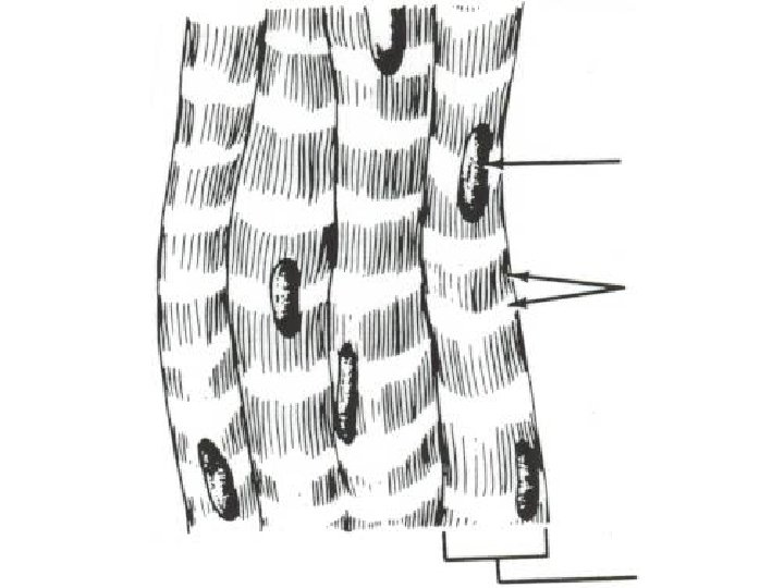
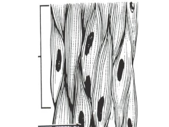
Skeletal Muscles • • • Skeletal muscles are usually attached to bones Skeletal muscles are responsible for such voluntary movements as typing on a computer keyboard, dancing, or winking an eye When viewed under a microscope at high magnification, skeletal muscle appears to have alternating light and dark bands called striations For this reason, skeletal muscle is sometimes called striated muscle Most skeletal muscles are consciously controlled by the central nervous system
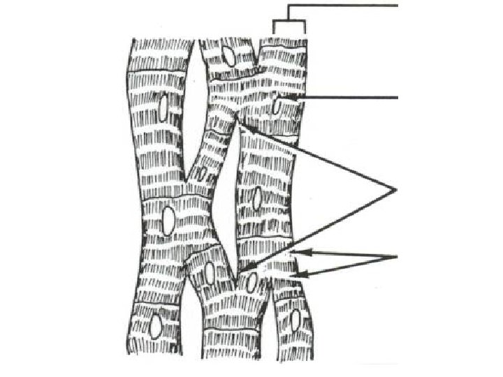
Skeletal Muscles • Skeletal muscle cells are large, have many nuclei, and vary in length from 1 millimeter to about 30 centimeters • Because skeletal muscle cells are long and slender, cells they are often called muscle fibers • Complete skeletal muscles consist of muscle fibers, connective tissues, blood vessels, and nerves • The figure at right shows the structure of a skeletal muscle in the leg
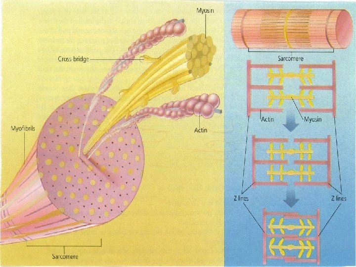
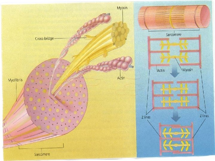
MUSCLE CELL • Muscle cell is also known as a fiber – Within each fiber are many smaller units called myofibrils (1, 000 to 2, 000) myofibrils • Each myofibril appears banded – Bands referred to as Z lines – Area between Z lines is called a sarcomere (functional unit sarcomere of contraction) » Striated appearance of a sarcomere results from the presence of two types of protein filaments, myosin and actin » The thicker myosin filaments have lateral extensions called cross-bridges which are attached to the thinner actin filaments that lie beside the myosin filaments
Skeletal Muscle Structure • Skeletal muscles are made up of bundles of muscle fibers, which in turn are composed of myofibrils • Each myofibril contains thin filaments made of actin and thick filaments made of myosin • Muscle fibers are divided into functional units called sarcomeres • What nervous system structures carry messages to skeletal muscles?
SKELETAL MUSCLE • Attached to the periosteum of bone – Either directly or by a tough connective tissue called a tendon • Striated: dark bands located at right angles to the long axis of the muscle • Voluntary: muscle contractions can be controlled • Nucleus in each cell but the separate cells are harder to see • Cell can sometimes be almost half a meter long
Skeletal Muscle Structure
Smooth Muscles • Smooth muscles are usually not under voluntary control • A smooth muscle cell is spindle-shaped, has one nucleus, and is not striated • Smooth muscles are found in the walls of hollow structures such as the stomach, blood vessels, and intestines • Smooth muscles move food through your digestive tract, control the way blood flows through your circulatory system, and decrease the size of the pupils of your eyes in bright light • Most smooth muscle cells can function without nervous stimulation – They are connected to one another by gap junctions that allow electrical impulses to travel directly from one muscle cell to a neighboring muscle cell
SMOOTH MUSCLE • Found in the walls of the stomach, intestines, and blood vessels • Not striated – Actin and myosin fibers are present but in the cytoplasm but not arranged in repeating units • Involuntary • Long spindle-shaped cells that contain a single nucleus
Cardiac Muscle • Cardiac muscle is found in just one place in the body—the heart – The prefix cardio comes from a Greek word meaning “heart” • Cardiac muscle shares features with both skeletal muscle and smooth muscle • Cardiac muscle is striated like skeletal muscle, although its cells are smaller • Cardiac muscle cells usually have one nucleus, but they may have two • Cardiac muscle is similar to smooth muscle because it is usually not under the direct control of the central nervous system and cardiac cells are connected to their neighbors by gap junctions • You will learn more about cardiac muscle in Chapter 37.
CARDIAC MUSCLE • Makes up the walls of the heart • Striated • Involuntary: movements cannot be controlled • Many mitochondria • Cell much shorter than skeletal cell • Cell nucleus is present, but separate cells are easier to see
Muscle Contraction • The muscle fibers in skeletal muscles are composed of smaller structures called myofibrils – Each myofibril is made up of even smaller structures called filaments • The striations in skeletal muscle cells are formed by an alternating pattern of thick and thin filaments – The thick filaments contain a protein called myosin – The thin filaments are made up mainly of a protein called actin • The filaments are arranged along the muscle fiber in units called sarcomeres, which are separated from each other by regions called Z lines
Muscle Contraction • During muscle contraction, the actin filaments slide over the myosin filaments, decreasing the distance between the Z lines
Muscle Contraction
Muscle Contraction • The tiny myosin and actin filaments are the forceproducing engines that cause a muscle to contract • A muscle contracts when the thin filaments in the muscle fiber slide over the thick filaments – This process is called the sliding-filament model of muscle contraction • For a muscle to contract, the thick myosin filament must form a cross-bridge with the thin actin filament – As the cross-bridge changes shape, it pulls on the actin filament, which slides toward the center of the sarcomere – The distance between the Z lines decreases – The cross-bridge detaches from the actin filament • The cycle is repeated when the myosin binds to another site on the actin filament
Muscle Contraction • When hundreds of thousands of myosin crossbridges change shape in a fraction of a second, the muscle fiber shortens with considerable force • The energy for muscle contraction is supplied by ATP • Because one molecule of ATP supplies the energy for one interaction between a myosin cross-bridge and an actin filament, the cell needs plenty of ATP molecules for a strong contraction • Recall that the cell can produce ATP in two ways—by cellular respiration and by fermentation
MUSCLE CONTRACTION • • Begins when a nerve impulse reaches a muscle (junction of a nerve branch and a muscle is called a motor end plate) Stimulus reaching the motor end plate causes a specialized membrane of the muscle to release calcium (Ca) into the muscle cytoplasm Before the nerve impulse (myosin cannot bind to actin) Attachment of calcium to muscle protein causes a change in their shapes allowing the binding of the myosin heads to the actin filament – Myosin heads then bend inward, pulling the actin filament toward the center of the sarcomere • Myosin is then released from the actin and uses the energy of ATP to bend the myosin heads back to their original position • Process is repeated with the myosin heads moving down the actin filament and reattaching to a new position – Z lines move closer to each other – The entire sarcomere contracts Synchronized shortening of sarcomeres along the full length of a muscle fiber causes the whole fiber, and hence the muscle, to contract Contraction process continues until calcium and ATP supplies are depleted or nerve stimulation stops When a single muscle fiber is stimulated, an all-or-none response occurs (no in-between state) – As more muscle fibers are activated, the force of the contraction increases
Control of Muscle Contraction • Skeletal muscles are useful only if they contract in a controlled fashion • Remember that motor neurons connect the central nervous system to skeletal muscle cells • Impulses from motor neurons control the contraction of skeletal muscle fibers
Control of Muscle Contraction • A neuromuscular junction is the point of contact between a neuromuscular junction motor neuron and a skeletal muscle cell • Vesicles, or pockets, in the axon terminals of the motor neuron release a neurotransmitter called acetylcholine – Acetylcholine molecules diffuse across the synapse, producing an impulse in the cell membrane of the muscle fiber – The impulse causes the release of calcium ions (Ca 2+) within the fiber – The calcium ions affect regulatory proteins that allow actin and myosin filaments to interact • From the time a nerve impulse reaches a muscle cell, it is only a few milliseconds before these events occur and the muscle cell contracts
Control of Muscle Contraction • A muscle cell remains contracted until the release of acetylcholine stops and an enzyme produced at the axon terminal destroys any remaining acetylcholine – Then, the cell pumps calcium ions back into storage, the cross-bridges stop forming, and contraction ends
Control of Muscle Contraction • What is the difference between a strong contraction and a weak contraction? • Each muscle contains hundreds of cells • When you lift something light, such as a sheet of paper, your brain stimulates only a few cells in your arm muscles to contract • However, as you exert maximum effort, almost all the muscle cells in your arm are stimulated to contract
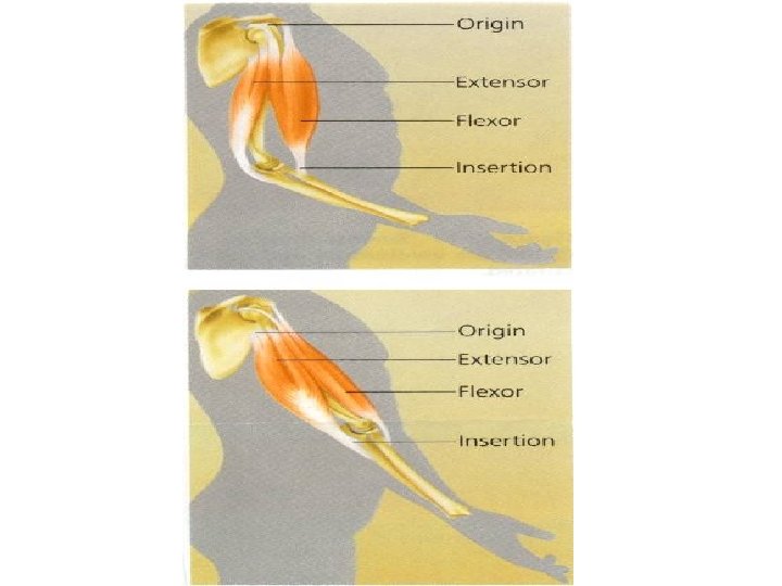
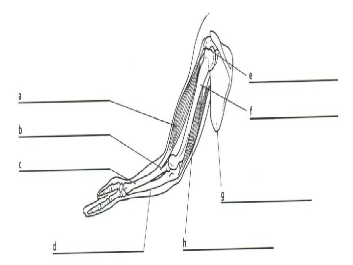
How Muscles and Bones Interact • Skeletal muscles generate force and produce movement by contracting, or pulling on body parts • Individual muscles can only pull in one direction • Yet, you know from experience that your legs bend when you sit and extend when you stand up • How is this possible?
How Muscles and Bones Interact • • • Skeletal muscles are joined to bones by tough connective tissues called tendons Tendons are attached in such a way that they pull on the bones and make them work like levers The joint functions as a fulcrum —the fixed point around which the lever moves The muscles provide the force to move the lever Usually, there are several muscles surrounding each joint that pull in different directions
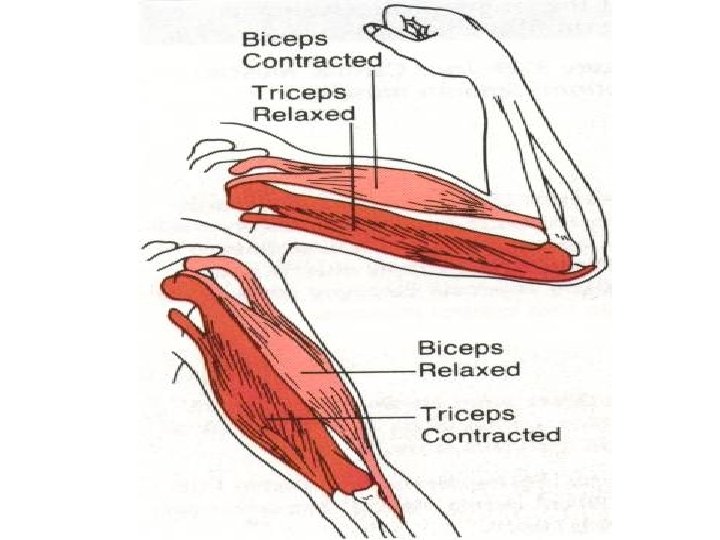
How Muscles and Bones Interact • • Most skeletal muscles work in opposing pairs – When one muscle contracts, the other relaxes The muscles of the upper arm shown in the figure a good example of this dual action When the biceps muscle contracts, it bends, or flexes, the elbow joint When the triceps muscle contracts, it opens, or extends, the elbow joint A controlled movement, however, requires contraction by both muscles To hold a tennis racket or a violin, both the biceps and triceps must contract in balance This is why the training of athletes and musicians is so difficult The brain must learn how to work opposing muscle groups in just the right ways to make the joint move precisely
Arm Movement • By contracting and relaxing, the triceps and biceps in the upper arm enable you to bend or straighten your elbow
Arm Movement
BONE MOVEMENT • • Skeletal muscle pulls at the base of a joint Point of attachment on the moving bone is called the insertion Point of attachment on the stationary bone is the origin Most skeletal muscles form antagonistic pairs – When one contracts, the other usually relaxes – One muscle in a pair moves a body part in one direction, and the other muscle moves it in the opposite direction • • Flexors: bend joints (biceps) Flexors Extensors: straighten joints (triceps) Extensors Abductors: move limb away from body (deltoid) Abductors Adductors: move a limb toward the body (pectorals) Adductors • Isometric contraction: exercise in which both antagonistic pairs contract (joint does not move)
Exercise and Health • Skeletal muscles generally remain in a state of partial contraction called resting muscle tone • Muscle tone is responsible for keeping the back and legs straight and the head upright, even when you are relaxed
Exercise and Health • Regular exercise is important in maintaining muscular strength and flexibility • Muscles that are exercised regularly stay firm and increase in size and strength by adding actin and myosin filaments – Muscles that are not used become weak and can visibly decrease in size
Exercise and Health • Aerobic exercises—such as running and swimming—cause the body's systems to become more efficient – For example, aerobic exercise helps your heart and lungs become more efficient • This, in turn, increases physical endurance—the ability to perform an activity without fatigue • Regular exercise also strengthens your bones, making them thicker and stronger • Strong bones and muscles are less likely to become injured
Exercise and Health • Resistance exercises, such as weight lifting, increase muscle size and strength • Resistance exercises also decrease body fat and increase muscle mass • Over time, weight-training exercises will help to maintain coordination and flexibility
The Integumentary System • “Good fences make good neighbors, ” wrote the American poet Robert Frost as he explained the importance of property boundaries • Living things have their own “fences, ” and none is as important as the skin— the boundary that separates the human body from the outside world
INTEGUMENTARY SYSTEM • Consist of the skin, hair, and nails • Protective barrier between the body and the outside world • Helps retain body fluids • Barrier against disease • Helps eliminate waste products • Helps regulates body temperature
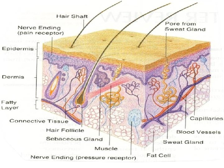
The Integumentary System • The skin, the single largest organ of the body, is part of the integumentary system • The word integument comes from a Latin word that means “to cover, ” reflecting the fact that the skin and its related structures form a covering over the entire body • Skin and its related structures—the hair, nails, and a variety of glands—make up the integumentary system
The Skin • The skin has many different functions, but its most important function is protection • The integumentary system serves as a barrier against infection and injury, helps to regulate body temperature, removes waste products from the body, and provides protection against ultraviolet radiation from the sun • Because the largest component of the integumentary system—the skin—contains several types of sensory receptors, it serves as the gateway through which sensations such as pressure, heat, cold, and pain are transmitted to the nervous system
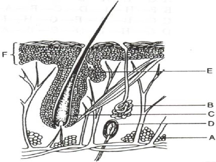
The Skin • The skin is made up of two main layers —the epidermis and the dermis • Beneath the dermis is a subcutaneous layer of fat (the hypodermis) and loose connective tissue that help insulate the body
SKIN • Largest organ of the human body • Two parts: – Epidermis – Dermis
EPIDERMIS • • Outer part of the skin Composed of many cell layers (epithelial cells) Protects the body from harmful UV light Outer layer of cells are dead and are constantly being shed and replaced by new cells from the rapidly dividing lower layer by mitosis – New cells are pushed toward the surface • Cells fills with a protein called keratin – Gives the skin its rough, leathery texture and its waterproof quality
Epidermis • The outer layer of the skin is the epidermis • The epidermis itself has two layers: – The outside of the epidermis—the part that comes in contact with the environment—is made up of dead cells – The inner layer of the epidermis is made up of living cells
Epidermis • Cells in the inner layer of the epidermis undergo rapid cell division, producing new cells that push older cells to the surface of the skin • As they move upward, the older cells become flattened and their organelles disintegrate • They also begin making keratin, a tough, fibrous protein
Epidermis • Eventually, the keratinproducing cells die and form a tough, flexible, waterproof covering on the surface of the skin • This outer layer of dead cells is shed or washed away at a surprising rate—once every four to five weeks
MELANIN • Pigment produced in the lower layers of the epidermis: – Gives color to the skin – Absorbs harmful ultraviolet light – Determines the color of the skin • Large amount: darker the skin • Small amount: lighter the skin • In some individuals exposure to sunlight increases the production causing the skin to become darker (tan)
Epidermis • • The epidermis also contains melanocytes Melanocytes are cells that produce melanin, a dark brown pigment Melanin helps protect the skin Melanin from damage by absorbing ultraviolet rays from the sun Although most people have roughly the same number of melanocytes in their skin, differences in skin color are caused by the different amount of melanin the melanocytes produce and where these cells are distributed
Epidermis • Look closely at the figure at right and you will see that there are no blood vessels in the epidermis • This explains why a slight scratch will not cause bleeding
Layers of the Skin • The skin has an outer layer called the epidermis and an inner layer called the dermis • What is the function of the dermis?
Layers of the Skin
DERMIS • • Inner layer of skin Thickest layer Composed of living cells Contains: – Nerves: receive environmental stimuli Nerves – Blood: release heat helping the body maintain Blood a comfortable temperature – Lymph vessels: helps the skin fight against Lymph vessels infection
DERMIS • Layer of fat cells (adipose tissue) below: – Enables the skin to store food for energy – Provides protection to the body – Insulates the body against heat loss
DERMIS • Location of hair follicles: – Cells at the base produce hair – Blood vessels surround the hair follicles nourishing the root hair – Shaft of hair that extends beyond the skin consist mostly of keratin and requires no nourishment
Dermis • The inner layer of the skin is the dermis • The dermis lies beneath the epidermis and contains collagen fibers, blood vessels, nerve endings, glands, sensory receptors, smooth muscles, and hair follicles
NERVES • Special sensory neuron receptors send a great deal of information to the brain from the environment • Types of sensory neuron receptors: – Pressure – Touch – Heat – Cold – Pain
Dermis • The skin interacts with other body systems to maintain homeostasis by helping to regulate body temperature • When the body needs to conserve heat on a cold day, the blood vessels in the dermis narrow, helping to limit heat loss • On hot days, the blood vessels widen, bringing heat from the body's core to the skin and increasing heat loss
Dermis • • The dermis contains two major types of glands: sweat glands and sebaceous, or oil, glands If your body gets too hot, sweat glands produce perspiration, or sweat – Sweat contains water, salts, and other compounds – When sweat evaporates, it takes heat away from your body – Sweat also gets rid of wastes from the blood, along with water – In this way, the skin acts as an organ of excretion • Sebaceous glands produce an oily secretion called sebum – Sebum spreads out along the surface of the skin and helps to keep the keratin-rich epidermis flexible and waterproof
SEBACEOUS GLAND • Oil gland • Secretes a substance called sebum – Production controlled by hormones • Exocrine gland (gland that releases fluid through a duct) • Next to each hair follicle • Oil from these glands help prevent the skin’s outer layer from drying and cracking keeping it soft and waterproof
SWEAT GLANDS • Function as excretory organs by releasing excess water, salts, and urea • Releasing excess water helps regulate the body temperature – When the body temperature rises, the circulation increases, and the skin becomes warm and flushed • Sweat glands then release sweat and as the water evaporates the skin is cooled • Helps maintain homeostasis by regulating heat loss
Skin Cancer • Excessive exposure to the ultraviolet radiation in sunlight can produce skin cancer, an abnormal growth of cells in the skin • You can help protect yourself from this dangerous disease by wearing a hat, sunglasses, and protective clothing whenever you plan to spend time outside • In addition, you should always use a sunscreen with a sun protection factor (SPF) of at least 15
The UV Index and Sunburn • Ultraviolet (UV) radiation is one type of energy from the sun • UV rays cause sunburn, some cataracts, and skin cancer • There are many factors that affect the amount of UV radiation to which you are exposed • These include the time of day, the season, the weather conditions, and your location • Recently, the National Weather Service, the Environmental Protection Agency, and the Centers for Disease Control agreed upon a national UV index • The UV index is issued daily to advise you of conditions in your region of the country
The UV Index and Sunburn
The UV Index and Sunburn • Describe the trend in the amount of time it takes to sunburn, from a minimal UV index level to a very high UV index level
The UV Index and Sunburn • Why do you think applying sunscreen is always recommended?
The UV Index and Sunburn • Why should a hat worn as protection against UV rays have a brim?
The UV Index and Sunburn • The minutes-to-burn data apply to most people • What variable could cause the time for a particular person to burn to be shorter or to be longer?
The UV Index and Sunburn • Use the data in the table to construct a bar graph • Place the UV index levels on the x-axis and the minutes to burn on the y-axis
Hair and Nails • The basic structure of human hair and nails is keratin • In other animals, keratin forms a variety of structures, including bull horns, reptile scales, bird feathers, and porcupine quills
Hair • Hair covers almost every exposed surface of the body and has important functions – Hair on the head protects the scalp from ultraviolet light from the sun and provides insulation from the cold – Hairs in the nostrils, external ear canals, and around the eyes (eyelashes) prevent dirt and other particles from entering the body
Hair • Hair is produced by cells at the base of structures called hair follicles • Hair follicles are tubelike pockets of epidermal cells that extend into the dermis • An individual hair is actually a large column of cells that have filled with keratin and then died – Rapid cell growth at the base of the hair follicle causes the hair to grow longer – Hair follicles are in close contact with sebaceous glands • The oily secretions of these glands help maintain the condition of each individual hair
Nails • Nails grow from an area of rapidly dividing cells known as the nail root • The nail root is located near the tips of the fingers and toes • During cell division, the cells of the nail root fill with keratin and produce a tough, platelike nail that covers and protects the tips of the fingers and toes • Nails grow at an average rate of 3 millimeters per month, with fingernails growing more rapidly than toenails—about four times as fast