MUSCLE PHYSIOLOGY MUSCLE INTRODUCTION Muscle cells like neurons


- Slides: 18

MUSCLE PHYSIOLOGY

MUSCLE INTRODUCTION • Muscle cells, like neurons, can be excited chemically, electrically, and mechanically to produce an action potential that is transmitted along their cell membranes. • Unlike neurons, they have a contractile mechanism that is activated by the action potential. • The contractile proteins actin and myosin are abundant in muscle, where they bring about contraction. • The actin-binding protein myosin and actin make up one of the molecular motors that converts the energy of ATP hydrolysis into movement of one cellular component along another.

• Muscle is generally divided into three types, skeletal, cardiac, and smooth, though smooth muscle is not a homogenous single category. • Skeletal muscle makes up the great mass of the somatic musculature. It has well - developed cross – striations, does not normally contract in the absence of nervous stimulation, lacks anatomic and functional connections between individual muscle fibers, and is generally under voluntary control. • Cardiac muscle also has cross-striations, but it is functionally syncytial and contracts rhythmically in the absence of external innervation owing to the presence in the myocardium of pacemaker cells that discharge spontaneously. • Smooth muscle lacks cross-striations. • The type found in most hollow viscera is functionally syncytial and contains pacemakers that discharge irregularly. The type found in the eye and in some other locations is not spontaneously active and resembles skeletal muscle.

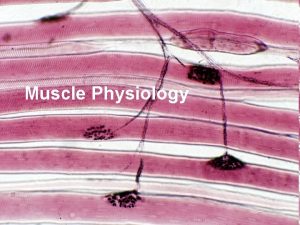
SKELETAL MUSCLE • Skeletal muscle forms the major part of the muscles in the body. They are attached to the bones. When they contract, they move a part of the body or the body as a whole. • They are under voluntary control and their contraction depends upon their nerve supply. Functional anatomy of skeletal muscle fibre Each skeletal muscle is an organised collection of slender bundles of muscle fibres known as fasciculi. A large muscle may have many fasciculi, whereas a small muscle contains fewer. Each muscle fibre is a single cell, multinucleated, long, cylindrical and surrounded by an elastic membrane, the sarcolemma.

Each muscle fibre contains several hundreds to several thousands of thread-like structures called myofibrils which often extend through the length of the muscle fibre. When viewed with a polarizing microscope, myofibrils exhibit a regular pattern of alternating light and dark bands. Each dark band is called an A band, because it is able to polarize light (i. e. anisotropic). The lighter band adjacent to each dark band is called an I band because it is non-polarized (i. e. isotropic). The functional unit of a myofibril is the sarcomere which is composed of one A band flanked on either side by one-half of an I band. The dark line bisecting each I band is called Z line. Therefore, one sarcomere is that section of a myofibril lying between two adjacent Z lines. The somewhat lighter band in the centre of the A band is an H band. In the centre of the H band, a narrow dark band called an M line can be seen.

The sarcomeres of each myofibril are built from even smaller units called myofilaments. A myofilament is an aggregation of proteins and stacking of myofilaments is responsible for the various bands and lines seen in skeletal muscle tissue. Myofilaments are the contractile elements that give muscle its remarkable ability to shorten. Within each sarcomere are two different types of myofilaments; thick (myosin) myofilament made up of many molecules of the protein myosin (mw 460, 000) and thin (actin) myofilament which contains a backbone made of the protein, actin (mw 43, 000). A myosin molecule has two globular heads and a long tail. The actin molecule is made up of two chains of globular units that form a long double helix. At intervals along the actin helix are active sites, which are points on the actin chains capable of binding to myosin.

Surrounding the actin helix are two other proteins important in muscle contraction. These are tropomyosin (mw 70, 000 and troponin (mw 18, 000 -35, 000). Tropomyosin is rod-like and lies in the groove between the chains of the actin molecule. When the muscle is at rest, tropomyosin covers the active sites located at intervals along the actin helix. Troponin is actually a complex of three globular proteins, namely, troponin T which binds other troponins to tropomyosin, troponin I, which inhibits the interaction of myosin with actin and troponin C which has a binding sites for ca 2+.

Neuromuscular (myoneural) junction: Motor endplate • The activity of the skeletal muscle inside the body is controlled by the central nervous system through the motor nerve. There is no cytoplasmic continuity between the nerve fibre and skeletal muscle fibre. • The point at which the axon ending of a motor neurone meets a muscle cell is the neuromuscular junction or the motor endplate. • At this junction, the plasma membrane of the axon ending remains separated from the muscle fibre sarcolemma by a small space, 20 -30 nm, called synaptic cleft.

Neuromuscular junction and events occurring at the NMJ which lead to an action potential in the muscle membrane.

• For a skeletal muscle to contract, the motor nerve controlling it must signal it to do so. • The signals sent to muscle fibres by the motor nerve are through chemical messengers called neurotransmitters. • The neurotransmitter released onto the skeletal muscle fibres at the neuromuscular junction is acetylcholine. • The acetylcholine in the axon endings is contained within membrane bound sacs known as synaptic vesicles. • The release of acetylcholine by axon ending onto the synaptic cleft is stimulated by the arrival of the action potential at the axon ending. Acetylcholine is released by the process of exocytosis.

• The acetylcholine released by a motor neurone diffuses across the synaptic cleft and binds to Ach receptors which are proteins embedded in the sarcolemma of the muscle fibre at the neuromuscular junction. • Acetylcholine receptors have two important properties; they contain binding sites to which acetylcholine can attach, and they are associated with a chemically-gated ion channel that allows Mg++ to diffuse through its centre when it is opened. • The binding of acetylcholine to its receptor sites opens the sodium channel.

CARDIAC MUSCLE • Cardiac muscle cell, like a skeletal muscle cell, possesses striations (A and I bands) that result from stack of thick and thin myofilaments. Myofibrils are not as distinct in cardiac muscle cells as in skeletal cells. • There is a single, central nucleus in a cardiac muscle cell, and the myofilaments separate to bypass it. • Large mitochondria are numerous between the myofibrils. • The sarcoplasmic reticulum is well developed, but it is closely associated with the T-tubules as in skeletal muscle. • cardiac muscle requires an increase in the level of Ca 2+ in the sarcoplasm for contraction. • The cardiac muscle cells are branched, also possess intercalated discs, which connect the ends of adjacent cells. • Each intercalated disc is a junction between two adjoining muscle cells and contains two types of cell-to-cell membrane junctions, each serving a distinct function. • The first is called desmosomes and are for strong attachment of one cell to another. The second is gap junctions which allow electrical signals to pass directly from cell to cell. • Cardiac muscle acts as functional syncytium, and enables excitation to reach every cell.

Excitation-contraction coupling • Like in the skeletal muscle, when an action potential passes over the cardiac muscle membrane, this leads to the release of calcium ions into the muscle’s sarcoplasmic reticulum. • This calcium ion is responsible for the chemical reaction that causes actin to slide over myosin (muscle contraction). • In addition to the Ca 2+ released from the cisternae of the sarcoplasmic reticulum, a large quantity of extra Ca 2+ diffuses into the sarcoplasm from the T-tubules themselves at the time of action potential. • Indeed, without this extra Ca 2+ from the T-tubules, the strength of the cardiac muscle contraction would be greatly reduced because the sarcoplasmic reticulum of cardiac muscle is less well developed unlike that of skeletal muscle and does not store enough Ca 2+ to provide full contraction. • However, the T-tubules of cardiac muscle have a diameter 5 times as great as that of the skeletal muscle tubules.

• The ends of the T-tubules open directly to the outside of the cardiac muscle cells, allowing the extracellular fluid to percolate through the T-tubules. • Therefore, the strength of contraction of the cardiac muscle depends to a great extent on the concentration of Ca 2+ in the extracellular fluids. • N. B: The process by which depolarization of the muscle fibre initiates contraction is called excitationcontraction coupling.

SMOOTH MUSCLE • Smooth muscle is found in the walls of hollow organs: blood vessels, the stomach, the intestines, the ducts of glands, respiratory passages, urinary ducts, the urinary bladder, the uterus, and genital ducts. • Contraction of smooth muscle in the walls of hollow organs cause the movement of food or fluids within the organs • Smooth muscle cells differ from the cells of skeletal and cardiac muscle in their shape and their lack of striations. • A smooth muscle cell is spindle-shaped, with a single nucleus in its wide central portion. • Individual smooth muscle cells lie side-by-side, forming sheets. • The contractile elements of smooth muscle cells are myofilaments containing actin and myosin, but the myofilaments are not arranged into sarcomeres with Z lines as in skeletal and cardiac muuscles.

Contraction of smooth muscle • The contractile mechanism of smooth muscle, like that of skeletal muscle appears to involve cross-bridge movement and sliding of myofilaments. • It is noted that the myofilament in smooth muscle are not regularly arranged into sarcomeres; this irregular arrangement may allow some cross-bridges to form even if the muscle is stretched to a point at which cross-bridge formation in skeletal muscle could not occur. • As in skeletal muscle, contraction in smooth muscle is triggered by an increase in Ca 2+ levels in the sarcoplasm. Rather than binding to the protein troponin, however, which is lacking in smooth muscle cells, Ca 2+ binds to a similar protein, calmodulin. • Smooth muscle contractions are slower and develop less force than skeletal muscle contrractions. • While a skeletal muscle twitch lasts a fraction of a second, a twitch in smooth muscle may last for several seconds.

Comparison of the different muscle types Characteristics Type of muscle Skeletal Multiunit smooth Single unit smooth cardiac Location Attached to skeleton Large blood vessels, eye, arrector pili muscle Walls of Heart only hollow organs in digestive, reproductive and urinary tracts Presence of striation Striated Non-striated Striated Innervation SNS ANS ANS Level of control Voluntary Involuntary involuntary Initiation of contraction Stimulation by nervous system Pacemaker activity of muscle fibres

Skeletal Multiunit smooth Single unit smooth Cardiac Role of nervous Initiates stimulation contraction Initiates contraction Modifies contraction Presence of gap Not present junctions Very few Present Requiring ATP for contraction Yes Yes Presence of actin and myosin Present Presence of Ttubules Present Absent Present (large) Presence of troponin and tropomyosin Present Absent Site of Ca 2+ regulation Troponin in thin filaments Myosin in thick Troponin in filament via thin filaments calmodulin Present
 Straw colored fluid
 Development of paranasal sinuses
 Proximal convoluted tubule
 Thyroid parafollicular cells
 Gametes vs somatic cells
 Why dna is more stable than rna?
 Chlorocruorin
 Prokaryotic versus eukaryotic
 Venn diagram for animal and plant cells
 Prokaryotic cell
 The organelle trail
 Masses of cells form and steal nutrients from healthy cells
 Younger cells cuboidal older cells flattened
 4 types of eukaryotic cells
 Which organisms are prokaryotes
 Chapter 8 cellular reproduction cells from cells
 Cells and life lesson 1 answer key
 Excitation contraction coupling
 Physiology of skeletal muscle
