The Skeletal System Mrs Hendricks Anatomy Physiology 1


 • Lacunae—spaces in matrix between lamella containing osteocytes •")


 • When calcium salts are deposited in the gellike matrix, bones")


 divisions: axial skeleton and appendicular skeleton")


 Suture Fontanel")
 - a recess, cavity, or channel •")

 divisions: axial skeleton and appendicular skeleton")

 sub-divisions: • Skull –Cranium (8 bones)")

 - Parietal (2) -")

: forehead bone - Parietal (2): form bulging topsides of cranium")

: small bones that form upper part of bridge of")

: means hammer for its shape Incus (2): means anvil")


 • Spine or vertebral column – Cervical (7 bones) –")
 Spine or vertebral column (continued) – Curves of the Spine")


 – Thorax • Composed of: – 12 pairs of ribs")
 • Bones in shoulder or pectoral")


 • Thigh bone—femur • Patella or")


 • Kinds of joints – Synarthroses (no movement)—fibrous connective tissue grows between")


 • Kinds of joints – Diarthroses (free movement)—most joints belong to this")

 • Kinds of joints (see class handout) – Types of freely movable")


- Slides: 79
The Skeletal System Mrs. Hendricks Anatomy & Physiology 1
What are the functions of the Skeletal System? Bones form the supporting framework. Joints or articulations function for movement Hard “boxes” protect delicate structures such as the brain Muscles are firmly anchored to them They maintain homeostasis of blood calcium - Safety deposit for calcium - When calcium amounts in the blood are above normal, calcium moves out of the blood into the bones - When blood calcium amounts decrease, comes out of the bone and moves back to the blood • Hemopoiesis: describes blood cell formation literally “to make blood” and blood cell formation is carried on in red bone marrow (soft connective tissue inside hard walls of some bones) • • •
How many and what types of bones are there? • There are four types of bones: long, short, flat and irregular • Long (as name suggests): example the humerus or upper arm bone) • Short: example carpals or wrist bones • Flat: example frontal or skull bone • Irregular: example vertebrae or spinal bones
Structure of Long Bones – Main Parts 1. Diaphysis or shaft – a hollow tube made of hard compact bone and light enough to permit easy movement. 2. Medullary cavity – hollow area inside diaphysis; contains soft yellow marrow, an inactive, fatty form of marrow found in adult skeletons 3. Epiphyses – the ends of the bone and red bone marrow fills in small spaces in spongy bone
Structure of Long Bones – Main Parts 4. Articular cartilage - thin layer of cartilage covering epiphysis; functions as a rubber cushion placed over bone ends where joint forms 5. Periosteum – strong fibrous membrane covering long bones except at joint surfaces instead covered by articular cartilage 6. Endosteum – fibrous membrane lining medullary cavity
Check for Understanding 1. What are some organs of skeletal systems? ANS: Bones and joints 2. What are the five major functions of the skeletal system? ANS: Support, protection, movement, storage and hemopoiesis 3. What are the four categories of bones in our skeleton? ANS: Long, short, flat and irregular 4. What are major features of a long bone? ANS: Diaphysis (shaft long tube), medullary cavity (hold yellow fatty marrow), epiphyses (bone end filled with red marrow in spongy areas), articular cartilage (covering each bone end – epiphyses), periosteum (strong covering of long bone except at joint), endosteum (fibrous membrane lining medullary cavity).
Let’s Look Inside the Bone and Cartilage • Skeletal system contains two major types of connective tissue: bone and cartilage • BONE FIRST!! • Bone has different textures and appearance • If outer layer is hard and dense it is known as dense or compact bone • Porous bone at ends of long bones are spongy bone, contains spaces and may be filled with marrow • Needlelike threads of spongy bone surrounding network of space are trabeculae
Microscopic Structure of Bone
Terminology to Learn • Compact or dense bone doesn’t contain network of spaces • Osteons or Haversian system: how the matrix is organized into numerous structural unit, which are circular and tubelike composed of calcified multiple layers resembling rings of an onion • Concentric lamella: the circular rings (like onion) • Central canal: surrounded by concentric lamellae and contain a blood vessel • Osteocytes: living bone cells
Terminology to Learn (cont. ) • Lacunae—spaces in matrix between lamella containing osteocytes • Canaliculi—canals or passageways that connect lacunae and all nutrients to reach osteocytes
• How are nutrients passed through this region? • ANSWER: By blood vessels in Haversian canals through canaliculi to the osteocytes • Worthy to note that numerous blood vessels from outer periosteum enter the bone and eventually pass through the Haversian canals • ONE MORE LOOK
Microscopic Structure of Bone
How is Cartilage Different? • ANSWER: resembles bone but consists more of intercellular substance (matrix) than of cells • Innumerable collagenous fibers reinforce matrix of both tissues, but in cartilage, fibers are embedded in a firm gel instead of in a calcified cement • Cartilage has flexibility of firm plastic rather than rigidity • Cartilage cells are also called chondrocytes and found in lacunae, but they are suspended in the matrix similar to air bubbles in a block of firm gelatin • Cartilage does not have blood vessels so nutrients diffuse through matrix to reach the cells • Because they lack blood cells, cartilage is slow to repair itself after an injury!!! Know this!
CARTILAGE vs. BONE
Discovery Education Movie Clip: Bones and Cartilage, 3: 07 • http: //player. discoveryeducation. com/index. c fm? guid. Asset. Id=AA 03350 B-070 D-4 CF 2 -95714 EF 95 A 7 A 8 C 59&bln. From. Search=1&productco de=US
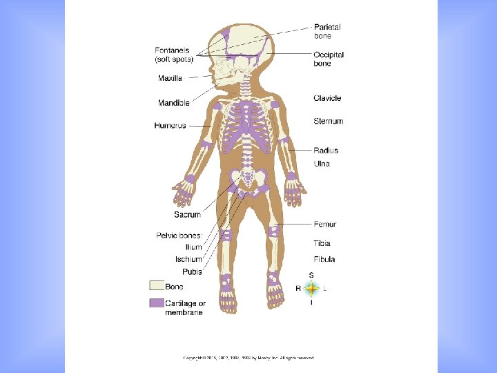
Bone Formation and Growth • When a child is born, there are no bones in the body yet, only cartilage and fibrous structures shaped like bones • Continuous activity of replacing the cartilage models with calcified bone matrix occur by bone-forming cells known as osteoblasts and bone-resorbing cells, osteoclasts • What does resorb mean? • Resorb (something) again: the ability to resorb valuable solutes from the urine- physiologically remove (cells, or a tissue or structure) by gradual breakdown into its component materials and dispersal in the circulation
Bone Formation (continued) • When calcium salts are deposited in the gellike matrix, bones are formed. This is an ongoing process of calcification that makes bones “hard as bone” (FYI… the stresses placed on certain bones during exercise increase the rate of bone deposition. For this reason, athletes or dancers may have denser, stronger bones than less active people. )
• In this process of “endochondral ossification”, osteoblasts and osteoclasts sculpts bones into their adult shape • Because of this process, bones can respond to stress or injury by changing size, shape and density due to bone-forming and bone
Endochondral Ossification
Endochondral Ossification • Figure. Endochondral ossification. A, Bone formation begins with a cartilage model. B and C, Invasion of the diaphysis (shaft) by blood vessels and the combined action of osteoblast and osteoclast cells result in cavity formation, calcification, and the appearance of bone tissue. D and E, Centers of ossification also appear in the epiphyses (ends) of the bone. F, Note the epiphyseal plate, an indication that this bone is not yet mature and that additional growth is possible. G, In a mature bone, only a faint epiphyseal line marks where the cartilage has disappeared and the centers of ossification have fused together.
• Endochondral Ossification: the way in which most bones are formed from a cartilage model • (A few of our flat bones are formed from another process called Intramembranous Ossification - example: skull bones) • Quick REVIEW: the long bones grow and become “ossified” from small center in both ends of the bone (epiphysis) and from the large center located in the shaft (diaphysis) • Important: as long as any cartilage, called an epiphyseal plate, remain between the epiphyses (plural) and diaphysis, growth will continue • When does growth cease? • ANSWER: When all epiphyseal cartilage is transformed into bone • How does a physician determine if a child will continue to grow? • ANSWER: Use an x-ray study on a child’s wrist to see if a layer of epiphyseal cartilage is still there; if not present, they have definitely attained their adult height
Endochondral Ossification
Check for Understanding 1. What is the basic structural unit of compact bone tissue called? ANSWER: Osteons or Haversian systems; each circular and tubelike osteon is composed of calcified matrix arranged in multiple layers resembling rings of an onion, and each ring is a concentric lamella and they surround the central canal containing the blood vessel. 2. What are osteocytes? Where would you find them in bone tissue? ANSWER: They are bone cells and lie between the hard layers of the lamellae in little spaces called lacunae. The tiny passageways or canals known as canaliculi connect the lacunae with one another and with the central canal in each Haversian system.
3. How does cartilage differ from bone? ANSWER: Cartilage resembles bone but consists of more intercellular substance than of cells, cartilage fibers are embedded in a firm gel instead of a calcified cement, and cartilage is flexible not rigid like bone. Also, cartilage doesn’t have blood vessels, as bone does. 4. What is ossification? What is the role of the osteoblast? ANSWER: Endochondrial ossification means formed in cartilage, as cartilage is first present and later replaced with bone. It also required continuous activity by bone-forming cells, osteoblasts, as cartilage is replaced with calcified bone matrix.
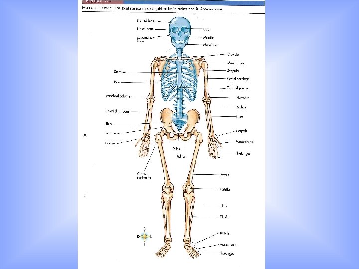
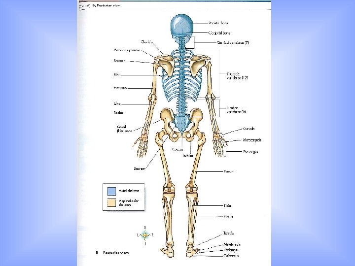
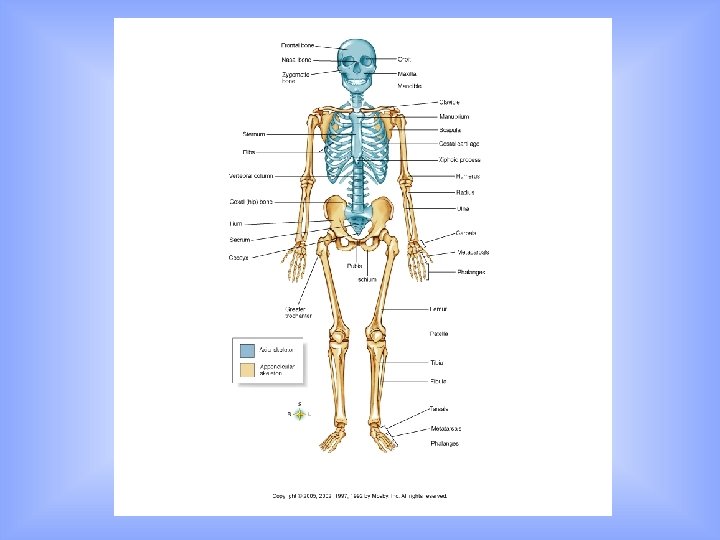
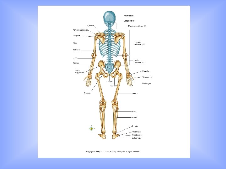
Divisions of Skeleton • Humans have two (2) divisions: axial skeleton and appendicular skeleton • Bones of center or axis make up axial skeleton and include skull, spine, chest and hyoid bone in neck • Bones of upper extremities (shoulder, pectoral girdles, arms, wrists and hands) and lower extremities (hip, pelvic girdles, legs, ankles and feet) or appendages make up appendicular skeleton • We are going to locate the parts of the axial and appendicular skeleton next……….
Main Parts of the Skeleton • Axial Skeleton • Appendicular Skeleton - skull • Upper Extremities - cranium - shoulder (pectoral) girdle - ear bones - arm - face - wrists - spine (vertebrae) - hands - thorax (ribs and sternum) • Lower Extremities - hyoid bone - hip (pelvic) girdle - legs - ankles - feet
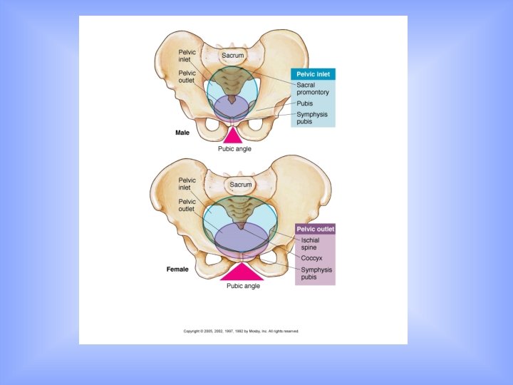
Differences Between a Male and Female Skeleton • Size difference: male skeleton is generally larger • Shape of pelvis: male pelvis is deep and narrow, female pelvis is broad and shallow • Size of pelvic inlet: female pelvic inlet is generally wider, normally large enough for baby’s head to pass through • Pubic angle: angle between pubic bones of female is generally wider
Bone Terms to Observe and Define • • Sinus (ex. paranasal sinus) Suture Fontanel Process Foramen Condoyle Tuberosity Malleoulus
Bone Terms • Sinus (ex. paranasal sinus) - a recess, cavity, or channel • Cranial Suture - the lines of junction between the bones of the skull • Fontanel - soft membranous gaps between the incompletely formed cranial bones of a fetus or infant. (Also called soft spot. ) • Process - a prominence or projection from a bone
Bone Terms • Foramen - a natural opening or passage in a bone • Condoyle - a rounded prominence at the end of a bone • Tuberosity - projection or protuberance at the end of a bone for the attachment of a muscle or tendon • Malleolus - A rounded bony prominence, such as those on either side of the ankle joint
Divisions of Skeleton • Humans have two (2) divisions: axial skeleton and appendicular skeleton (approx. 206 bones total) • Bones of center or axis make up axial skeleton (80 bones) and include skull, spine, and chest • Bones of upper extremities (shoulder, pectoral girdles, arms, wrists and hands) and lower extremities (hip, pelvic girdles, legs, ankles and feet) or appendages make up appendicular skeleton (126 bones) • We are going to locate the parts of the axial and appendicular skeleton next……….
Main Parts of the Skeleton • Axial Skeleton • Appendicular Skeleton - skull • Upper Extremities - cranium - shoulder (pectoral) girdle - ear bones - arm - face - wrists - spine (vertebrae) - hands - thorax (ribs and sternum) • Lower Extremities - hip (pelvic) girdle - legs - ankles - feet
Axial Division of Skeleton Axial skeleton (80 bones) sub-divisions: • Skull –Cranium (8 bones) –Face (14 bones) –Ear (6 bones)
Axial Skeleton- Skull
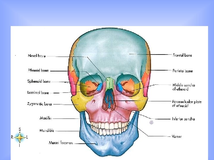
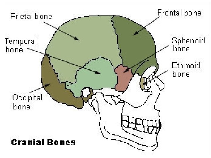
Bones of the Skull • Cranial Bones - Frontal (1) - Parietal (2) - Temporal (2) - Occipital (1) - Sphenoid (1) - Ethmoid (1) • Face Bones - Nasal (2) - Maxilla (2) - Zygomatic (2) - Mandible (1) - Lacrimal (2) - Palatine (2) - Inferior concha (2) - Vomer (1) • Ear Bones - Malleus (2) - Incus (2) - Stapes (2)
Axial Skeleton- Skull • 8 bones form the cranium
Cranial Bones Frontal (1): forehead bone - Parietal (2): form bulging topsides of cranium - Temporal (2): form lower sides of cranium - Occipital (1): forms back of skull - Sphenoid (1): forms central part of cranial floor - Ethmoid (1): bone that helps form floor of cranium, side walls and roof of nose -
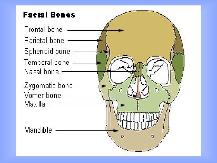
• 14 bones form the face
Facial Bones - Nasal (2): small bones that form upper part of bridge of nose - Maxilla (2): upper jawbones (form roof of mouth) - Zygomatic (2): cheek bones - Mandible (1): lower jawbones - Lacrimal (2): small bones forming medial wall of eye socket and side wall of nasal cavity - Palatine (2): form back part of roof of mouth - Inferior concha (2): form curved “ledge” along inside of side wall of nose - Vomer (1): forms lower, back part of nasal septum
• 6 tiny bones form the middle ear
• • Malleus (2): means hammer for its shape Incus (2): means anvil for its shape Stapes (2): means stirrup for its shape All are found in middle of the cavity of the temporal bone
SKULL • Sinuses: found within the skull are spaces or cavities inside the cranial bones - There are four pairs found in the frontal, maxillary, sphenoid and ethmoid bones - They have opening into the nose and are referred to as paranasal sinuses - We have trouble with sinuses when the mucous membrane lining them becomes inflamed, swollen and painful Sutures: immovable joints that provide shape to the bulging topside of the skull - Fontanels: there are six “soft spots” on a baby’s skull and areas where ossification is incomplete at birth
Vertebrae Bone Formations
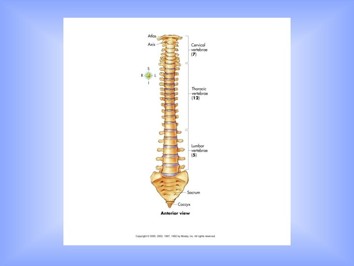
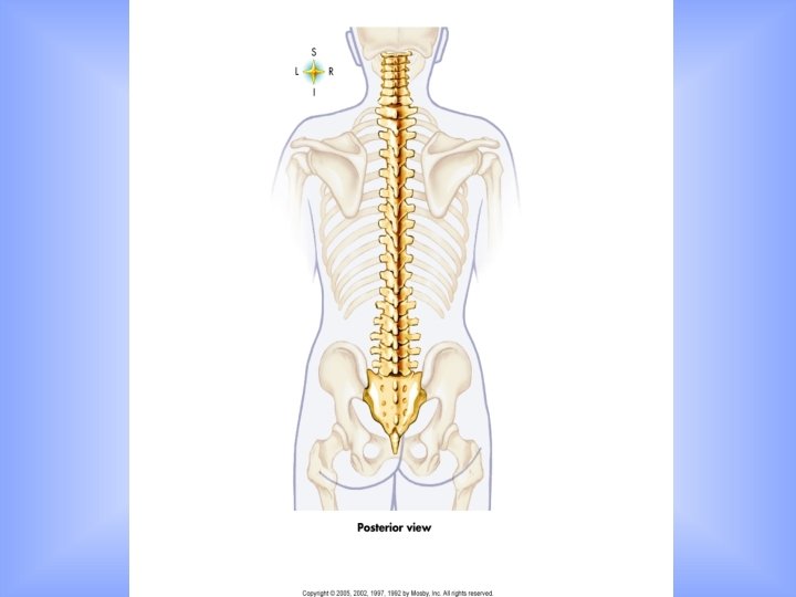
Axial Skeleton Sub-Divisions (continued) • Spine or vertebral column – Cervical (7 bones) – Thoracic (12 bones) – Lumbar (5 bones) – Sacrum (1 bone) – Coccyx (1 bone)
Axial Skeleton Sub-Divisions (continued) Spine or vertebral column (continued) – Curves of the Spine » Four normal curves • Cervical • Thoracic • Lumbar • Pelvic » Three abnormal curves A. Lordosis or “swayback” B. Kyphosis or “hunchback” C. Scoliosis
Normal Curves of Spine
Three abnormal curves A. Lordosis or “swayback” B. Kyphosis or “hunchback” C. Scoliosis
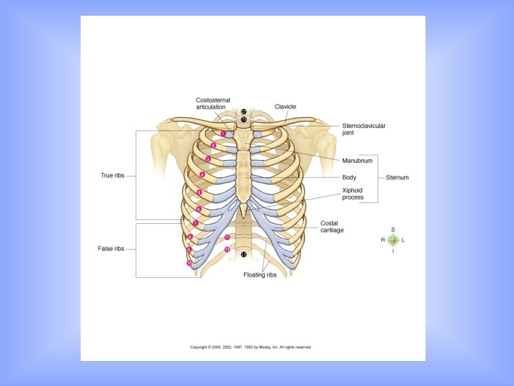
Axial Skeleton Sub-Divisions (continued) – Thorax • Composed of: – 12 pairs of ribs – Sternum or breastbone – Thoracic vertebrae • Ribs – True ribs—rib pairs 1 through 7 – False ribs—rib pairs 8 through 10 – Floating ribs—rib pairs 11 and 12
Divisions of Skeleton – Appendicular skeleton (126 bones) • Bones in shoulder or pectoral girdle connect bones of upper extremity (arm, forearm, wrist, and hands) to axial skeleton • Bones in hip or pelvic girdle connect bones of lower extremity (thigh, leg, ankle, and foot) to axial skeleton
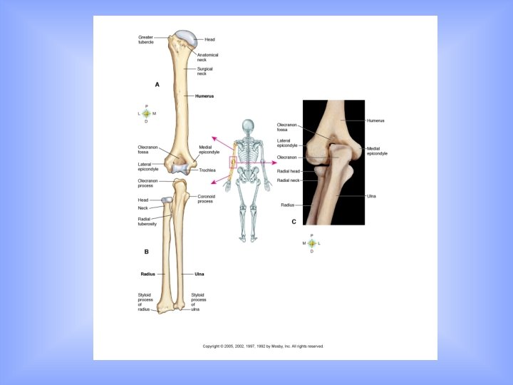
Sub-Divisions of Appendicular Skeleton – Upper extremity • Shoulder or pectoral girdle formed by: – Scapula – Clavicle (frequently fractured) • Arm—humerus • Forearm—radius and ulna • Wrist— 8 carpal bones • Hand— 5 metacarpal bones • Fingers— 14 phalanges or finger bones
Hand
Sub-Divisions of Appendicular Skeleton – Lower extremity • Hip or pelvic girdle formed by the two coxal or pelvic bones (one on each side) with sacrum and coccyx behind – Each coxal bone in infant consists of separate ilium, ischium and pubic bones—bones are fused into a single coxal bone in the adult – Acetabulum is cup shaped socket—articulates with head of femur
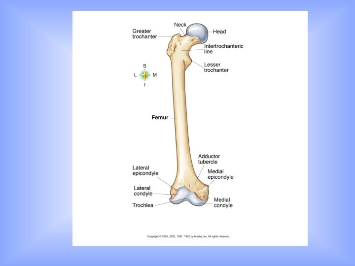
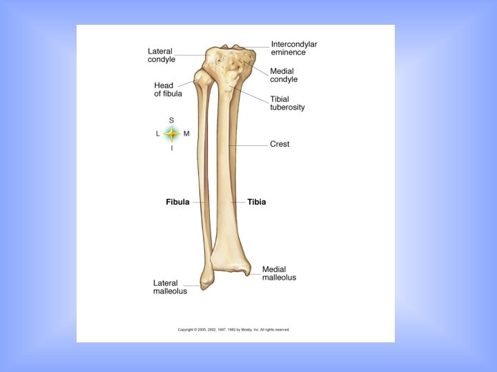
Sub-Divisions of Appendicular Skeleton – Lower extremity (continued) • Thigh bone—femur • Patella or knee cap articulates with femur and tibia • Lower leg—tibia (“shinbone”) and fibula
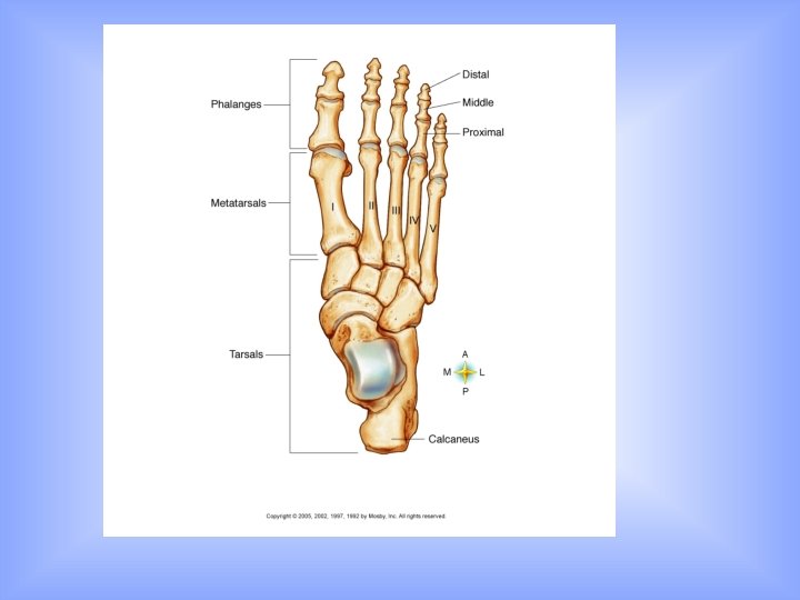
Sub-Divisions of Appendicular Skeleton – Foot • 5 metatarsal bones • 7 tarsal bones (calcaneus or heel bone is largest tarsal) • 14 phalanges or toe bones • 3 arches of foot—two longitudinal (medial and lateral) and a transverse or metatarsal arch— if weakened, result is “flat feet”
Foot Arches
Joint (Articulations) • Kinds of joints – Synarthroses (no movement)—fibrous connective tissue grows between articulating bones (e. g. , sutures of skull) – Amphiarthroses (slight movement)— cartilage connects articulating bones (e. g. , symphysis pubis)
Synarthroses
Amphiarthroses
Joint (Articulations) • Kinds of joints – Diarthroses (free movement)—most joints belong to this class • Structures of freely movable joints—joint capsule and ligaments hold adjoining bones together but permit movement at joint • Articular cartilage—covers joint ends of bones and absorbs jolts • Synovial membrane—lines joint capsule and secretes lubricating fluid • Joint cavity—space between joint ends of bones
Diarthroses
Joint (Articulations) • Kinds of joints (see class handout) – Types of freely movable joints—ball-and-socket, hinge, pivot, saddle, gliding, and condyloid
Herniated Disc
Bibliography • http: //pathwiki. pbworks. com/f/pagets%20 diagram. jpg • Thibodeau and Patterson, The Human Body in Health and Disease, 3 rd Edition, Mosby Inc. , St Louis, MO, 2002. • http: //player. discoveryeducation. com/index. cfm? guid. Asset. Id =AA 03350 B-070 D-4 CF 2 -95714 EF 95 A 7 A 8 C 59&bln. From. Search=1&productcode=US • http: //www. learnbones. com/wpcontent/uploads/Ear_bones. jpg&imgrefurl=http: //www. learn bones. com/middle-ear-bones-anatomy&usg • http: //www. arthursclipart. org/medical/senseorgans/ear%20 bones. gif • http: //www. webbooks. com/e. Library/Medicine/Physiology/Skeletal/cranial_b ones. jpg • http: //upload. wikimedia. org/wikipedia/commons/thumb/7/7 7/Illu_facial_bones. jpg/200 px-Illu_facial_bones. jpg