The Skeletal System Medical Terminology to the body

The Skeletal System Medical Terminology

________to the body. This framework helps protect _________ and furnishes attachment points for ______, _________ and ______. Bones also store minerals and contain ___________bone marrow.

Bone is a specialized form of __________tissue containing about _____% solid matter and ______% water.

Bone consists of a hard outer shell called ______ bone and an inner spongy structure called ________ or __________ bone

The bone surfaces are covered by a tough fibrous vascular membrane called the ___________.
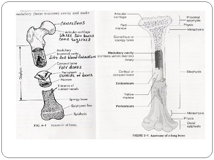

The _____bones grow in length at the ‘__________’. This is what the ends of the developing bones is called.
 Medullary cavity epiphysis")
Diaphysis (shaft) Medullary cavity epiphysis

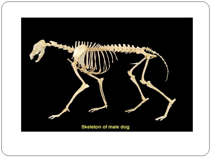
Bones are classified according to their shape. _____________

The skeleton is divided into two main parts: ________: includes the skull, vertebral column, ribs and sternum. ________: including the limbs
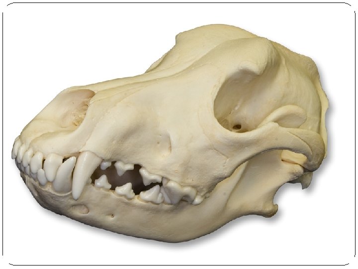
 and the _________. All skull")
The skull includes two major segments: _______: (brain case) and the _________. All skull bones are ______except the ________. The skull bones are united by _______. Within the bones of the skull are hollows called ________. The function of the sinuses include, lessening the _________, providing chambers for _________and moistening and ________air.

Facial bone
- 7 * Thoracic")
Vertebrae Vertebral Anatomy The horse has 37 vertebrae: * Cervical (neck)- 7 * Thoracic (chest) - 18 * Lumbar (lower back) - 6 (except in Arabs - 5) * Sacral (pelvis) - 5 (fused) The dog has 31 vertebrae: * Cervical (neck)- 7 * Thoracic (chest) - 13 * Lumbar (lower back) - 7 * Sacral (pelvis) - 3 (fused) The cat has 31 vertebrae: * Cervical (neck)- 7 * Thoracic (chest) - 13 * Lumbar (lower back) - 7 * Sacral (pelvis) - 3 (fused)

Cervicle vertebrae

C-1 and C-2 are called the _____ and the ______. The words atlas (holding up the world) and axis (what the world spins on) come from Greek mythology. There can be an instability in this area in large dogs that will cause neurologic problems. The cervical vertebrae are quite flexible, for obvious reasons.
.")
As the cervical vertebrae become the _______ vertebrae they go past the shoulder (S). The nerves that come off this cervicalthoracic junction at the shoulder are called the _________(you cannot see nerves on a plain radiograph). They innervate the front legs on each side. Each of the thoracic vertebrae corresponds to a _____ (R) on each side of the chest

As we continue down the thoracic vertebrae you can visualize how high their dorsal ________________are. Also notice how these processes start to get smaller as we get closer to the _______vertebrae

Moving towards the end of the thoracic vertebrae we come to what is termed the ___________(T-L) junction. It is a very common area to have VSC disease. As we pass into the _________ vertebrae we have now made our way into the lower back.
. The ______ sacral")
The 7 ________ vertebrae eventually lead into the _________ vertebrae (S). The ______ sacral vertebrae are hard to visualize because they are within the __________. After the sacrum we are at the ____.

ribs

Forelimbs Front legs
 In the feline: a small flat bone attached to the ______ In")
________: (collarbone) In the feline: a small flat bone attached to the ______ In the canine: a _____ bone that is not attached to any other bone and may be _____ in some dogs.
")
Scapula (shoulder blade)
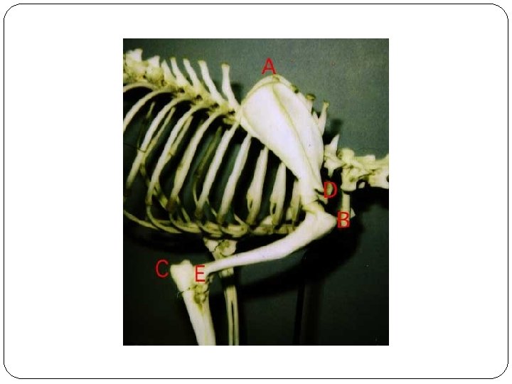

_______ the long bone extending from the ______to the ______. The ________ articulates with the _______ and the radius/ulna
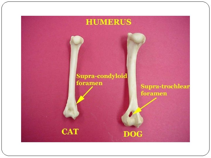

humerus

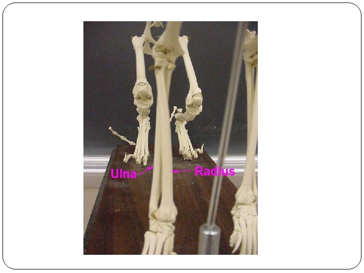
______ /_____: the _____ bone that articulates with the ______at the olecranon. _______ : the _____bone that articulates with the _______ and _____at the elbow.

_______ Is composed of 7 – 8 ______ shaped bones in two rows. This joint is called the _____ in humans

Carpus

Metacarpus

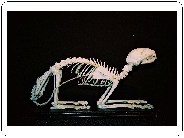
Equine skeleton

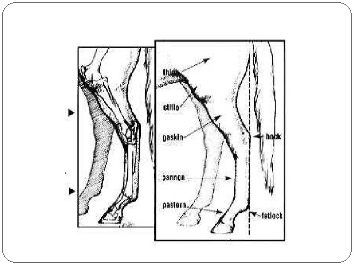
Equine forelimb

Pelvis illeum ichium pubis

Femur femur

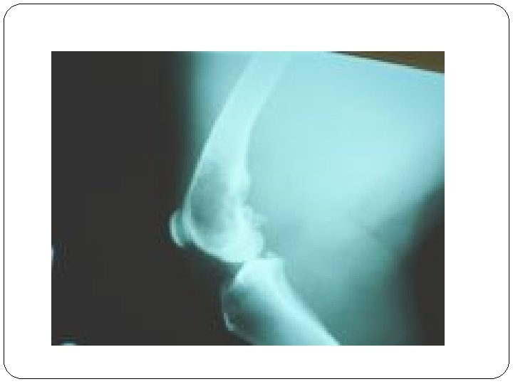
Knee Joint

Tibia/Fibula tibia fibula

Tarsus

Metatarsus

A _____is an _______ between bones, or between bones and cartilage. They are classified by the degree of _______they permit.
")
__________: allow no movement (Fibrous tissue)
")
____________ allow slight movement. (Cartilaginous tissue)
 _____________________ ___________")
_________ freely permit movement. (Synovial joints) _____________________ ___________

Joint capsule

___________: lines the joint capsule and secretes ________ fluid.

__________: the space between opposing surfaces of ________.

Articular cartilage and disks

_________: sacs of connective tissue lined with synovial membrane filled with synovial fluid
- Slides: 54