Hemostasis Hemostasis Hemostasis or haemostasis from the Ancient


- Slides: 57

Hemostasis

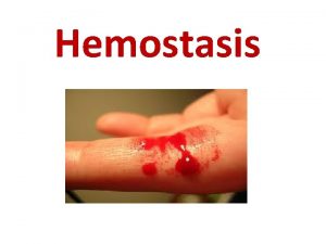
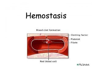
Hemostasis • Hemostasis or haemostasis (from the Ancient Greek: αἱμόστασις haimóstasis ) is a process which causes bleeding to stop. • Purpose : to keep blood within the damaged blood vessel • It is the first stage of wound healing • The opposite of hemostasis is hemorrhage).

Platelets

Platelets (Thrombocytes) • Is small, non-nucleated, granulated bodies • Reddish purple granules with Leishman stain • Size is between 2 and 4. • May be circular or oval shaped. • Normal count 1. 5 – 4 lakhs/cu mm of blood.

Platelets (Thrombocytes) • Life span is about 10 days. • The cells are produced by the megakaryocytes present in red bone marrow. • Thrombopoietein- regulate the synthesis of platelets • Site of destruction is mainly spleen, which contains reticulo-endothelial system (RES).

Thrombopoisis Pluripotent stem cell Commited stem cells Megakaryoblast Promegakaryocyte Megakaryocyte Platelets

Platelets Structure Leishman Stain

Platelets Structure

Platelets Structure • Cell membrane : Glycoprotein & Phospholipids • Membranes contain receptors for ØCollagen ØADP ØVon Willibrand factor ØFibrinogen • Microtubules – Ring of microtubules below the cell membrane – Provides structural support

Platelets Structure Cytoplasm– • Proteins: – – – • • Actin Myosin Thrombosthenin Lysosomes Granules

Platelets Structure • 2 types of granules 1. Dense granules • Serotonin & ADP, calcium • Secreted in response to platelet activation 2. Alpha granules • Clotting factors • Platelet Derived Growth Factors (PDGF) • Von Willebrand factor • Platelet Activating Factor

Functions of Platelets 1. 2. 3. 4. 5. Hemostasis (Platelet Plug Formation) Blood coagulation Clot retraction Repair of capillary endothelium. Phagocytosis.

Hemostasis • Hemostasis is the spontaneous arrest of bleeding. • By the process of forming clots in the walls of damaged blood vessels and maintaining blood in a fluid state in the vascular system

Hemostasis • Hemostasis is brought about by three mechanisms : 1. Vasoconstriction 2. Platelet plug formation 3. Coagulation of blood

Vasoconstriction • Constriction of the blood vessel is a local phenomenon • The collagen fibers are exposed in a damaged blood vessel • Platelet Activation : The platelets adhere to the collagen and get activated (accelerated by von Willebrand factor) • Activated platelets secrete serotonin and other vasoconstrictors

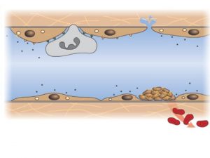
Platelet plug formation • Platelet Adhesion • Platelet Activation • Platelet Aggregation

Platelet plug formation

Platelet plug formation • Adherence of platelets to the collagen secretes ADP and ‘thromboxane A 2 which attract more platelet and activate them. • Aggregation of platelets is accelerated by platelet activating factor (PAF) • These platelets aggregate and form a temporary loose plug to prevent blood loss.

Platelet plug formation

Blood coagulation or clotting • Blood in circulation is in fluid state. • The process of conversion of fluid blood into semisolid jelly like mass is known as coagulation or clotting of blood. • At the site of injury, clotting occurs over the temporary haemostatic plug. • So by the process of coagulation, The temporary plug is converted to permanent sealing of the injured area which is a must for permanent arrest of bleeding.

Blood coagulation or clotting Why should clotting occur on hemostatic plug? • Hemostatic plug is not stable one. • If clotting doesn’t take place on this plug, the hemostatic plug may get washed away. • This results in prolonged bleeding • Seen in certain coagulation disorder like hemophilia.

Reactions involved in hemostasis

Clotting 1. Formation of Prothrombin Activator 1. Intrinsic system 2. Extrinsic system 2. Conversion of prothrombin to thrombin 3. Conversion of fibrinogen to fibrin and clot formation

CLOTTING FACTORS I Fibrinogen II Prothrombin III Tissue thromboplastin IV Ionized calcium (Ca 2+) V Proaccelerin VII Proconvertin VIII Antihemophilic factor A IX Antihemophilic factor B; plasma thromboplastin component (PTC); Christmas factor X Stuart–Prower factor XI Plasma thromboplastin antecedent(PTA) XII Hageman factor XIII Fibrin-stabilizing factor (FSF) – Prekallikrein (PKK); Fletcher factor – High-molecular-weight kininogen(HMK); Fitzgerald factor • Kallikrein • Platelet Phospholipid

Formation of Prothrombin Activator Stimulus for thrombus formation • Intrinsic System – Exposure of blood to collagen fibres underlying endothelium – Change in blood constituents – Electro negatively charged wettable surfaces such as glass (In vitro) Activates Factor XII • Extrinsic system – Injury to blood vessel wall – Injury to other body tissue Activates Factor III

Formation of Prothrombin Activator • When clotting occurs in the body, it involves both the intrinsic and extrinsic systems and these systems will complement each other. • During the process of clotting, the inactive factors are converted into active forms (denoted by adding letter ‘a’ ) • Their enzymatic actions produce successive reactions in a cascading manner.

Cascade

III Cascade reaction

Intrinsic pathway Endothelial damage + Collagen exposure HMW Kininogen, Plasma kallikrein XIIa XII Platelets XIa XI Ca IX IXa VIII Phospholipids VIIIa, Ca Xa X V Va, Ca Prothrombin activator Prothrombinase complex

Extrinsic pathway Tissue thromboplastin VIIa VII X Xa Phospholipids, Ca, III V Va, Ca Prothrombin activator

Conversion of prothrombin to thrombin • Intrinsic pathway Extrinsic pathway Prothrombin activator complex XIII Prothrombin Thrombin XIIIa Fibrinogen Fibrin (monomer) Fibrin(polymer) Fibrin clot

Conversion of fibrinogen to fibrin and clot formation Soluble fibrinogen Proteolysis THROMBIN Fibrin monomer polymerization Fibrin polymer XIIIa, Ca Fibrin clot

Clot

Clot retraction • Clot retraction-within 20 -60 min • Platelet contractile proteins (actin-myosin like filaments) cause strong contraction of platelet spicules attached to the fibrin • Decrease in the size of clot can be to the extent of 40 to 60% of the original size. • As the clot retracts edges of broken blood vessels are pulled together contributing for the ultimate state Hemostasis

Negative feedback mechanisms (Limiting Reactions) Inhibits the actions of thrombin They are: 1. Once the fibrin is formed it adsorbs thrombin on to fibrin. 2. Antithrombin III is present in the plasma, which neutralizes thrombin and factors IX, X, XI and XII. 3. Thrombin combines with thrombomodulin and forms a complex. This complex activates protein C and S which in turn inhibit factor V and VIII. 4. Thrombaxane A 2 and Prostacyclin

Positive feedback mechanisms during coagulation Positive feed back mechanism comes into play to hasten the process. They are: 1. When once thrombin is formed it brings about conversion of prothrombin to thrombin by autocatalytic reaction. 2. Thrombin also activates factors V, VIII, IX, X, XII and also XIII.

Role of Calcium • Ca++ are essential in almost all steps

Role of platelets in clotting • Platelets contribute for coagulation of blood by releasing substance like phospholipids • Phospholipids provide a surface for the cascade reactions • Required formation of prothrombin activator by intrinsic system.

Role of Vitamin K • Vitamin K is essential for the synthesis of coagulation factors II, VII, IX and X by the liver. • Post-translational γ-carboxylation of glutamate residues of these factors

Intravascular prevention of blood clotting 1. 2. 3. 4. 5. 6. Movement of blood Smoothness of endothelium Layer of glycocalyx-on the endothelium Secretion of prostacyclin Fibrin threads -85% of the thrombin adsorbed Antithrombin III-Heparin-blocks activity of thrombin & factor IX, X, XII. 7. Thrombomodulin on the surface of the endothelium-binds to thrombin

The fibrinolytic system • Fibrinolysis is a process by which the clot is broken down. It is essential to prevent clogging or blocking of capillary. • After the formation of clot, the fibrinolytic system becomes active. • It involves many factors that are present in circulation in inactive form and get activated during fibrinolysis.

The fibrinolytic system

Plasmin • Plasmin is generated by proteolytic cleavage of plasminogen, a plasma protein synthesized in the liver. • This cleavage is catalyzed by tissue plasminogen activator (t-PA), which is synthesized and secreted by endothelium. • Plasmin is fibrinolytic • It proteolytically cleaves fibrin into fibrin degradation products that inhibit excessive fibrin formation.

Anticoagulants • Anticoagulants are substances that prevent clotting of blood. • They also have important role to play in maintaining the fluidity of blood in circulation. • They can be classified into in vivo and in vitro anticoagulants.

Anticoagulants In vivo anticoagulants are used to prevent coagulation of blood inside the body. • – Eg: heparin and dicoumarol. In vitro anticoagulants are used to prevent coagulation of blood outside the body. • – Eg: heparin, inorganic salts of sodium, potassium and ammonium.

Heparin • • • Produced by the mast cells & basophils. Mechanism of action Combines with Antithrombin III and it neutralizes the action of thrombin Prevents aggregation of platelets Therapeutically used for preventing intra vascular thrombosis, during dialysis, myocardial infarction etc

Dicoumarol • • • Exogenous administration. It can be taken orally. It acts as vitamin K antagonist. Vitamin K is essential for the synthesis of coagulation factors II, VII, IX and X by the liver. Therapeutically dicoumarol is used to prevent intra vascular clotting in long standing bedridden patients.

In vitro anticoagulants • • Heparin Sodium citrate solution (3. 8 %) Double oxalate (ammonium and potassium) EDTA (ethylene diamino tetra acetic acid). • Mechanism : The inorganic salts will remove the ionic calcium by precipitating calcium ions • In vitro anticoagulants are used - outside the body in laboratories and in blood banks.

Tests for hemostasis • • • Bleeding time It’s the time interval from oozing of blood till arrest of bleeding. Normal duration is 1 to 6 min. Clotting time Time interval from oozing of blood till formation of clot Normal duration is 2 to 8 min.

Prothrombin time • • • Prothrombin time is the time interval required to convert prothrombin to thrombin and final clot formation. This measures the extrinsic system involved in blood coagulation. Normal time is about 12 – 16 sec. Prothrombin time increases in liver diseases and forms one of the liver function tests. Prothrombin time is also increased in vitamin K deficiency, as this vitamin is essential for the synthesis of prothrombin

Hemophilia • • • Hemophilia is a disorder of coagulation of blood. It is sex linked inherited disease Males suffer the symptoms and females are the carriers. The gene responsible is a recessive gene and is present on the X chromosome (X linked recessive) It occurs due to lack of formation of prothrombin activator Ø Ø Coagulation time is prolonged BT is normal

Hemophilia A • Factor VIII is deficient • 85% of people with hemophilia are affected by this type. Hemophilia B • Factor IX is deficient • 15% of people with hemophilia are affected by this type. Its also called “Christmas disease”

Hemophilia

Hemophilia Complications • Deep internal bleeding • Joint damage from haemarthrosis • Transfusion transmitted infection • Intracranial haemorrhage

Purpura • • Here the bleeding time is prolonged but clotting time is normal. Thrombocytopenic purpura– due to deficiency of platelets Thromboesthenic purpura– due to abnormal platelets Idiopathic thrombocytopenic purpura.

Others • • Atherosclerosis Thrombus Emboli DIC

THANK YOU
 Ivy method
 Blood clotting factor
 Primary hemostasis
 Virchow triad
 Thrombopoisis
 Hemotasis
 Skema hemostasis
 Hemostasis definition
 Hemostasis
 Primary hemostasis
 What is hemostasis
 1ry hemostasis prescribed by:
 Hemostasis definition
 Hemostasis
 Ancient and modern means of communication
 Ancient india vs ancient china
 Môn thể thao bắt đầu bằng chữ f
 Hát kết hợp bộ gõ cơ thể
 Sự nuôi và dạy con của hươu
 Dot
Dot điện thế nghỉ
 Thế nào là sự mỏi cơ
 Trời xanh đây là của chúng ta thể thơ
 độ dài liên kết
 Chó sói
 Thiếu nhi thế giới liên hoan
 Phối cảnh
 Một số thể thơ truyền thống
 Thế nào là hệ số cao nhất
 Ng-html
 Hệ hô hấp
 Số nguyên là gì
 đặc điểm cơ thể của người tối cổ
 Các châu lục và đại dương trên thế giới
 Mật thư anh em như thể tay chân
 Tư thế worm breton
 ưu thế lai là gì
 Tư thế ngồi viết
 Thẻ vin
 Bàn tay mà dây bẩn
 Các châu lục và đại dương trên thế giới
 Bổ thể
 Từ ngữ thể hiện lòng nhân hậu
 Tư thế ngồi viết
 Thế nào là giọng cùng tên
 Thể thơ truyền thống
 101012 bằng
 Bài hát chúa yêu trần thế alleluia
 Hổ đẻ mỗi lứa mấy con
 Diễn thế sinh thái là
 đại từ thay thế
 Vẽ hình chiếu vuông góc của vật thể sau
 Công thức tiính động năng
 Tỉ lệ cơ thể trẻ em
 Thế nào là mạng điện lắp đặt kiểu nổi
 Lời thề hippocrates
 Vẽ hình chiếu đứng bằng cạnh của vật thể
 Quá trình desamine hóa có thể tạo ra
