Hemostasis Red very important Green only found in


 of bleeding from ruptured blood vessels.")


 � Platelets are small “biconvex” disc")

")
 Activation: Platelet adhesion o Exposed collagen attracts")
 When platelets are activated they secrete: 1) 5 HT")
 3 - Blood Coagulation: secondary hemostasis � CLOT is a")

 mechanism (steps) � A series of biochemical reactions leading to")


 � The trigger is the activation")
 �")
 Intrinsic Pathway (also known as contact activating")

 � 4 - Fibrinolytic system (Fibrinolysis) Formed blood clot can")


 Excessive bleeding can result from: • Platelet defects: deficiency in")


 • •")


 3. Blood coagulation(clot formation) What is it Clotting")


- Slides: 38
Hemostasis Red: very important. Green: only found in males’ slides. Purple: only found in females’ slides. Gray: notes. Physiology Team 436 – Foundation block lecture 12 1 Lecture: If work is intended for initial studying. Review: If work is intended for revision.
Objectives o o o o 2 Recognize different stages of hemostasis. Describe formation and development of platelets. Explain the role of platelets in hemostasis. Recognize different clotting factors & cascade of clotting. Describe the intrinsic, extrinsic and common pathways. Recognize the role of thrombin in coagulation. Explain the process of fibrinolysis and function of plasmin.
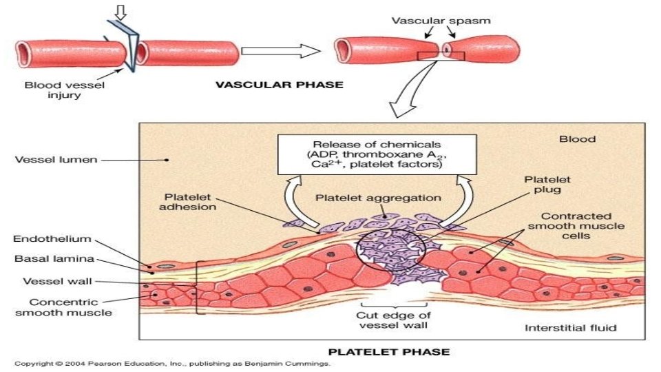
Hemostasis o It is the spontaneous arrest (stop) of bleeding from ruptured blood vessels. Its mechanism (steps of function): 1) Vessel wall (Vascular spasm). 2) Formation of platelet plug. 3) Blood coagulation & clot retraction. 4) Fibrinolytic system (Fibrinolysis). o 3
Steps of Hemostasis
Hemostatic Mechanism 1 -Vessel wall. � Immediately after injury there is a localized Vasoconstriction (vascular spasm, in order to decrease blood flow) � Mechanism of vasoconstriction (causative Factor): o Local myogenic spasm (systemic release of adrenaline). o Local release of Thromboxane A 2 [TXA 2] & 5 HT” serotonin” by platelets. (humoral factors) o Nervous factors “stimulation for nerve impulses + nerve reflex is vasocontraction”. (reflex action involves simple nervous pathway) � Importance: Crushing injuries > intense spasm > no lethal loss of Notes: Causative Factor: meaning that it is “a cause” blood. Vasoconstriction is produced by vascular smooth muscle cells, and is the blood vessels first response to injury. The damaged vessels will constrict (vasoconstrict) which reduces the amount of blood flow through the area and limits the amount of blood loss. Myogenic contraction refers to a contraction initiated by the myocyte cell. (muscle cell) Platelets release cytoplasmic granules which contain, ADP, 5 HT (serotonin) and thromboxane A 2 (TXA 2), all of which, increase the effect of vasoconstriction. *will be explained in the next slides* 6
7
� 2 -Formation of platelet plug (primary hemostasis) � Platelets are small “biconvex” disc shaped cells. Platelet � (Remember: RBCs are biconcave discs) � Platelet count: 150 x 103 – 300 x 103/ml. � Life span: 8 – 12 days. � Contain high calcium content & rich in ADP. � Active cells contain contractile protein. � Thrombocytes are fragments of megakaryocytes in the bone marrow. shape: � Regulation of thrombipoiesis is done by thrombopoietin. � Site of formation: bone marrow, steps: Stem cell > Megakaryoblast > Megakaryocyte > Platelets. � Importance: enough to stop bleeding from small vascular damage. ﺯﻱ Nucleus ﻣﺎﻓﻴﻬﺎ ﺑﺲ ﻓﻴﻬﺎ RBCs ﺍﻟـ Enzyme It also has pores that enable movement of substances from granules to circulation. Remember: Regulation of Erythropoiesis by Erythropoietin (Erythropoietin is formed in kidney & liver , while Thrombopoietin in liver) 8 Bone marrow Megakaryocyte
Platelet Cont. Platelet Charactersitics: shape: minute round or oval discs size: 1 -4 um in diameter location: 80% in blood & 20% in spleen Contractile, adhesive, cell fragments. Store coagulation factors & enzymes Surface Binding sites: GP Ib (Glyco Protein Ib or 1 b on cell membrane of platelets) is a component of GP Ib-IX-V complex that functions as a receptor of v. W factor. FUNCTIONAL CHARACTERISTICS: • Motile: Actin And Myosin Molecules (for muscle contraction) • Active: Endoplasmic Reticulum, Golgi Apparatus & Mitochondria • Enzymes Systems For Synthesis Of Prostaglandins • Granules Postglandins: any of a group of compounds with varying hormone-like effects 9 Platelet Receptors: -GP Ia, GP VI (adhesion to Collagen) -GP Ib-IX-V (receptor for v. W Factor) -TPα (for TXA 2) -GP IIb-IIIa (for Fibrinogen, v. W Factor) -P 2 Y 12 (for ADP) Dense or δ granules contain: • Serotonin • ADP • Ca++ α granules contain: • Coagulation Factors Fibrinogen • PDGF (Platelet-derived growth factor)
Platelet Plug Formation (Importance → enough to stop bleeding from small vascular damage. ) Secrete prostacyclin (PGI 2) NO ADP phosphatase 10 Intact (normal) endothelium secret prostacyclin and NO which inhibit aggregation.
Platelet Function: Begins With Platelet (Thrombocytes) Activation: Platelet adhesion o Exposed collagen attracts platelets, o Platelets stick to exposed collagen (to the sub endothelial layer) (Von Wille. Brand Factor) released from the damaged endothelial cells in vessel wall or underlying damaged endothelial cells in vessel wall. Shape change Activation and Aggregation* Release reaction/Secretion Clot retraction* o Activated platelets stick together and activate new platelets to form a mass called a platelet plug. o Activated platelets (activated o Myosin and actin by adhesion) extend filaments in projections (protrusions) to platelets are make contact with each other stimulated to and release Serotonin, ADP contract during (activating other platelets) aggregation o Plug reinforced ﻣﺜﺕ by & thromboxane A 2 (TXA 2). further fibrin threads formed reinforcing the during clotting o Serotonin & TXA 2 are plug and help process. ** vasoconstrictors decreasing release of blood flow through the injured granule contents. o *Platelet aggregation: vessel. The clumping together o *Clot retraction: is of platelets in the o 1. ADP & TXA 2: increase the shrinking of a blood. stickiness of platelets clot over a ﺗﻤﺴﻜﻬﺎ ﺍﻟﺸﺒﻜﺔ ﺯﻱ ﺗﺴﻮﻱ **ﺧﻴﻮﻁ number of days. o 2. aggregation is increased. Activated Platelets* ( ﺍﻟﺴﺪﺍﺩﻩ )ﺯﻱ ﺍﻟﺪﻡ ﻳﻮﻗﻒ ﻋﺸﺎﻥ o 3. cut vessel is plugged. 11 Video ; - https: //www. youtube. com/watch? v=9 QVTHDM 9
Activated Platelets (during shape change) When platelets are activated they secrete: 1) 5 HT > causes vasoconstriction. 2) ADP (Adenosine Di-Phosphate) > stickiness 3) Platelet phospholipid (PF 3) > clot formation. 4) Thromboxane A 2 (TXA 2) > a prostaglandin formed from arachidonic acid, and it is inhibited by aspirin. (its function is vasoconstriction and platelet aggregation) � ﻳﻤﻨﻊ ﺗﻜﻮﻥ ﺍﻟﺠﻠﻄﺎﺕ ﻟﻤﻘﺪﺭﺗﻪ ﻋﻠﻰ ﺟﻌﻞ ﺍﻟﺪﻡ ﻓﻲ ﺣﺎﻟﺔ ﺳﺎﺋﻠﺔ (Found in the males’ slides) Further explanation of the process: platelets adhere to damaged endothelium to form a platelet plug (primary hemostasis) and then degranulate Platelets play one of the biggest roles in the hemostatic process. When platelets come across the injured endothelium cells, they change shape, release granules and ultimately become ‘sticky’. Platelets express certain receptors, some of which are used for the adhesion of platelets. When platelets are activated, they express receptors that interact with other platelets, producing aggregation and adhesion. Platelets release cytoplasmic granules such as (ADP), serorotin and thromboxane A 2. Adenosine diphosphate (ADP) attracts more platelets to the affected area, serotonin is a vasoconstrictor and thromboxane A 2 assists in platelet aggregation, vasoconstriction and degranulation. More chemicals are released more platelets stick and release their chemicals; creating a platelet plug and continuing the process in a positive feedback loop. Platelets alone are responsible for stopping the bleeding of unnoticed wear and tear of our skin on a daily basis. This is referred to as primary hemostasis 12
Hemostatic Mechanism (cont. ) 3 - Blood Coagulation: secondary hemostasis � CLOT is a meshwork of fibrin fibers running in all directions entrapping blood cells, platelets and plasma. � Blood clotting is the transformation of blood (soluble fibrinogen) from a liquid into a solid gel form (insoluble fibrin strands) this fibrin will strengthen the previous Platelet Plug. � Clotting Cascade Pathways: Intrinsic and Extrinsic � Begins to develop in 15 -20 sec → MAJOR/SEVERE TRAUMA 1 -2 min → MINOR TRAUMA 13 Factors Names I (1) II (2) III (3) IV (4) V (5) VII (7) VIII (8) IX (9) X (10) XI (11) Fibrinogen Clotting Factors Prothrombin Circulate in plasma Thromboplastin in inactive state Calcium *most of them synthesized by the liver Labile factor Stable factor Antihemophilic factor A Antihemophilic factor B Stuart-Power factor Plasma thromboplastin antecedent (PTA) Hagman factor Fibrin stablizing factors XII (12) XIII (13)
14 Found in the males’ slides
Blood coagulation (clot formation) mechanism (steps) � A series of biochemical reactions leading to the formation of a blood clot � 1. Formation of Prothrombin activator complex (Xa+Ca+PF-3+V) by Extrinsic & Intrinsic Pathways leading to Common Pathway. � 2. This reaction leads to the activation of thrombin enzyme from inactive form prothrombin (Conversion of prothrombin (clotting factor II) into thrombin) � Prothrombin (inactive thrombin) is activated by a long intrinsic or short extrinsic pathways � 3. Thrombin will change fibrinogen (plasma protein) to fibrin (insoluble protein) (Conversion of fibrinogen into fibrin) � 4. Fibrin converts to stable fibrin polymer Explanatio n: ﻳﺘﻨﺸﻂ ﺍﻟـ 1 Coagulation system Intrinsic or short ﻳﺘﻨﺸﻂ ﻋﻦ ﻃﺮﻳﻖ Prothrombin (inactive form) ﺍﻟـ 2 ﺍﻟﻰ ﺍﻳﺶ ؟ ﺍﻟﻰ Extrinsic pathways Thrombin (active form enzyme) Fibrogen (plasma protien+soluble in plasma) ﻳﺨﻠﻲ ﺍﻟـ Thrombin ﻳﺠﻲ ﺍﻟـ -3 ﺍﻟﻤﻮﺟﻮﺩ ﺑﺎﻟﺪﻡ ﻳﺘﺤﻮﻝ ﺍﻟﻰ 15 Fibrin ( insoluble protein) Blood clot -4 Clotting Cascade Found in the males’
Thrombin � Thrombin changes fibrinogen to fibrin � Activates factor V (proaccelerin or labile factor) and XIII (fibrin stabilizing meshwork) � Thrombin is essential in platelet morphological changes to form primary plug. � Thrombin stimulates platelets to release ADP & thromboxane A 2; both stimulate further platelets aggregation. � Explanation: As part of its activity in the coagulation cascade, thrombin also promotes platelet activation and aggregation via activation of receptors on the cell membrane of the platelet. 16
ACTION OF THROMBIN ON FIBRONOGEN TO FORM FIBRIN ROLES OF THROMBIN IN HEMOSTASIS Acts as positive feedback to facilitate its own formation Found in the males’ slides 17
Platelet Haemostatic Plug Formation 18 Platelet plug formation primary blood coagulation secondary
19
Intrinsic pathway (intrinsic mechanism for initiating clotting) � The trigger is the activation of factor XII (12) (Hagman factor). This occurs when blood comes in contact with foreign surface (different from normal) such as glass, injured blood vessel and exposed collagen or endothelium. � Activated factor (XIIa) will activate XI (11) � Xla will activate IX (9) � IXa + VIII (8) + platelet phospholipid + Ca activate X Following this step the pathway is common for both � Trauma to the blood itself or exposure of the blood to collagen (from a traumatized blood vessel wall), foreign surface/glass Note: In the blood coagulation pathway, thrombin acts to convert factor XI to Xia (activated form) and VIII to VIIIa, V to Va, fibrinogen to fibrin, and XIIIt o XIIIa. 20 Found in the males’ slides Colours are for your understanding ONLY.
Extrinsic pathway � • Triggered by material released from damaged tissues (tissue thromboplastin) � • tissue thromboplastin + VII (7) + Ca activate X Common pathway � • Xa + V +PF 3 + Ca ( prothrombin activator) it is a proteolytic enzyme* activate prothrombin to change into thrombin. Xa: main factor. V: enhances its activity. � Thrombin act on fibrinogen and changes it into insoluble thread like fibrin � Factor XIII (13) (fibrin stabilizing factor) + Ca strong fibrin (strong clot) � TF or tissue thromboplastin** includes phospholipids from the membranes of the tissue plus a lipoprotein complex that functions mainly as a proteolytic enzyme . *noun: protease: enzymes that breaks down protein. (Protein here is thromboplastin it broken down. **Thromboplastin is a plasma protein catalyzes conversion of prothrombin to 21 thrombin Colours are for your understanding ONLY.
� Activation of Blood Coagulation: (2 ways) Intrinsic Pathway (also known as contact activating pathway): all clotting factors present in the blood. (initiated by injury to the blood vessel) � Extrinsic Pathway (also known as tissue factor pathway): triggered by tissue factor (initiated by trauma to tissue) � � Both activate Final Common Pathway ﻣـــــــﻬﻢ Intrinsic ﺍﻻﺧﺘﻼﻑ ﺍﻟﺘﺸﺎﺑﻪ 22 LONGER Extrinsic FASTER&SHORT ER Both will be stimulated at the SAME TIME. Both will activate ( Factor 10 ) and the result is the formation of Thrombin which then converts Fibrinogen into Fibrin. P* = phospholipid from platelets
Blood trauma Contact with collagen Blood clot 23
Hemostatic mechanism (cont. ) � 4 - Fibrinolytic system (Fibrinolysis) Formed blood clot can either become fibrous or dissolve • Fibrinolysis (dissolving) = Break down of fibrin by naturally occurring enzyme plasmin therefore prevent intravascular blocking. • There is balance between clotting and fibrinolysis Examples of imbalance – Excess clotting blocking of Blood Vessels – Excess fibrinolysis tendency for bleeding 24
Plasmin is present in the blood in inactive form plasminogen � Plasmin is activated by tissue plasminogen activators (t-PA) in blood. � Plasmin digest intra & extra vascular deposit of Fibrin fibrin degradation products (FDP)* � Unwanted effect of plasmin is the digestion of clotting factors � • Plasmin is controlled by: – Plasminogen-Activator Inhibitor (PAI) – Anti-plasmin from the liver. (inhibits the activation of plasminogen) � – Tissue Plasminogen Activator (t-PA) used to activate plasminogen to dissolve coronary and cerebral clots (activates the activation of plasminogen) � *As a cut heals, the clotting slows down. Eventually the clot is broken down and dissolved by plasmin. When the clot and fibrin net dissolve, fragments of protein are released into the body. These fragments are 25 fibrin degradation products or FDPs. � LYSIS OF BLOOD CLOTS BY PLASMIN Plasminogen (Profibrinolysin) ↓Antiplasmin from the liver ↑ T-PA (Tissue Plasminogen Activator) Plasmin (Fibrinolysin) Lysis of clot
We’ll talk about retraction in the next slides. 26
Bleeding disorders (coagulation defects) Excessive bleeding can result from: • Platelet defects: deficiency in number (thrombocytopenia) or defect in function. Coagulation factors defect: Deficiency in coagulation factors (e. g. hemophilia). • Hemophilia: (To be discussed later – slide#31) • Vitamin K deficiency Prothrombin, Factor VII, Factor IX, & Factor X require vitamin K for their synthesis. • • Thrombocytopenia � Low number of platelets • Vitamin K deficiency. 27 Hepatic (Liver) Disease (Almost all coagulation factors are synthesized in the liver. ) e. g. Hepatitis, Cirrhosis - Decreased formation of clotting factors - Increased clotting time
Only found in males’ slides. ROLE OF CALCIUM IONS IN CLOTTING No Ca++ → No Clotting (Needed in many steps) CLOT RETRACTION. When clot retracts (contracts), it expresses most of the fluid (serum) out from the clot within 2060 min. Blood samples are prevented from clotting by: Citrate ions → Deionization of Ca++ Oxalate ions → Precipitate (creation of a solid from solution) of the Ca++ Serum cannot clot. Role of platelets in clot formation & retraction: they are contractile (capable of or producing contraction. ) Vitamin K: helps blood clotting by combining with oxygen to activate clotting cascade. 28 Coagulation inhibition Heparin molecule: is not active by itself but increases the effect of antithrombin 100– 1000 -fold, with the added effect of removing activated factors XII, X and IX. (Inhibition of thrombin) Warfarin (anticoagulant): decrease production of Factors VII, IX and X by liver. (vitamin Kdependent factors) -> Vitamin K antagonist. EDTA → chelates (binds) calcium ions
NATURAL INTRAVASCULAR ANTICOAGULANTS � 1. Endothelial Surface Factors � The smoothness of endothelial cell surface. (which prevents contact activation of the intrinsic clotting system) � Glycocalyx Layers (which repels clotting factors and platelets) � Thrombomodulin protein: � binds to thrombin � thrombomodulin-thrombin complex activates a plasma protein, protein C, that Produced by: acts as an anticoagulant by inactivating Factors V and VIII � Increases the formation of plasmin � 2. Anti-thrombin action of Fibrin and Antithrombin III � 85 -90 % Thrombin binds with Fibrin � 10 -15 % Thrombin binds with Antithrombin III � Antithrombin III is a circulating protease blocking clot factors (It blocks your blood clotting mechanism by turning off the major clotting protein “thrombin. ”) 29 3. Heparin is a highly negatively (-) charged conjugated polysaccharide Function: as anticoagulant by increasing the effectiveness of Antithrombin III. 1. Mast cells 2. Basophil cells Most widely used anticoagulant clinically e. g. in stroke 4. Alpha 2 – Macrogobulin Acts as a binding agent for sever coagulation factors. Only found in males’ slides.
Role of Thrombin in Hemostasis 30
Only found in males’ slides. THROMBOCYTOPENIA Diagnosis: Disorder in which there is a decrease of platelets (PLT or thrombocytes. ) � Platelet count up to 50, 000 ul thrombocytopenia that requires emergency care. � Bleeding time increases. � Less than 10, 000 Fatal. ETIOLOGY: Decreased production of platelets due to: � Aplastic anemia � Leukemia � Drugs � Infections (HIV, Measles) 31 Increased destruction due to: ITP (immune thrombocytopenia purpura) Drugs Infections Clinical Features • Easy brusability • Epistaxis • Gum bleeding • Hemorrhage after minor trauma • Petechiae/Ecchumosis Rx • • Rx of the underlying cause PLT concentrates Fresh whole blood transfusion Splenectomy Extra
Hemophilia (Rare bleeding disorder in which the blood does not clot normally) • • • ↑ bleeding tendency. Genetic disorders X-linked disorder. Transmitted by female chromosome as recessive trait. Occurs exclusively (affects) in males Females are carriers Clinical features: Easy bruising, massive bleeding after trauma or operation, hemorrhages in joints. -Rx Hemophilia A: (Classic) 85% of cases Deficiency (and therefore we inject) of factor VIII (hemophilia A , 1/10, 000) 32 Hemophilia B: 15% of cases Deficiency (and therefore injection) of factor IX (hemophilia B, 1/100, 000)
33
haemostasis Hemostasis mechanism What is it? It is the spontaneous Arrest of bleeding from ruptured blood vessels. 1. vessel wall 2. Platelet plug 3. Blood coagulation Bleeding disorders 4. Fibrinolytic system hemophilia – ↑ bleeding tendency. – X-linked disease. – Affects males. – 85% due to FVIII deficiency (hemophilia A) - 15% due to FIX deficiency (hemophilia B). 34 Vitamin k deficiency and liver disease – Almost all coagulation factors are synthesized in the liver. – Prothrombin, FVII, FIX, & FX require vitamin K for their synthesis. Platelet defect Defect in function Deficiency in number (thrombocyto penia)
Hemostasis Mechanism 1. vessel wall Componen ts of blood vessel Endothelial cells Smooth muscle Localized vasocon striction Collagen 1. 35 It happen immediate ly after injury. Release of adrenaline 2. Platelets (thrombocytes) Mechanism 1. 2. Nervous factors 3. Local release of thromboxa ne A 2 and 5 HT by platelets To help vessel contraction Structure and shape - Biconvex disc - Life span 8 -12 days - Do not have nucleus - Contain high calcium & rich in ADP. Platelet Receptors What is it ? It is the fragments of megakaryoc ytes -GP Ia, GP VI Collagen -GP Ib-IXV (v. W Factor) -(TPα) TXA 2 -GP IIb-IIIa (Fibrinoge n, v. WF) Regulation of platelets -(P 2 Y 12) ADP By thrombo p-oietin which is formed in liver function - Adhesion - Activation - Aggregation - Secretion Steps of formation Site of formation In bone morrow 4. platelets 3. megakaryocyte 2. megakayoblast 1. Stem cell
Hemostasis Mechanism 4. Fibrinolytic system (fibrinolysis) 3. Blood coagulation(clot formation) What is it Clotting factors - it is a series of chemical reactions leading to the formation of a blood clot. Chemical reactions Activate prothrombin (inactive form) by long intrinsic or short extrinsic pathways To thrombin enzyme (active form) Circulate in plasma in inactive state I (1) Fibrinogen II (2) Prothrombin VIII (8) Antihemophilic factor A IX (9) Antihemophilic factor B XII (12) Hagman factor XIII (13) Fibrin stablizing factors Coagulation pathways Extrinsic pathway What is it? Intrinsic pathway activation of factor XII “ 12” (XIIa) will activate XI tissue thromboplastin + VII + Ca will activate X • Xla will activate IX • IXa + VIII + platelet phospholipid + Ca activate X - change fibrogen to fibrin - Activate factor V “ 5” Common pathway : - Stimulate platelets to release ADP and thromboxane A 2 • Xa + V +PF 3 + Ca will activate prothrombin to give thrombin enzyme - Essential in platelet morphological changes to form primary plug • Thrombin act on fibrinogen and change it to give fibrin (insoluble thread) 36 • Factor XIII + Ca will make the fibrin strong (strong clot) It is the break down of fibrin by naturally occurring enzyme plasmin therefore prevent intravascular blocking Steps of fibrinolysis • Plasmin is present in the blood in inactive form plasminogen • Plasmin is activated by tissue plasminogen activators (t-PA) • Plasmin digest intra & extra vascular deposit of Fibrin to give fibrin degradation products (FDP)
Online Quiz https: //www. onlinequizcreator. com/hemostasis/quiz-221751 37
Thank you!. ﻋﻤﻼ ﺃﺤﺴﻦ ﻣﻦ ﺃﺠﺮ ﻳﻀﻴﻊ ﻻ ﺍﻟﻠﻪ ﺃﻦ ﺗﻌﻠﻢ ﺃﻨﺖ ﻭ ﺍﻋﻤﻞ ، ﺩﻣﻌﺔ ﻟﺘﻤﺴﺢ ﺍﻋﻤﻞ ، ﺑﺴﻤﺔ ﻟﺘﺮﺳﻢ ﺍﻋﻤﻞ The Physiology 436 Team: Fahad Al Fayez Ibrahim Al Deeri Rana Barasain Hassan Al Shammari Heba Alnasser Abdullah Al Otaibi Munira Aldofayan Abdullah Al Subhi Sara Alshamrani Al Subaei Sundus Alhawamda Omar Al Babteen Foad Fathi Ruba Ali Faisal Al Fawaz Rehab Alanazi Muhammad Al Norah Alshabib Aayed Muhammad Al Mutlaq Nouf Alaqeeli Nasser Abu Dujeen Buthaina Almajed Waleed Al Asqah Alaa Alaqeel Lina Alwakeel 38 Team Leaders: Qaiss Almuhaideb Lulwah Alshiha Contact us: Physiology 436@gmail. com @Physiology 436