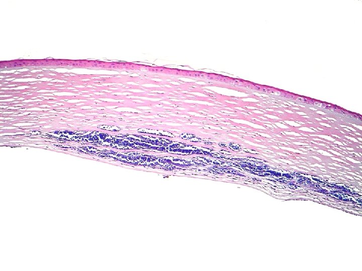
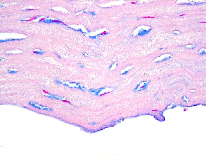
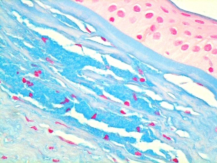
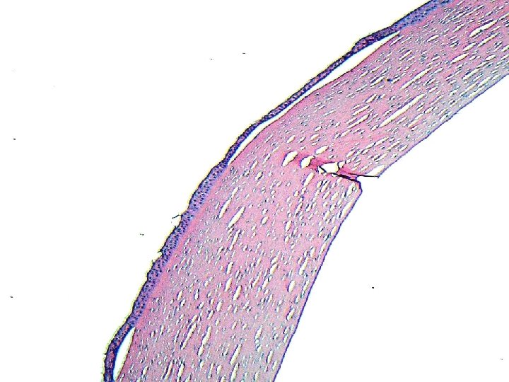
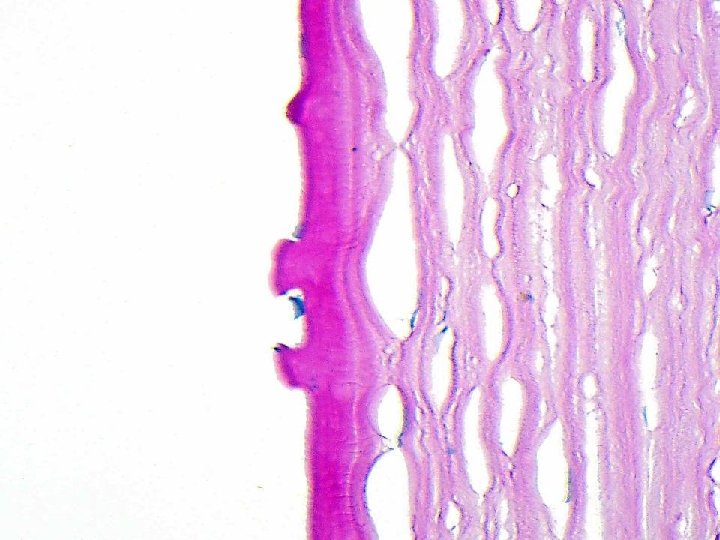
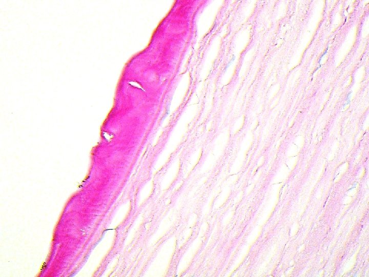
NORMAL CORNEA HISTOLOGY 1 2 3 1 4


")


- Slides: 123
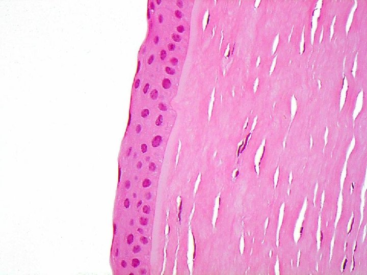
NORMAL CORNEA HISTOLOGY
1 2 3 1 4 5
epithelium 5 cells Stratified Non-keratinising Bowmans-12 microns thick Non-secretory
STROMA keratocytes ARTEFACTUAL CLEFTS
Endothelial cells Descemet’s
CORNEAL REACTION PATTERNS
Atrophy and Oedema
Epithelial hyperplasia
Bullous lifting
Excessive intraepithelial basement membrane
Band keratopathy Dystrophic calcification Epithelial hyperplasia
Breaks in Bowman’s
Stromal thinning
Neutrophils in stroma. Acute keratitis
Chronic inflammationchronic keratitis Blood vessels Plasma cells
Foamy macrophages lipid keratopathy
Infective agent-bacteria
protozoa
Dystrophic deposits
Dystrophic deposits
Dystrophic deposits
GUTTAE
Endothelial Cell Loss
Ruptured Descemet’s
Host-donor interface scar
Corneal pathology
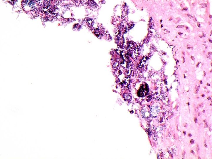
Case 1
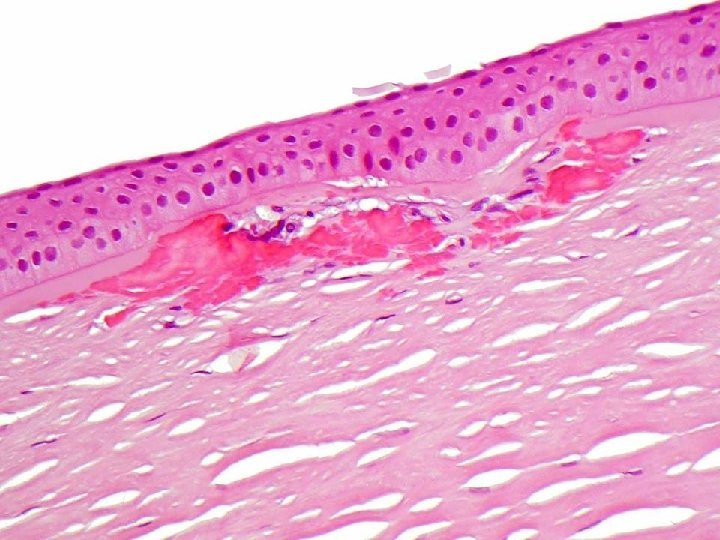
Acute inflammation
Bacterial colonies. Gram + cocci
Diagnosis ?
Bacterial acute keratitis Predisposing factors: adnexal infection, entropion, exposure, dry eyes, contact lens, bullous keratopathy, trauma etc G + cocci-s aureus, S. epidermidis, S pneumoniae, S pyogenes, S viridans G – cocci-N gonorrhoeae, M meningitidis G + bacilli-C Diphtheriae, diphtheroids G- bacilli- Moraxella, Acinebacter, E-coli, K pneumoniae, proteus, psuedomanas G+ filamentous bacteria
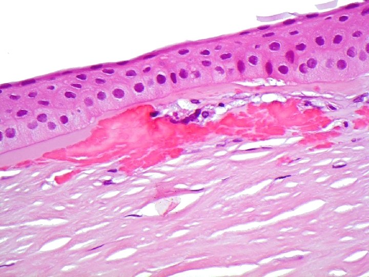
Case 2
History Topical steroids after PK. Drop in vision……………….
Gram + cocci without inflammation
INFECTIOUS CRYSTALLINE KERATOPATHY Elaboration of biofilm by bacteria-protects them from immune system-therefore no inflammation, but also means poor response to antibiotics. Commonest bugs-strep viridans and staph epidermidis Aso can be caused by fungi and protozoa.
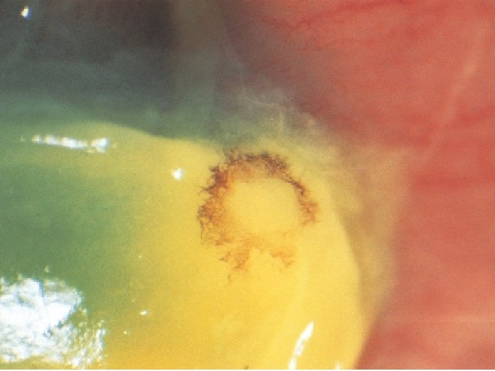
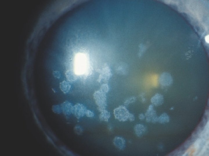
CASE 3
Fungal hypha -filamentous branching septate spores Fungal keratitis Penetrate Descemet’s without any problem
Common causes of fungal keratitis Trauma with organic material Humid warm conditions Exogenous or endogenous(immunocompromised) Aspergillus Candida Fusarium Sabaraud’s or equivalent medium for culturing Immunocompromised, steroids
CASE 4
cyst Trophozoite
DIAGNOSIS ?
AMOEBIC KERATITIS Amoebic keratitis-cysts and trophozoites-little inflammation Loss of keratocytes PERIODIC ACID SCHIFF (PAS) + GIEMSA + Can use immunohistochemistry Differential: Acanthamoeba, Hartmannella Vahlkampfia, Naegleria
Amoeba 10 -50 microns Replicate by binary fission Exist as trophozoites and cysts Trophozoites are active, infectious and feed by phagocytosing. Cysts from under hostile conditions and have a double layer.
Corneal epithelial trauma predisposes to infection Trophizoites attach to damaged epithelium, multiply and cause cytolysis. Migrate to stroma-elicit inflammation. Trigger keratoneuritis (inflammation follows corneal nerves).
Diagnosis Culture-corneal scrapes, biopsies, keratoplasty specimens. Contact lens, cases and solutions. Non-nutrient agar inoculated and seeded with Ecoli-food source for the amoeba. Wet-mount examination of contact lens solution. Can use PAS, calcofluor white, silver stains, immunohistochemistry, EM
CASE 5
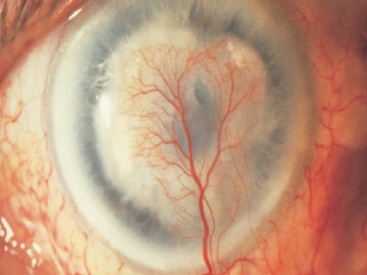
Stromal thinning
chronic Inflammation With giant cells. Bowman’s loss due to ulceration
Chronic inflammation Scarring vascularisation
Secondary lipid keratopathy Cholesterol clefts=leaky vessels
DIAGNOSIS ?
Herpes simplex chronic DISCIFORM keratitis
HSV DNA VIRUS Type 1 usually, occasionally type 2 Diagnosis-Electron microscopy of affected cells, aspirate from blister, viral cultures, staining paraffin sections with monoclonal antibodies to HSV, PCR on corneal biopsy.
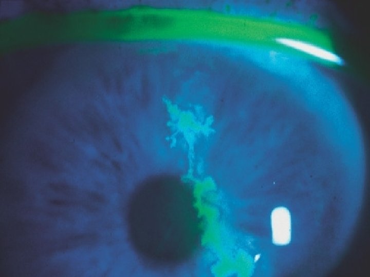
HSV Primary infection-self-limiting periocular vesicles and crusting, follicular and papillary blepharconjunctivitis, punctate epithelial keratopathy. Virus lives in trigeminal ganglionreactivation Dendritic ulcer
HSV Geographic ulcer Trophic keratitis Stromal infiltrative keratitis Disciform keratitis-type 4 hypersensitivity reaction-immune response to parasitized corneal stromal keratocytes-sets up vicious circle of inflammation-scarringinflammation.
Complications Uveitis Glaucoma Episcleritis Secondary bacteria infection Perforation Recurrence in corneal graft.
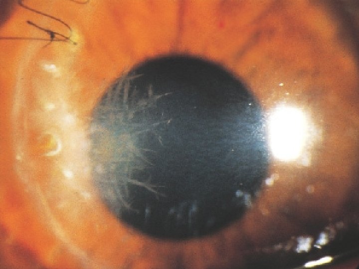
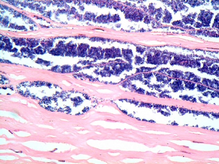
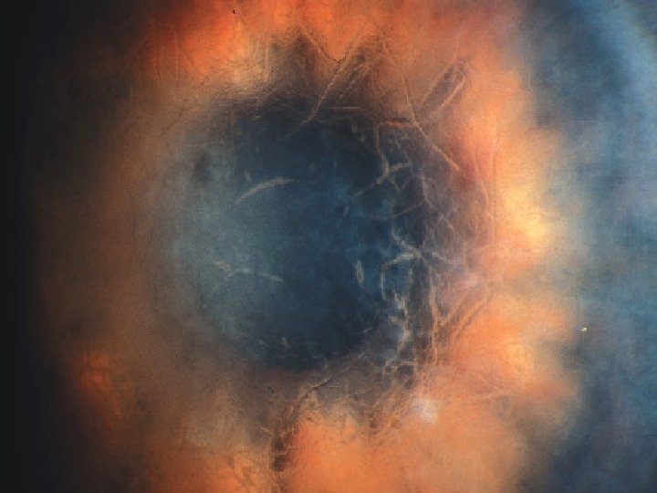
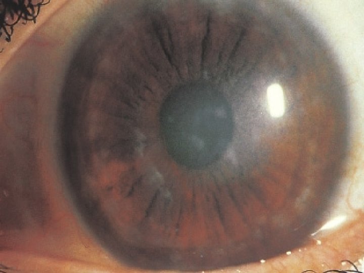
CASE 6
Angulated Bowman’s breaks
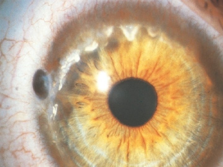
Perl’s stain shows intraepithelial iron deposits-Fleischer’s ring
DIAGNOSIS ?
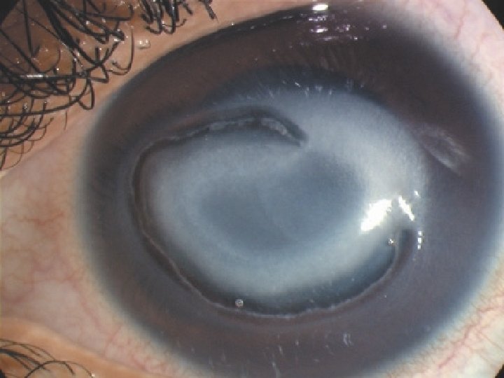
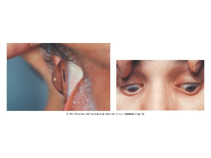
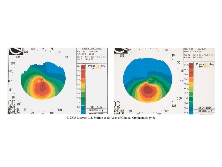
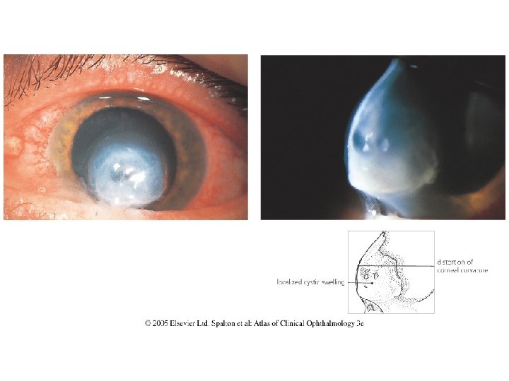
KERATOCONUS Associations: Atopy Down’s syndrome Turner’s syndrome Marfan’s syndrome Ehlors-Danlos syndrome Aniridia Retinitis pigmentosa Ectopia Lentis Microcornea Non-specific systemic collagen abnormalities Chronic eye rubbing. Cause of prominent corneal nerves.
Ruptured Descemet’s-KC Hydrops-PAS stain
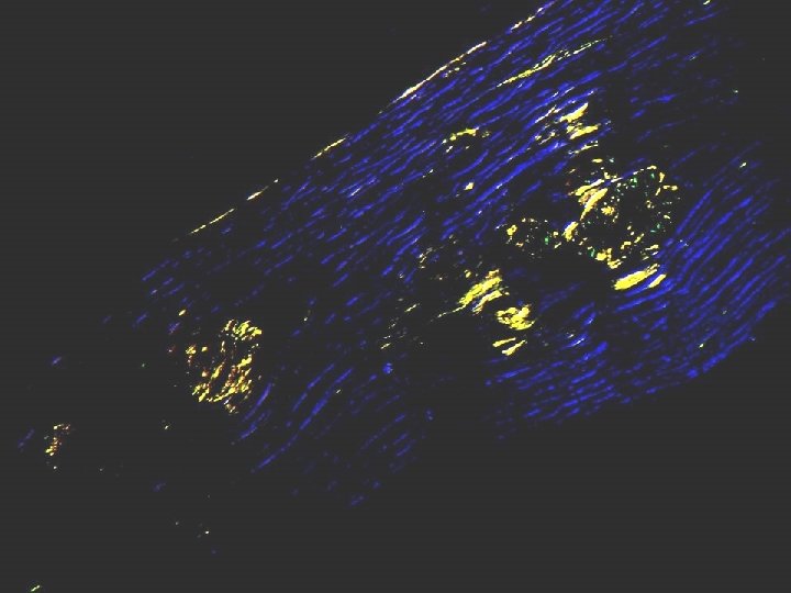
CASE 7
Masson’s trichrome stain-deep pink, non-birefringent hyaline bodies in anterior stroma
DIAGNOSIS ?
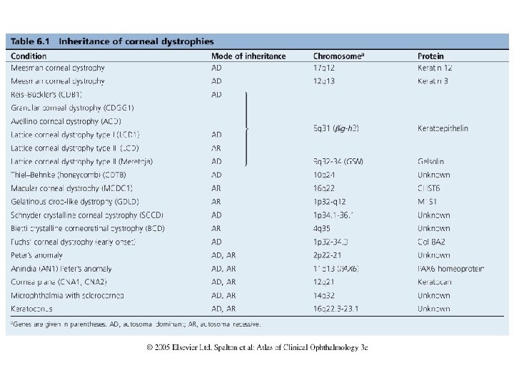
GRANULAR DYSTROPHY Masson’s trichrome positive hyaline deposits. Mutations in BIG H 3 /TGF-B 1 gene-encodes keratoepithelin protein. Exclude Avellino dystrophy (combined Lattice and Granular dystrophy)-by doing a Congo Red. Can recur in corneal graft-due to migration of host keratocytes into donor stroma, with elaboration of abnormal keratoepithelin
CASE 8
3
DIAGNOSIS ?
LATTICE DYSTROPHY Multiple, discrete, spindle shaped amyloid deposits in superficial, mid and deep stroma. Apple green birefringence of Congo red positive amyloid deposits when cross polarised Type 1, 2 and 3 Mutations in BIG H 3 / TGF-B 1 gene Exclude Avellino by doing Masson’s trichrome stain Other amyloid stains: Thioflavine T, Immunohistochemisty using antibodies to amyloid, Sirius Red. Recurs in graft because of migration of host keratocytes into donor stromaelaboration of amyloid in donor graft.
CASE 9
DIAGNOSIS?
MACULAR DYSTROPHY Alcian blue positive, deposits. Present in all layers except epithelium. Deposits in keratocytes and between collagen lamellae Material is mucopolysaccharide. Can recur in graft
Summary of corneal stains Lattice dystrophy-amyloid-use Congo Red / Sirius red and view under cross polarised light-apple green birefringence Granular dystrophy-hyaline material-use Masson’s trichrome Avellino dystrophy-use both Congo Red and Masson’s trichrome Macular dystrophy-mucopolysaccharide-use Alcian Blue or Hale’s colloidal iron stains or PAS Iron-use Perl’s / Prussian Blue stain- BLUE colour Calcium in band keratopathy- Alizarin Red- Red colour Basemant membranes, Descemet’s, Fungi- PAS stain- great for guttata. Bugs-Gram (bacteria), PAS (Fungi and Amoeba), Grocott silver stains for fungi.
CASE 10
Epithelial bullous lifting. Thinned epithelium over bulla Epithelium loses polarity
Excessive intraepithelial basement membrane-indication of chronic corneal oedema
Endothelial cell loss Thickened Descemet’s -implies chronic endothelial cell loss No obvious guttata
Patient had a cataract operation 1 year ago
DIAGNOSIS ?
Pseudoaphakic Bullous Keratopathy
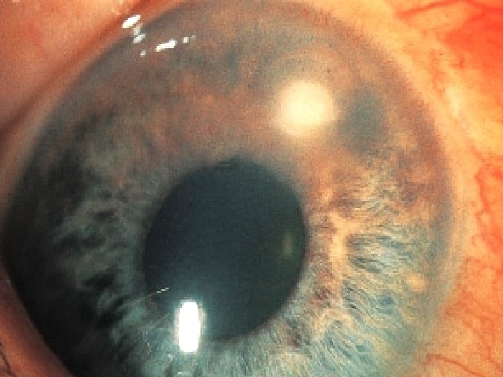
CASE 11
HISTORY 65 YEAR OLD MALE Recent cataract operation Early corneal decompensation. No better PK
Fuch’s Endothelial Dystrophy Axial diffuse guttae or excrescences Endothelial cell loss Thickened-multilayered Descemet’s Burried guttata Can get non-guttate forms, with just very thickened Descemet’s. With chronicity, fibrous degenerative pannus formation under epithelium.
CASE 12
Multilayered cellsretrocorneal surface No previous surgery or trauma
Cytokeratin positive Multilayered cells
DIAGNOSIS ?
POSTERIOR POLYMORPHOUS DYSTROPHY Autosomal dominant, but can be recessive Circumscribed or total opacities in childhood Cells assume epithelial characteristics (stain for cytokeratin 7) Histological differential diagnosis-epithelial downgrowth, ICE syndrome, CHED (these conditions express cytokeratins)
CASE 13
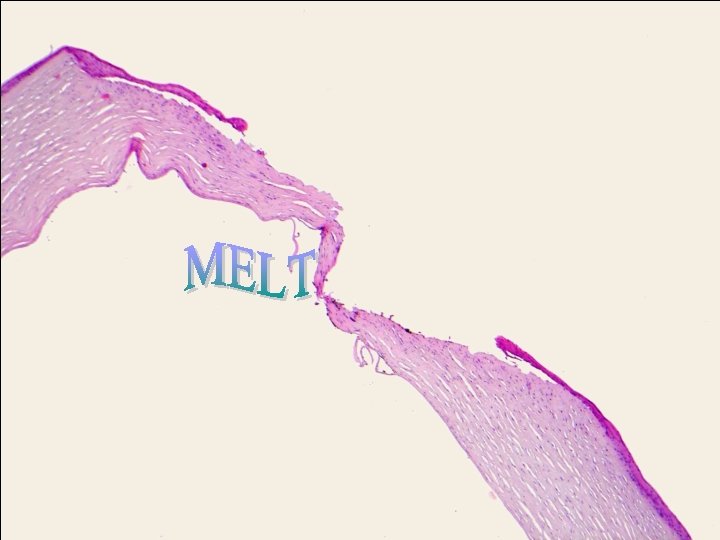
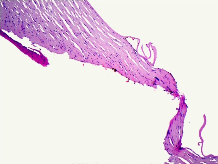
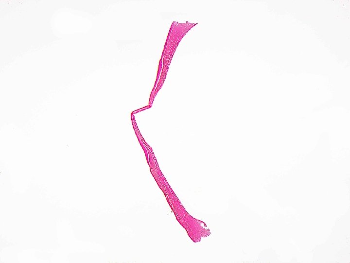
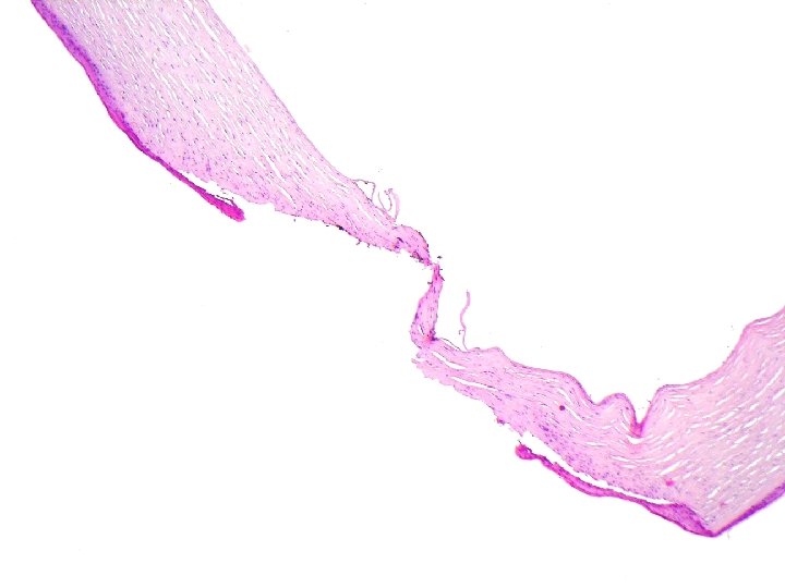
Ruptured and recoiled Descemet’s Shelved / sloping stroma Pauciinflammatory perforation
DIAGNOSIS ?
Rheumatoid Corneal melt Rheumatoid arthritis Systemic lupus erythematosis Scleroderma Churg-Strauss. Wegener’s granulomatosis Polyarteritis nodosa Giant cell arteritis Relapsing polychondritis Rosacea Dysentery Leukaemias Above are associated with peripheral corneal ulcers and melt Other causes of peripheral corneal ulceration: Marginal, Mooren’s Terrien’s. Imbalance between matrix metalloproteinases and tissue inhibitors of metalloproteinases Enzymes released by keratocytes and epithelial cells to cause dissolution of stromal collagen.

 Normal cornea histology
 Normal pancreas histology
 Folia cerebellum
 Hiperplasia cornea de la pezuña
 Corneal transparency is maintained by
 Refractive apparatus of the eye
 Cornea concava
 Distrofia di fuchs
 Retinae commotio
 Cornea light reflex test
 Eye scar
 Cornea homály
 Cornea
 Keratoconus oil droplet sign
 Fornices of eye
 Senile furrow degeneration cornea
 Thick clear jelly that helps give the eyeball its shape
 Bcop test
 Capa cornea de la piel
 Trabeculodysgenesis meaning
 Semmelweis szemészeti klinika
 Bowman's capsule
 Corneal preservation
 Cornea guttata
 Sheep eye dissection
 Latin accent rules
 Dr maria reinoso
 Keratoglobus vs keratoconus
 Print pushing eye exercise
 5 layers of cornea
 Carcinoma de celulas escamosas
 Lidia rutkowska sak
 It's normal to be normal
 Orcein festés
 Meissner’s corpuscles
 Practice histology lab practical
 Hyperemia
 Practice histology lab practical
 Holocrine gland
 Intestine
 Tunica intima histology
 Pogil epithelial tissue histology
 Pancreatic tissue
 Epithelial tissue
 Hepatocytes function
 Nyu histology
 Serosa vs adventitia
 Mechanical parts of microscope
 Ductus intercalaris
 Histology
 Anatomy histology slides
 Falciform ligament
 Histology
 Endometrial histology menstrual cycle
 Histology of small intestine
 Hair sheath
 Hypothalamus
 Histology job
 Continuous capillaries
 Histology of large intestine
 Spleen histology
 Cells
Cells Esophageal stomach junction histology
 Scdc b12
 Mount and hume classification
 Digital histology vcu
 Respiratory epithelium
 Mitosis histology
 Histology of liver
 Glioblastoma multiforme
 Introduction to histology
 Perisinusoidal
 Hyperplasia
 Histology hair
 Anatomy of the respiratory system
 Dr gallatz katalin
 Colloid thyroid histology
 White lesions
 Nidus parotideus
 Lactose intolerance histology
 Agranulocytes
 Corpus ciliare histology
 Tunica intima
 Vibs 243
 Minute colorless anucleate corpuscles found in the blood
 Epithelial tissue
 Respiratory system histology
 Follicular epithelium
 Nephron histology
 Loop of henle histology
 Histology
 Oligodendrocyte histology
 Cells in stratum spinosum
 Nodus lymphaticus histology
 Intestine
 Anetoderma histology
 Epithelium
 Mucus salivary gland
 Digestive histology
 Vulva blood supply
 Larynx histology
 Fundus of stomach histology
 Histology
 Feather histology
 Bronchiloes
 Renal papilla
 Http://www.biologycorner.com/anatomy/histology/
 Macrofage
 Histology of compact bone
 Outer plexiform layer
 Histology
 Dictalie
 Caseating vs noncaseating granulomas
 Tendon histology
 Histology of digestive system
 Osteon histology
 Characteristics of squamous epithelium
 Juxtacrin
 What is this
 Histology of the digestive tract
 This is a connective tissue with a "cobwebby" appearance.
 Interalveolar septa
 Bone matrix anatomy
 Lamellae


