
Liver Embryology Anna Robson Ruben Vilela Primitive gut
Liver Embryology Anna Robson & Ruben Vilela
Primitive gut Made from the endoderm and visceral mesoderm Foregut: Oropharyngeal membrane liver bud. Coeliac A. Oesophagus, stomach, liver, biliary apparatus, pancreas & ½ duodenum Midgut: Liver bud 2/3 rds transverse colon. SMA. Distal ½ of duodenum jejunum, ilium, caecum, verifrom appendix, ascending colon & right 2/3 rds of TC Hindgut: 2/3 rds TC cloacal membrane. IMA. Left 1/3 rd TC , descending colon, sigmoid colon, rectum and anal canal
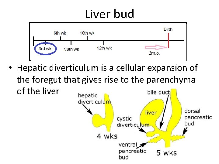
Liver bud • Hepatic diverticulum is a cellular expansion of the foregut that gives rise to the parenchyma of the liver
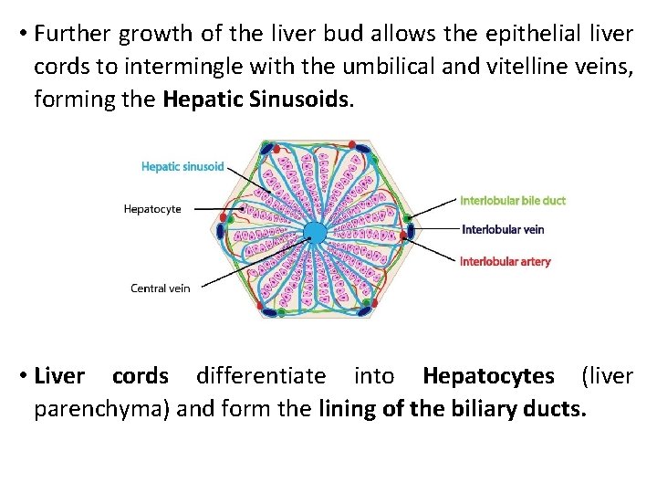
• Further growth of the liver bud allows the epithelial liver cords to intermingle with the umbilical and vitelline veins, forming the Hepatic Sinusoids. • Liver cords differentiate into Hepatocytes (liver parenchyma) and form the lining of the biliary ducts.
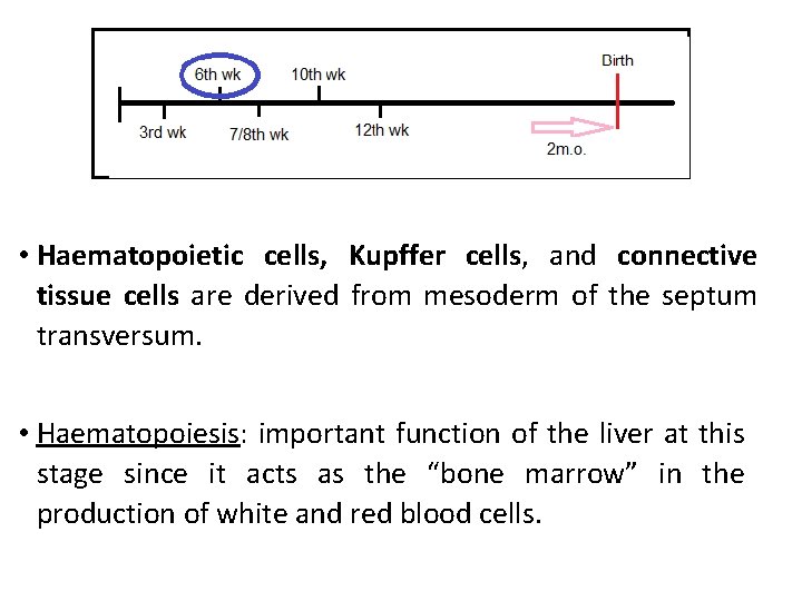
• Haematopoietic cells, Kupffer cells, and connective tissue cells are derived from mesoderm of the septum transversum. • Haematopoiesis: important function of the liver at this stage since it acts as the “bone marrow” in the production of white and red blood cells.
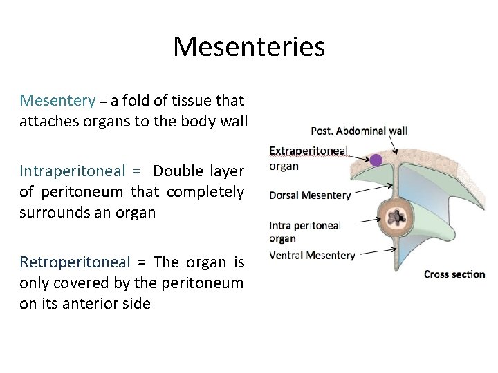
Mesenteries Mesentery = a fold of tissue that attaches organs to the body wall Intraperitoneal = Double layer of peritoneum that completely surrounds an organ Retroperitoneal = The organ is only covered by the peritoneum on its anterior side
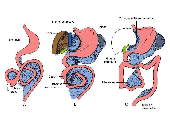
Ventral mesentery Occurs only in the foregut Derived from the septum transversum Foregut has dorsal + ventral mesenteries Midgut and hindgut only have a dorsal mesentery
Ventral mesentery The liver divides the ventral mesentery. Falciform ligament to the anterior abdominal wall Free edge contains the umbilical vein the round ligament (ligamentum teres) after birth Lesser omentum to the ventral borders of the stomach and the duodenum Free edge contains hepatic artery, portal vein + bile duct

Bare area of liver Bare area = the only area of the liver that is not covered by the ventral mesentery

Falciform ligament
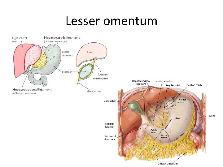
Lesser omentum
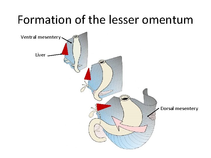
Formation of the lesser omentum Ventral mesentery Liver Dorsal mesentery
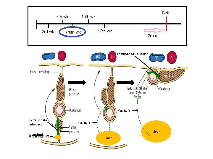
Entrance of Co. Bile duct Co. B. D Extrahepatic bile duct Liver bud Co. B. D Liver
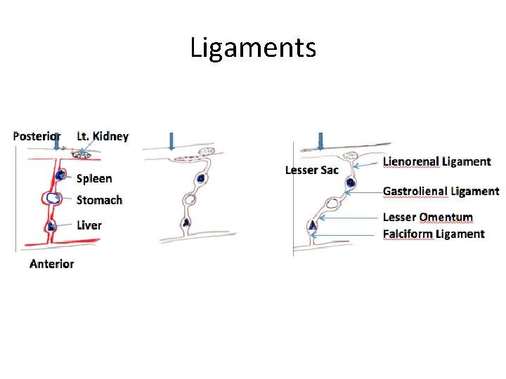
Ligaments
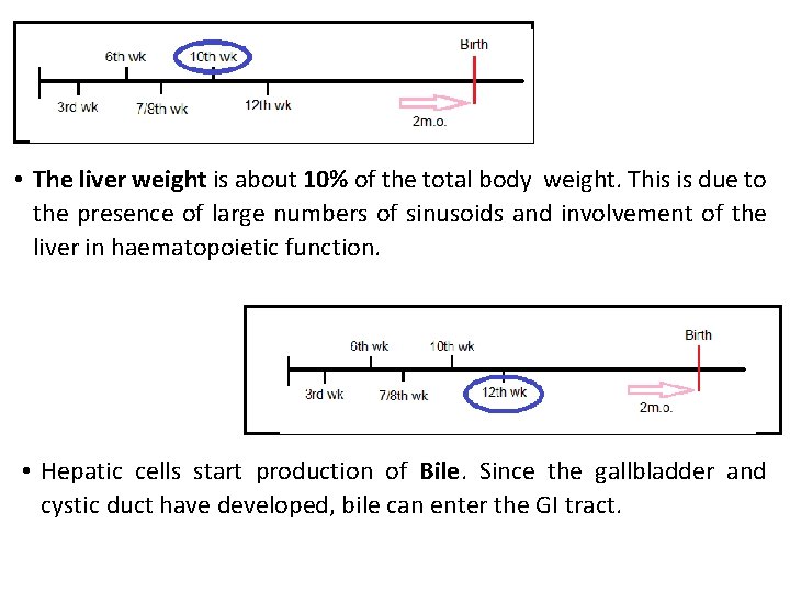
• The liver weight is about 10% of the total body weight. This is due to the presence of large numbers of sinusoids and involvement of the liver in haematopoietic function. • Hepatic cells start production of Bile. Since the gallbladder and cystic duct have developed, bile can enter the GI tract.
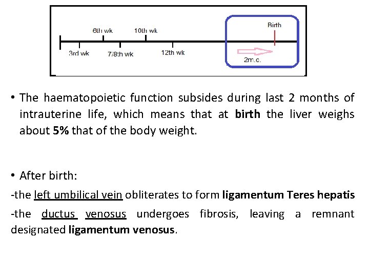
• The haematopoietic function subsides during last 2 months of intrauterine life, which means that at birth the liver weighs about 5% that of the body weight. • After birth: -the left umbilical vein obliterates to form ligamentum Teres hepatis -the ductus venosus undergoes fibrosis, leaving a remnant designated ligamentum venosus.
Key points • 3 rd week: endodermal growth from Foregut • Narrowing of the connection = Bile Duct • 5 th week: Ventral outgrowth will form the Cystic Duct and Gallbladder • Epithelial cords + umbilical & vitelline veins = Hepatic Sinusoids • 6 th week: active haematopoietic function • Ventral mesentery divided by liver into Lesser Omentum and Falciform Ligament • 7/8 th week: Rotation of the foregut shifts entrance of bile duct to a posterior position • 12 th week: production of bile and its entrance to the GI tract • Left umbilical vein Ligamentum teres hepatis • Ductus venosus Ligamentum Venosum

Questions? ?
CONGRATULATIONS GUYS… … you made it through!!! Check out “Awkward Yeti” XD
- Slides: 20