Histology of Respiratory System AlMustansiriya University Collage of


- Slides: 18

Histology of Respiratory System Al-Mustansiriya University Collage of Pharmacy Lecturer Dr Samah A. Jassam BSc MSc Ph. D MRSB

Role of RESPIRATORY SYSTEM The complex of organs and tissue which are necessary to exchange blood carbon dioxide (CO 2) with air oxygen (O 2). Function: Act of breathing, which includes inhaling and exhaling air in the body; the absorption of oxygen from the air in order to produce energy; the discharge of carbon dioxide, which is the by product of the process.

The parts of the respiratory system The respiratory system is divided into two parts: Upper respiratory tract: This includes the: nose, mouth, and the beginning of the trachea.

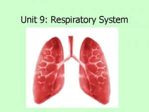
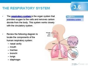
Lower respiratory tract It includes the trachea, the bronchi, broncheoli and the lungs. The trachea : It connecting the throat to the bronchi. The bronchi : It divides into two bronchi (tubes). The broncheoli : the bronchi branches off into smaller tubes called broncheoli which end in the pulmonary alveolus. The Lungs: The structure of the lungs includes the bronchial tree – air tubes branching off from the bronchi into smaller and smaller air tubes, each one ending in a pulmonary alveolus.

The Nasal Cavities The nasal cavities are lined by a ciliated pseudostratified columnar epithelium containing the cell bodies of bipolar nerve (olfactory) cells. of these olfactory cells contain proteins that act as odorant receptors.

The mucosa of the nasal cavities has: • olfactory nerves • olfactory glands that secrete onto the epithelial surface a proteinaceous substance, that keeps the surface moist and provides a trap for aromatic substances.

The pharynx contains mucous glands and it is lined by stratified squamous epithelium, that is continuous with this type of epithelium at the proximal end of the larynx.

The respiratory epithelium is a tissue that lines the respiratory system. Role of the respiratory epithelium are: 1 - It serves as a protective barrier and, 2 - It also provides moisture.

There are eight (8) different cell types within the respiratory system epithelium 1. Ciliated Cells: These are the most abundant airway epithelial cells. They are found from the trachea to respiratory bronchioles and contain approximately 200 -300 cilia per cell. 2. Goblet (Mucous) Cells: This cell type, which is present in the trachea and bronchi, has a wide, extended apical region, these cells contribute to the mucous secretion lining the airways 3. Basal (Short) Cells These cells, which are found in the trachea and bronchi, do not reach the airway lumen and have nuclei that are close to the basal lamina, thereby giving the epithelium a pseudostratified appearance.

There are eight (8) different cell types within the respiratory system epithelium 4. Clara Cells (Bronchiolar Epithelial Cell) : These non-ciliated columnar epithelial cells are found in bronchioles. These cells play a major role in the metabolism of exogenous agents (e. g. , atmospheric pollutants), and act as progenitor cells for bronchiolar epithelium following lung injury. 5. Brush Cells: This non-ciliated columnar cell, found in trachea and bronchi, is distinguished by a dense population of long, straight, blunt microvilli on the luminal surface and epitheliodendritic (afferent) synapses near the cell base. The function of these cells is unknown, although a chemoreceptor and sensory function is suspected.

There are eight (8) different cell types within the respiratory system epithelium 6. Dense core granule cells (small granule cell, neuroendocrine cell)These cells are found throughout the airways, either as isolated cells or clusters called neuroepithelial bodies (often found near airway junctions). Their role in regulating lung function is incompletely understood. 7. Serous Cells: These non-ciliated secretory cells are found predominantly in the trachea and bronchi. They secrete glycoproteins and lysozymes and probably contribute to the low viscosity periciliary fluid covering the bronchial epithelium.

There are eight (8) different cell types within the respiratory system epithelium 8. Intermediate: These non-ciliated columnar cells are immature and replace cells cast off from the epithelium. They may differentiate into mucous secreting Goblet cells or ciliated cells. These cells may be difficult to distinguish from brush cells.

Epithelial transitions The respiratory system provides beautiful examples of epithelial transitions. • The pseudostratified ciliated columnar epithelium of the trachea and bronchi gives way to a simple columnar ciliated epithelium in the bronchioles and then to the simple squamous epithelium of the alveolar ducts and alveoli. • The ciliated cells undergo a gradual reduction in height from trachea to terminal and respiratory bronchiole.

Change of airway wall structure at three principal levels in the lung. The epithelium (EP) gradually reduces from pseudostratified to cuboidal and then to squamous, but retains its organization as a mosaic of lining and secretory cells. The smooth-muscle layer (SM) disappears in the alveoli.

Histology of the Alveolar Region Five major cell types are present in the alveolar region of the lung. 1. Alveolar Type I Cell (Squamous alveolar epithelial cell) These elongated thin cells line the alveoli and cover a large surface area (approximately 95% of the alveolar surface) due to extreme flattening and marked cytoplasmic attenuation. These cells form an extended, continuous surface of minimal thickness that is permeable to gases and is the major location of gas exchange. 2. Alveolar Type II Cell (Great alveolar cell, granular pneumocyte): These cells form tight junctions with Type 1 cells, and are often positioned in alveolar corners (Figure 4) and at alveolar septal junctions. These secretory cells protrude into the alveolar lumen

Histology of the Alveolar Region 3. Capillary Endothelial cell : The pulmonary capillary bed is the largest vascular bed in the body--covering a surface area of 70 m 2. It receives the entire cardiac output. Endothelial cells are specialized for both gas 4. Alveolar macrophages These large cells wander freely in the alveoli. Located in the aqueous hypophase of the surfactant layer, they move over the alveolar surface ingesting microorganisms and inhaled particulate matter.

Histology of the Alveolar Region 5. Interstitial cells a progenitor cell is noted during lung development that is capable of differentiation into fibroblasts or smooth muscle cells. Interstitial cells of the alveolar region are primarily fibroblasts with ramified cytoplasmic extensions.

Exercise Q 1: Name the 8 cell types in the airway epithelium and briefly state their function. Q 2: Cell types are present in the alveolar region of the lung.
 Bronchus histology
 Respiratory system histology
 Respiratory system histology
 Conducting zone of the respiratory system
 Digestive system circulatory system and respiratory system
 How respiratory system work with circulatory system
 Circulatory system and respiratory system work together
 Digestive system histology
 Histology of the digestive tract
 Ppt
Ppt Digestive system histology
 Digestive system histology
 Cells in stratum spinosum
 Renal corpuscle
 Urinary bladder histology
 Respiratory system bozeman
 Unit 9 respiratory system
 Diagnostic test of respiratory system
 Respiratory system
