Histology of the digestive systemreview Anna L Kiss

 Mucosa a. ) l. epithelialis")


 intracellular canaliculi+microvilli")


: submucosa")
")

 villi Lieberkühn cripts (glands!!)")
 epithelium propria Mucosa muscularis mucosae Submucosa Muscularis Serosa circular")


 cells) modifyed enterocytes: antigén presenting cells")
 muscularis mucosae")

 cripts submucosa muscularis ext. serosa")
")


 • a. hepatica propria branches connective")
: wall of the bile canaliculi:")


")


: Histology")
- Slides: 39
Histology of the digestive system-review Anna L. Kiss Department of Human Morphology and Developmental Biology Semmelweis University, Budapest 2019
General histology of the digestive tract layers 1. ) Mucosa a. ) l. epithelialis b. ) propria (limphoreticular conn. tissue) c. ) muscularis mucosae (smooth muscle) 2. ) Submucosa: loose connective tissue submucosus plexus (Meissneri): innervation of the a mucosa (villi) lymphatic tissue (lymphocytes, follicles: solitaer) 3. ) T. muscularis: smoothe muscle inner: circular outer: longitudinal myentheric plexus (Auerbach): peristaltic movement 4. ) T. serosa vs. adventitia
Esophagus
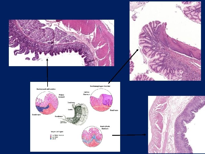
Histology of the stomach
Histology of the stomach t. serosa t. muscularis t. submucosa t. mucosa
Histology of the stomach chief cells parietal cells
Histology of the stomach mitochondria parietal cells (EM) intracellular canaliculi+microvilli
Histology of the stomach chief cells: pepszinogen
Histology of the stomach Enteroendocrine cells dense secretory granules in the basal cytoplasm
Histology of the digestive tract Structures increasing the surface: plicae circulares (Kerkring foldes): submucosa villi intestinales: mucosa microvilli (brush border): finger-like projections of the apical plasma membrane of the enterocytes
Plicae circulares (Kerkring redők)
Kerkring foldes The GI tractus wall: 4 concentric layerts: mucosa submucosa muscularis externa (the external muscle layer) adventitia or serosa Z S M P
Villi (Scanning Electron Microscopy) villi Lieberkühn cripts (glands!!)
villi Lieberkhün cripts (intestinal glands) epithelium propria Mucosa muscularis mucosae Submucosa Muscularis Serosa circular longitudinal
microvilli
Lieberkühn cripts Paneth cells: antibacterial enzymes: lysozime
Enteroendocrine cells dividing cells
M (microfold) cells) modifyed enterocytes: antigén presenting cells
Duodenum villusok Brunner glands: alcalic secretum: p. Hshift propria Lieberkühn cripts (glands) muscularis mucosae Brunner glands (submucosa) submucosa
Jejunum muscularis mucosae villi epithelium of the mucosa Kerkring folds mucosa submucosa tunica muscularis + serosa
Ileum villi Peyer plaques (aggregated lymphatic follicles) cripts submucosa muscularis ext. serosa
pl. myentericus (Auerbach)
Mucosa of the colon
Colon: • No villi • deep Lieberkühn cripts • many goblet cells • No Paneth-cells • taeniae • adipose tissue in the submucose and subserose layer • only solitaer lymphatic follicles (folliculi lymphatici solitarii)
Histology of the colon epithelium mucosae tunica muscularis mucosae lamina propria mucosae (+ Lieberkühn krypták) Tela submucosa Tunica muscularis + Serosa Tenia coli
Histology of the colon epithelium mucosae tela submucosa tunica muscularis mucosae
Colon: simple columnar epithelium Junctio anorectalis Transition: stratified columnar or cuboidal epithelium Cutis: stratified keratinized squamouos epithelium Sphincter ani internus Folliculi lymphatici Sphincter ani externus
Portal triad • a v. portae branch (es) • a. hepatica propria branches connective tissue • bile duct • lymph vessels • nerve fibers v. portae branch bile duct a. hepatica branch
Bile canaliculi Neighbouringh surface of the hepatocytes (apical surface): wall of the bile canaliculi: plasma membrae of the hepatocytes!!!) Zonula occludens (tight junction) NO communication between sinusoids and bile canaliculi!!! Interconnecting network. At the periphery: short canaliculi: Hering-canal (epithelium!!!!) stem cells! Bile canaliculi hepatocytes Epekapilláris km (EM) Tight junction
Bile canaliculi
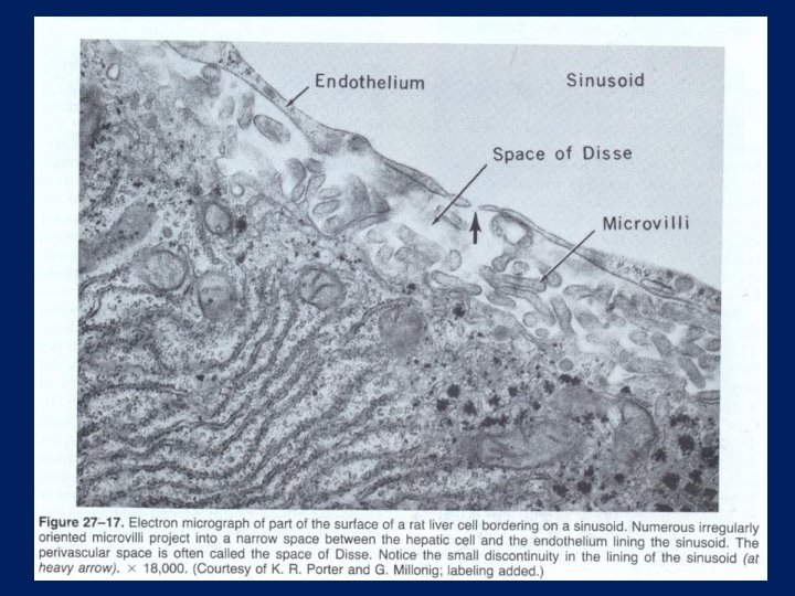
Sinusoidsk • wide capillaries, connect the branches of v. portae ágait with vv. Hepaticae through the central vein • wall: endothelium is not continouos, NO basement membrane (fast exchange between blood and hepatocytes!) • Kupffer-cells: stellate-shaped makrophages (monocyte/makrophage system). • Disse space: between hepatocytes and sinusoids’ endothel. (reticular fibers, collagen type III). • Ito-cells: in space of Disse. (Vitamin A storage, reticular fiber synthesis). Inflammation, cirrhózis: activated, differentiated into myofibroblasts
Kupffer cells
kollagen III: reticular fibbers in the liver (space of Disse)
A májsejt és környezetének sémás rajza cell r. ER glycogen space
Histology of the pancreas Langerhans islands
Histology of the pancreas Ductus intercalaris
Schematic drawings and microphotographs were taken from the following textbook: P. Röhlich (editor): Histology (in Hungarian), Semmelweis Budapest, 2006