Histology of Endocrine System Ch 20 Hypophysis p


 • Adenohypophysis • Rathke’s pouch • Oral ectoderm • Loses")


 • 75% of pituitary • Two major cells types upon staining")
 • Affects for")
;")


")


 virilizing and an anabolic effect")


- Slides: 71
Histology of Endocrine System Ch 20 Hypophysis p. 403 Ch 21 Other endocrine p. 413
Major Mechanisms for Signaling • Endocrine hormones - small molecules released into the circulation to effect target cells at distant sites from the original release point. • Paracrine hormones - small molecules released in a local area which has an effect only on cells within that local area of the body • Neurotransmission - synaptic transmission
Comparison of the three
Our Focus Today • Endocrine Organ Histology • General schema of Endocrine signaling • A General overview of cell biology of signal transduction
Endocrine System • The endocrine system is a major regulatory system • The endocrine system is a global system that works through release of chemical messages called hormones
Hormone Signaling • Hormone produced by cells in endocrine glands or organs • Hormone is released into the circulation • Hormone affects function of target cells – Target cells have receptors – Receptors begin a process called signal transduction
Endocrine Tissues • Work in association with nervous system • Work in association with the immune system • Groups of cells participating – Isolated cells – enteroendocrine cells – Mass of endocrine cells in exocrine organ – Islets of Langerhans in pancreas – Endocrine organs
Endocrine Glands
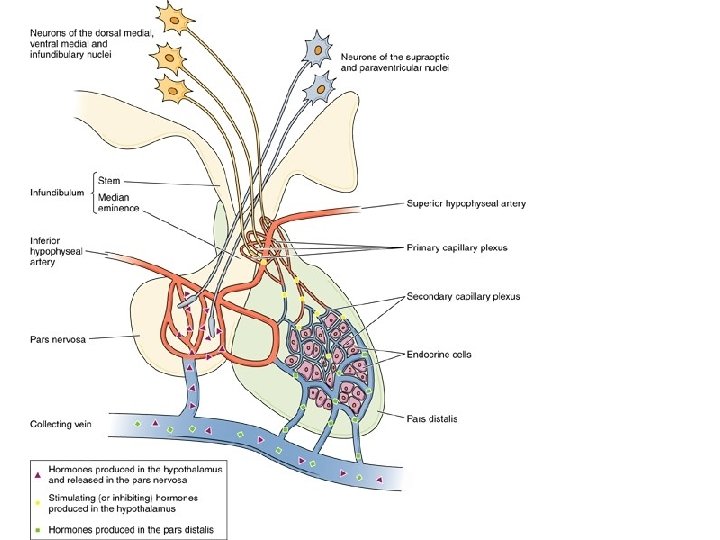
Origins of Pituitary (Hypophysis) • Adenohypophysis • Rathke’s pouch • Oral ectoderm • Loses attachment with oral cavity • Neurohypophysis • Neuroectoderm • Outgrowth from floor of diencephalon • Remains attached to brain (HT) via infundibulum
Pituitary Development
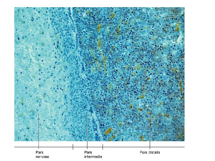
Hypophysis or Pituitary • Divisions – Adenohypophysis • Pars distalis • Pars intermedia • Pars tuberalis – Neurohypophysis • Pars nervosa • Infundibulum
Pituitary Gland Infundibulum Pars tuberalis Pars Nervosa Pars Distalis Pars Intermedia
Acidophils, Chromophobes & Basophils of Adenohypophysis
Secretory Cell
Adenohypophysis (pars distalis) • 75% of pituitary • Two major cells types upon staining – Chromophobes – resist stain (undiff. Cells) – Chromophils – important cells • Acidophils – eosinophilic • Basophils – basophilic
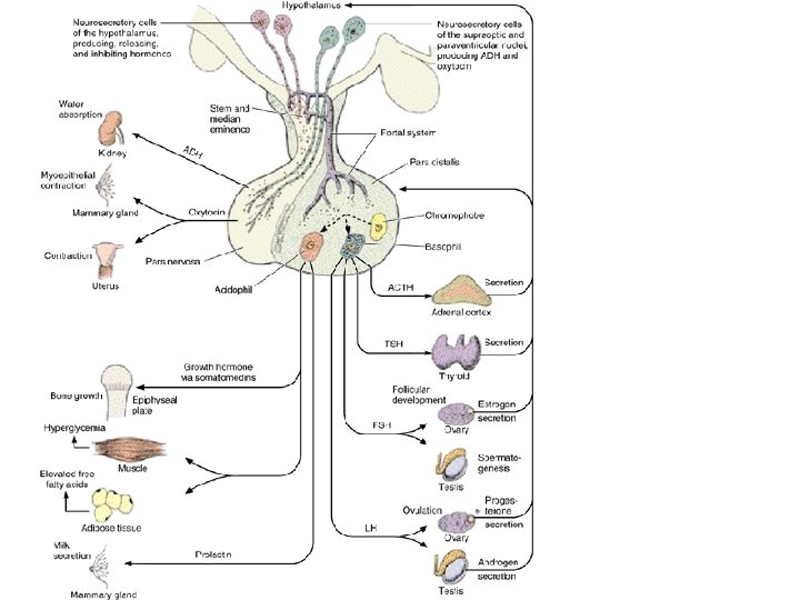
Acidophils • Important secretions produced – Somatotropes – somatotropin (h. GH) • Affects for example epiphyseal plates of long bones • Human growth hormone (h. GH) also coordinates growth in many other areas – Mammotropes – prolactin • Stimulates milk secretion from mammary glands
Basophils • General classes of secretion – Thyrotrophes – secrete thyroid stimulating hormone (TSH); causes thyroid to release T 3 and T 4 (thyroid hormones) setting basal metabolic rate – Gonadotropes – see next slide – Corticotropes – secrete Adrenocorticotropic hormone (ACTH) promotes growth of adrenal cortex and stimulates release glucocorticoids and gonadocorticoids
Gonadotropes • Gonadotropes of adenohypophysis – FSH – females stimulates development of ovarian follicles ; in males stimulates Sertoli cells to produce androgen binding protein – LH – in females promotes maturation of follicle and ovulation and maintains corpus luteum ; in males called interstitial cell secreting hormone (ICSH) promotes secretion of testosterone by Leydig cells
Regulation of Adenohypophysis • Secretions of adenohypophysis are all regulated by Hypothalamic Releasing Hormones – These are given names based on the hormones they are encouraging the release of • Example Hormones of the thyroid gland are released upon release from the adenohypophysis of TSH is not released until stimulation of the thyrotrophes by thyroid releasing hormone (TRH) See general scheme on next slide.
Note: The releasing hormones are actually produced by neurosecretory cells. These cells function as receivers of action potential and they release hormones instead of neurotransmitters. Note also that this is a negative feedback system. The T 3 and T 4 released tend to inhibit both the release of TRH and TSH.
An overview of Pituitary Function • See the scheme for TRH and TSH release can be generalized to other large groups of hormones release by other endocrine glands • Study the next slide for an overview of how this works • Please note the pivotal role of Hypothalamus – Hypophysis (Nervous – Endocrine connection)
Neurohypophysis • Hypothalamo-hypophyseal tract – – Neurosecretory cells axon processes – At the end of these axons are the • Herring bodies the swollen axon knobs • These distended tips are bigger than most axon knobs • Release either hormone or a releasing hormone
Herring Bodies (pars nervosa)
Neurosecretory Hormomes • Releasing Hormones • Two other hormones – Oxytocin – paraventricular nucleus • Stimulates smooth muscle of uterus and myoepithelial cells of mammary glands – Antidiuretic hormone (ADH or Vasopressin) • Supraoptic nucleus release • Acts on distal and collecting tubules of kidney making them more permeable to water; generates more hypertonic urine
Blood Supply to Pituitary • Inferior hypophyseal arteries to neurohypophysis • Hypothalamo-hypophyseal portal system – Delivers hypothalamic regulating hormones to adenohypophysis (releasing hormones)
Releasing Hormones • Produced in HT by neurosecretory cells – Examples on previous slide (#24 above) – All are release into circulation that goes to adenohypophysis – Releasing hormones trigger stimulating hormones for other endocrine organs
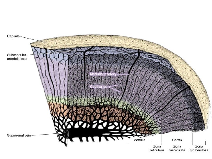
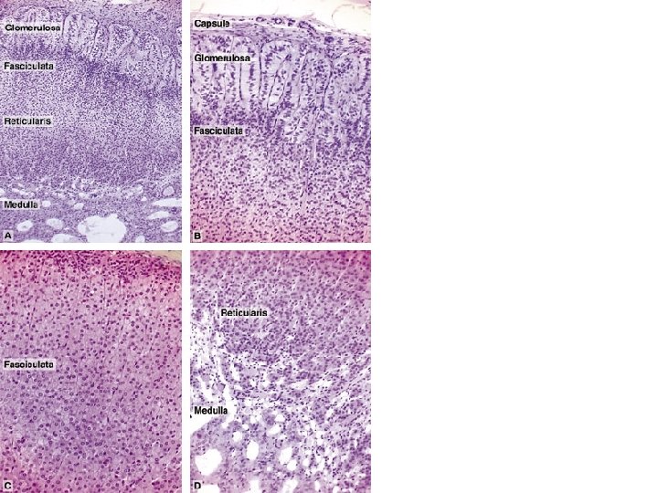
Adrenal glands • Paired glands superior to kidney – Suprarenal gland • Two glands in one – Cortex – Medulla
Adrenal Glands
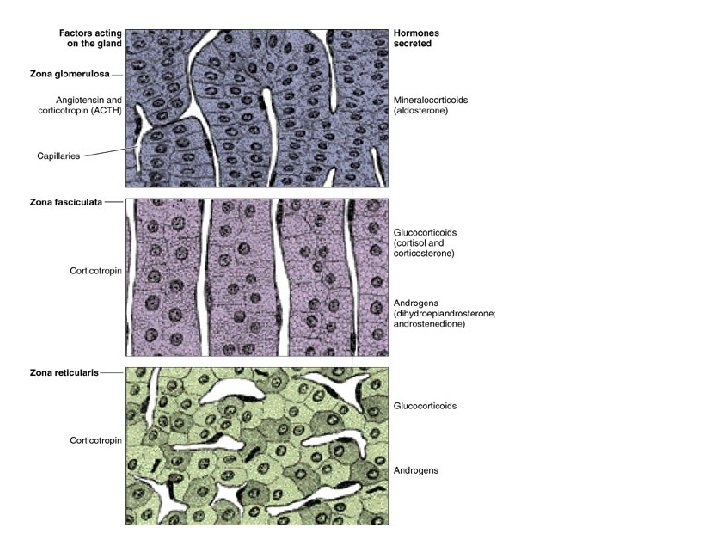
Adrenal Cortex • Derived form mesoderm • Three layers – Zona Glomerulosa (15% of cortex) – Zona Fasciculata (78% of cortex) – Zona Reticularis (7% of cortex)
Adrenal Cortex Hormones • Cortex utilizes – Cholesterol and acetate to synthesize steroid hormones
Zona Glomerulosa • Secretes mineralcorticoids – Aldosterone – Maintenance of electrolyte and water balance – Regulated by renin-angiotensin system – Unaffected by ACTH of pituitary
Zona Fasciculata • Secrete glucocorticoids – cortisol – Prepares body for maximal immediate energy demands; part of “fight or flight” stress response – Depresses immune function and inflammatory response – Regulated by ACTH of pituitary
Zona Reticularis • Secretes gonadocorticoids – – Dehydroepiandrosterone (DHEA) virilizing and an anabolic effect – Negligible amount compared to amount released from testis – Regulated by ACTH
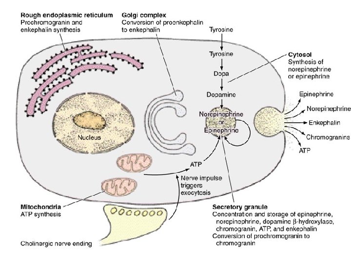
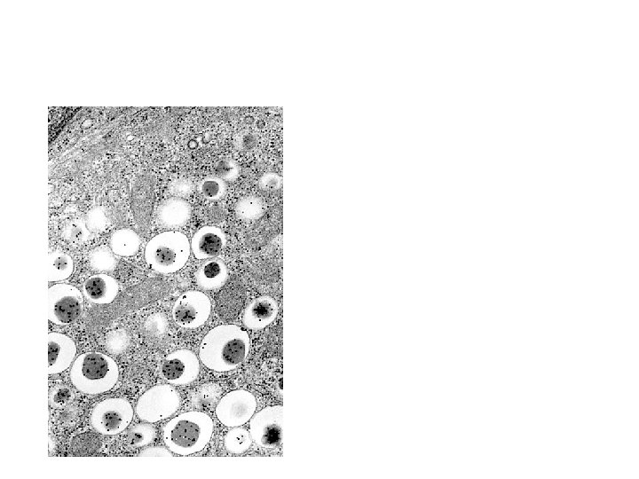
Adrenal Medulla • Derived form neural crest • Modified sympathetic ganglion – Important cell type • Chromaffin cells – modified post-ganglionic neurons • These cells secrete epinephrine (80%) and norepinephrine (20%) • These cells regulated by preganglionic sympathetic neurons
Adrenal Gland Atrophy & Hypertrophy
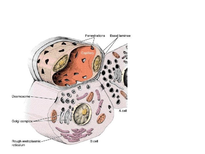
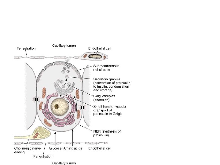
Pancreas • Mixed Exocrine and Endocrine Gland • Islets of Langerhans have endocrine – Islets 2% of entire gland – Alpha cells secrete glucagon; [blood glucose] – Beta cells secrete insulin; [blood glucose] – Delta cells secrete somatostatin; inhibits secretion of glucagon and insulin
Islets of Langerhans
a and b cells b-cells secret insulin a-cells secret glucagon
a-cells stained
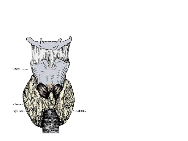
Thyroid Gland • Gross structure – Two lobes – Connected by isthmus – Connective Tissue Capsule
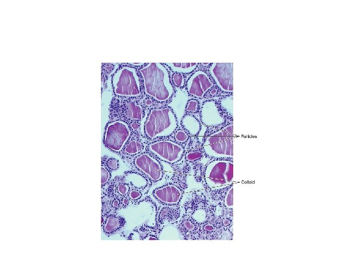
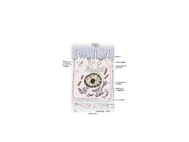
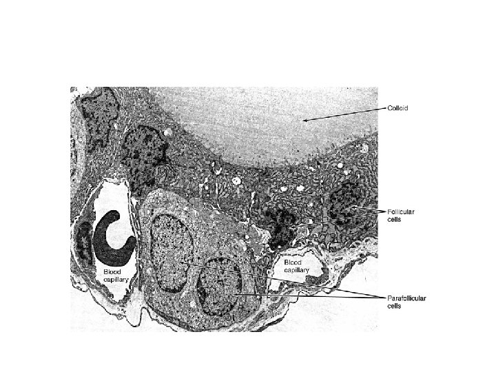
Follicles of Thyroid • Structural and functional units of gland – Follicular cells simple cuboidal epithelium – Colloid collects in follicles • Precursor of Thyroid hormones – Thyroxine (T 4) – Triiodothyronine (T 3) • Thyroglobulin secretory path built from tyrosine and iodinated to become T 3 and T 4 • Only endocrine gland to store its secretions; storage extracellular as colloid
Thyroid follicles
Thyroid Gland Regulation • Release of T 3 and T 4 stimulated – By TSH from pituitary
Parafollicular Cells • AKA Interfollicular cells – Secrete calcitonin = inhibits osteoclast activity and stimulates osteoblast activity adding Ca+2 to boney matrix – Regulated by blood Ca+2 levels – Works in opposition to parathyroid hormone
Thyroid junctions w Parathyroid
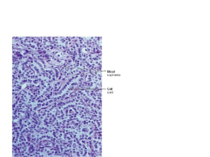
Parathyroid glands • Structure – Two pair – Embedded on posterior of thyroid gland – Cords of cells • Chief cells • Oxyphil cells – function unknown
Parathyroid Gland
Chief cells of parathyroid gland • Secrete parathyroid hormone – Low blood calcium stimulates secretion – Parathyroid hormone stimulates • Osteoclast increase in number and • Osteoclast to degrade bone and raise blood Ca+2 – Also decreases blood level of phosphate by decreasing resorption in kidney tubules, promoting excretion – Most important regulation blood Ca+2
Pineal Gland • AKA epiphysis cerebi • Pinealocytes secrete melatonin – Involved in diurnal rhythms – Innervated by neurons of the ANS • Brain Sand – Crystallized deposits of calcium carbonates and calcium phosphates