
1 Respiratory System Define five activities of the

- Slides: 63
1
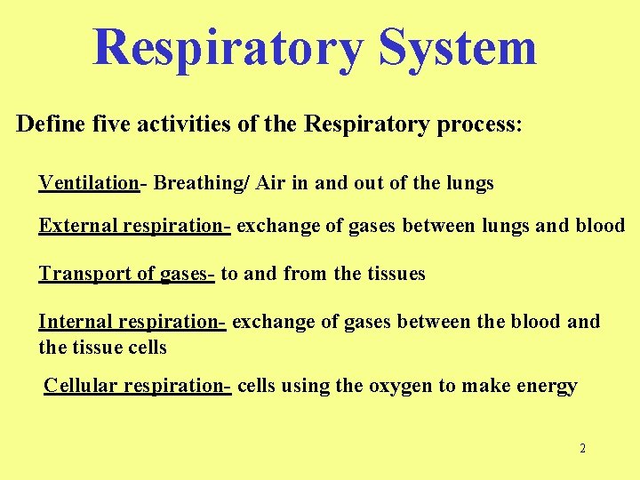
Respiratory System Define five activities of the Respiratory process: Ventilation- Breathing/ Air in and out of the lungs External respiration- exchange of gases between lungs and blood Transport of gases- to and from the tissues Internal respiration- exchange of gases between the blood and the tissue cells Cellular respiration- cells using the oxygen to make energy 2
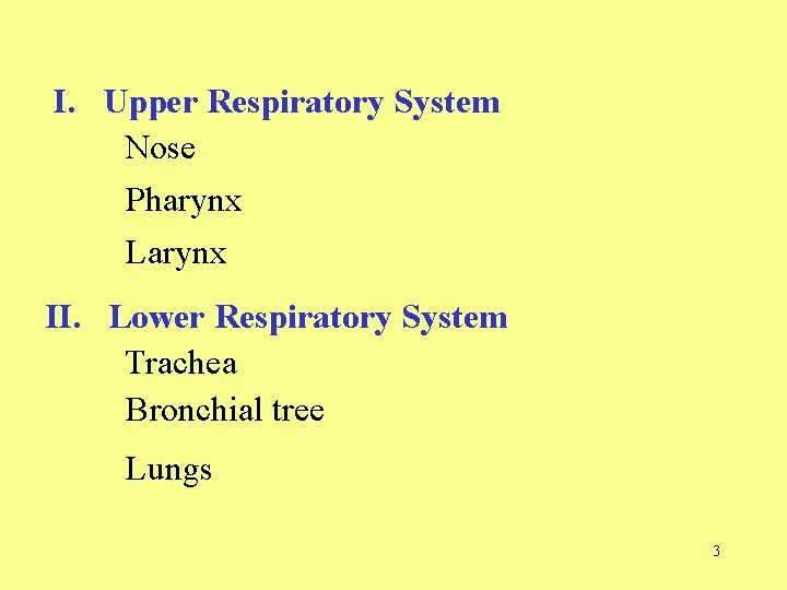
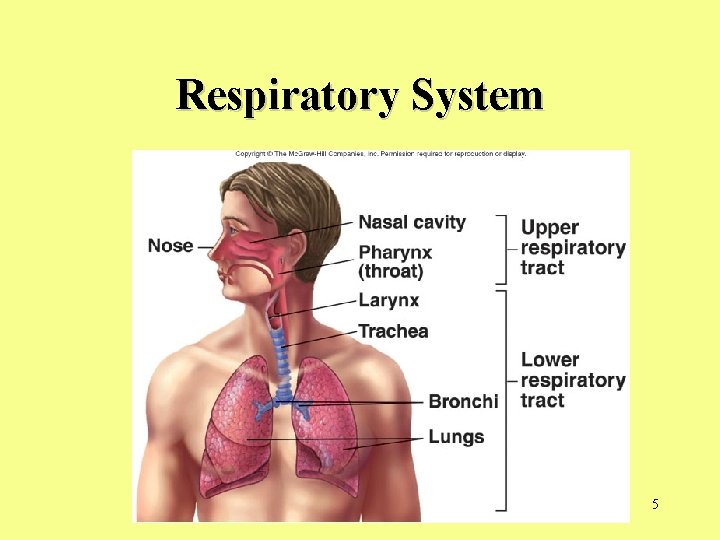
I. Upper Respiratory System Nose Pharynx Larynx II. Lower Respiratory System Trachea Bronchial tree Lungs 3
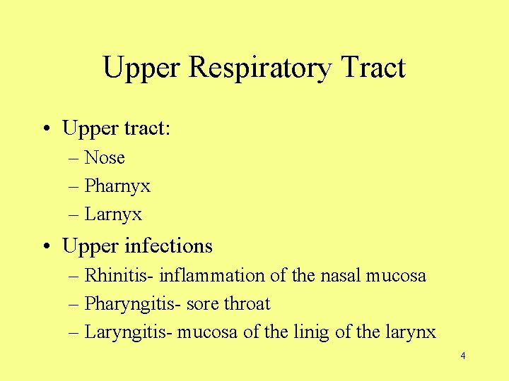
Upper Respiratory Tract • Upper tract: – Nose – Pharnyx – Larnyx • Upper infections – Rhinitis- inflammation of the nasal mucosa – Pharyngitis- sore throat – Laryngitis- mucosa of the linig of the larynx 4
Respiratory System 5
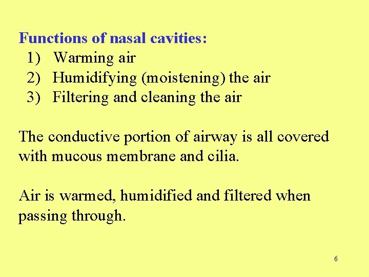
Functions of nasal cavities: 1) Warming air 2) Humidifying (moistening) the air 3) Filtering and cleaning the air The conductive portion of airway is all covered with mucous membrane and cilia. Air is warmed, humidified and filtered when passing through. 6
Lower Respiratory Tract • Lower tract – Always open to the outside and are lined with mucous membranes – Some regions have hairs to help filter air • Trachea • Bronchial tree • lungs 7
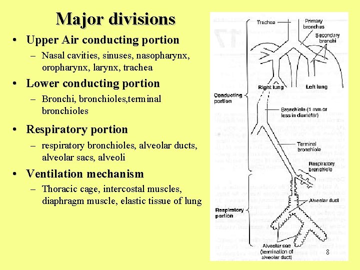
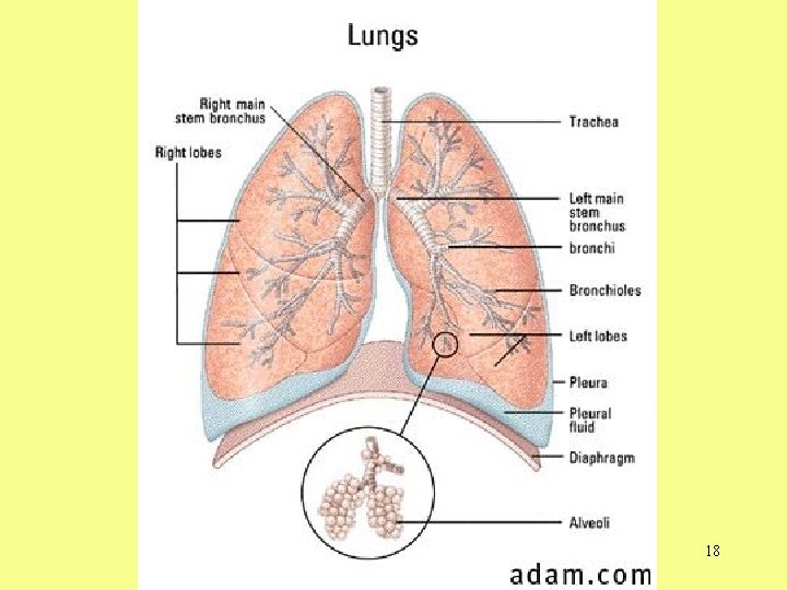
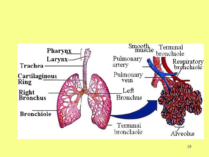
Major divisions • Upper Air conducting portion – Nasal cavities, sinuses, nasopharynx, oropharynx, larynx, trachea • Lower conducting portion – Bronchi, bronchioles, terminal bronchioles • Respiratory portion – respiratory bronchioles, alveolar ducts, alveolar sacs, alveoli • Ventilation mechanism – Thoracic cage, intercostal muscles, diaphragm muscle, elastic tissue of lung 8
Nose • Nose – External nose – Nasal cavity • Functions – Passageway for air – Cleans the air – Humidifies, warms air – Smell – Along with paranasal sinuses are resonating chambers for speech
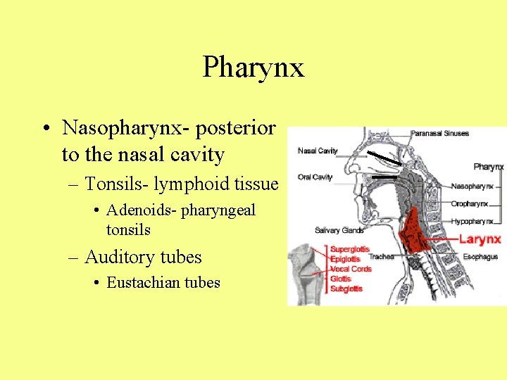
Pharynx • Nasopharynx- posterior to the nasal cavity – Tonsils- lymphoid tissue • Adenoids- pharyngeal tonsils – Auditory tubes • Eustachian tubes
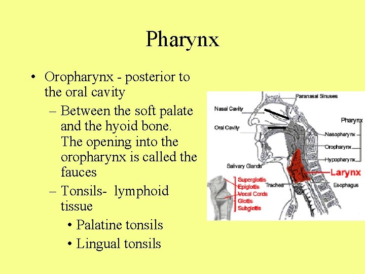
Pharynx • Oropharynx - posterior to the oral cavity – Between the soft palate and the hyoid bone. The opening into the oropharynx is called the fauces – Tonsils- lymphoid tissue • Palatine tonsils • Lingual tonsils
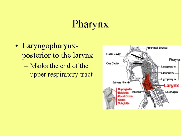
Pharynx • Laryngopharynx- posterior to the larynx – Marks the end of the upper respiratory tract
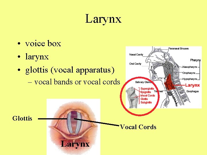
Larynx • voice box • larynx • glottis (vocal apparatus) – vocal bands or vocal cords Glottis Vocal Cords Larynx
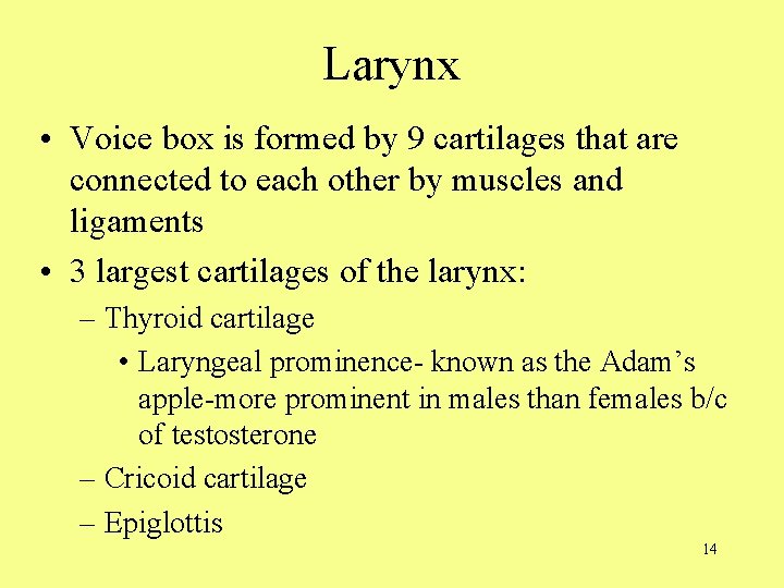
Larynx • Voice box is formed by 9 cartilages that are connected to each other by muscles and ligaments • 3 largest cartilages of the larynx: – Thyroid cartilage • Laryngeal prominence- known as the Adam’s apple-more prominent in males than females b/c of testosterone – Cricoid cartilage – Epiglottis 14
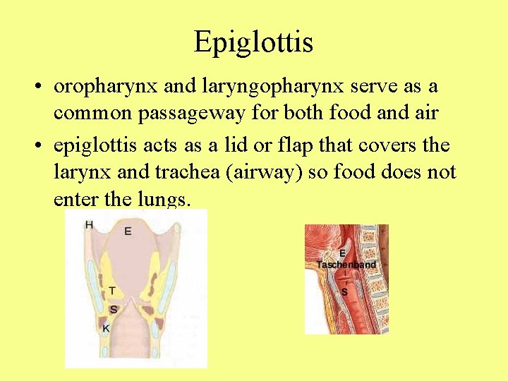
Epiglottis • oropharynx and laryngopharynx serve as a common passageway for both food and air • epiglottis acts as a lid or flap that covers the larynx and trachea (airway) so food does not enter the lungs.
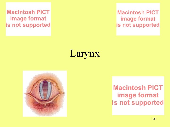
Larynx 16
Folds of the Larynx • The upper pair of folds are the vestibular folds • The lower pair are the true vocal cords and the opening – Males- longer and thicker & vibrate slower • Epiglottis- flap that folds over the glottis when you swallow • Between the vocal cords is the glottis – Opening to the larynx 17
18
19
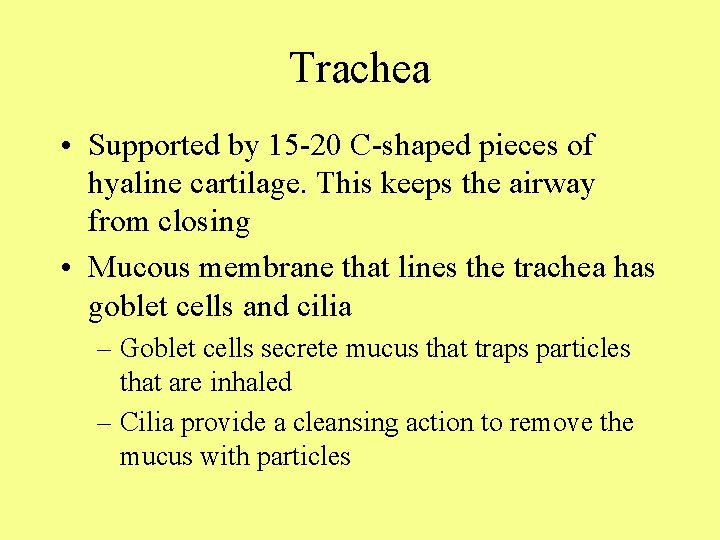
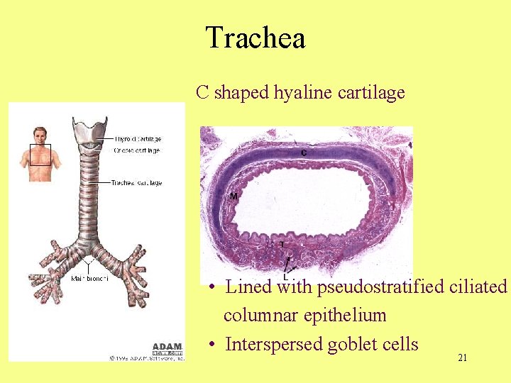
Trachea • Supported by 15 -20 C-shaped pieces of hyaline cartilage. This keeps the airway from closing • Mucous membrane that lines the trachea has goblet cells and cilia – Goblet cells secrete mucus that traps particles that are inhaled – Cilia provide a cleansing action to remove the mucus with particles
Trachea C shaped hyaline cartilage • Lined with pseudostratified ciliated columnar epithelium • Interspersed goblet cells 21
22
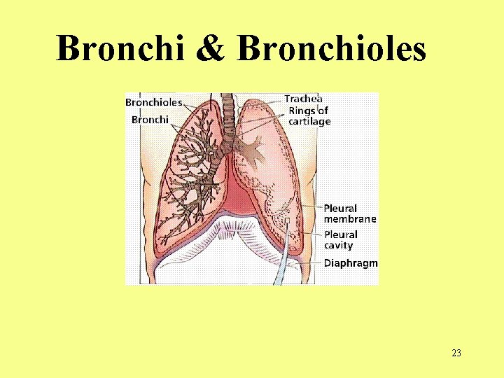
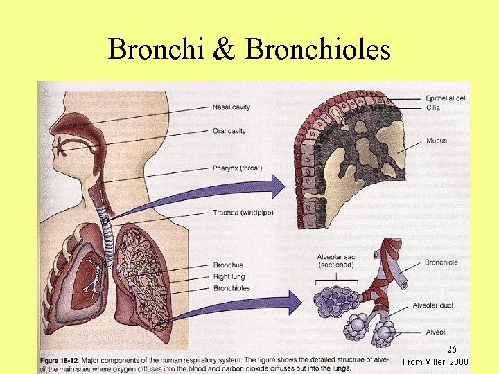
Bronchi & Bronchioles 23
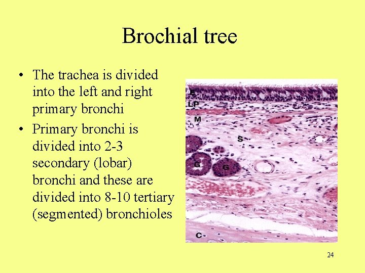
Brochial tree • The trachea is divided into the left and right primary bronchi • Primary bronchi is divided into 2 -3 secondary (lobar) bronchi and these are divided into 8 -10 tertiary (segmented) bronchioles 24
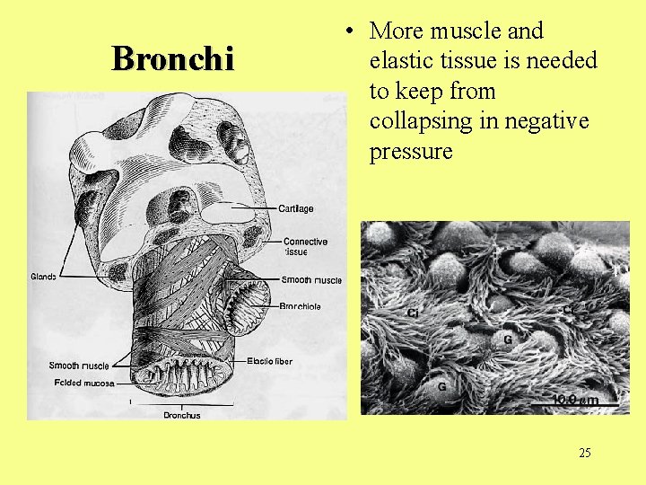
Bronchi • More muscle and elastic tissue is needed to keep from collapsing in negative pressure 25
Bronchi & Bronchioles 26
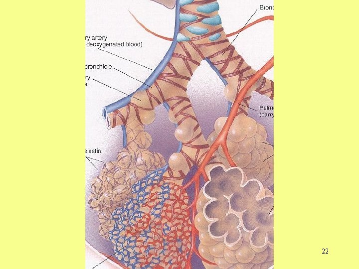
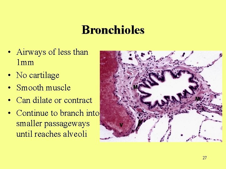
Bronchioles • Airways of less than 1 mm • No cartilage • Smooth muscle • Can dilate or contract • Continue to branch into smaller passageways until reaches alveoli 27
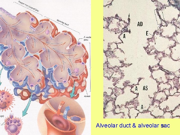
Alveoli • Consist of simple squamous epithelium which permits rapid diffusion of oxygen and carbon dioxide • . 2 to. 5 mm in size • 150 million little sacs • 70 square meters • Very low blood pressure around the alveoli
29 Alveolar duct & alveolar sac
Right vs. Left Lung • Right lung is shorter, broader and has greater volume than the left lung • Right lung is divided into 3 lobes; the left lungs has 2 lobes • The left lung has an indentation called the cardiac notch, for the apex of the heart. 30
Lungs Right-3 lobes Left-2 lobes trachea
Pleura • • • each lung enclosed in pleura parietal pleura (inside the chest cavity wall) visceral pleura (outside lining of the lung) pleural space or pleural cavity lubricating fluid
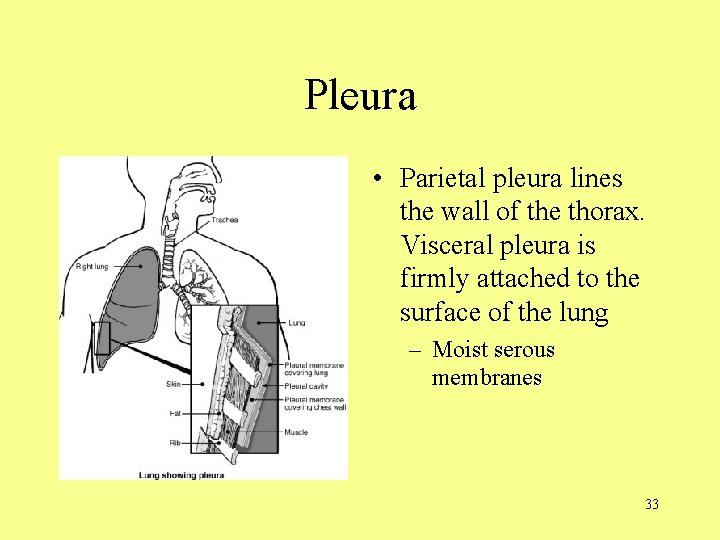
Pleura • Parietal pleura lines the wall of the thorax. Visceral pleura is firmly attached to the surface of the lung – Moist serous membranes 33
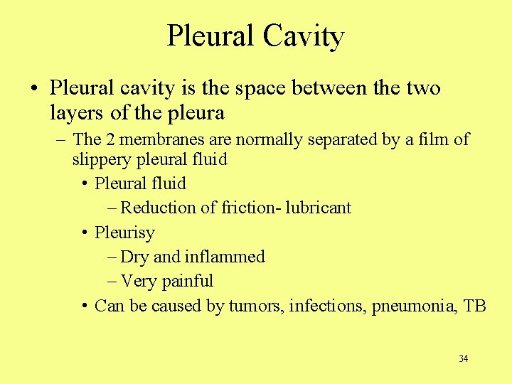
Pleural Cavity • Pleural cavity is the space between the two layers of the pleura – The 2 membranes are normally separated by a film of slippery pleural fluid • Pleural fluid – Reduction of friction- lubricant • Pleurisy – Dry and inflammed – Very painful • Can be caused by tumors, infections, pneumonia, TB 34
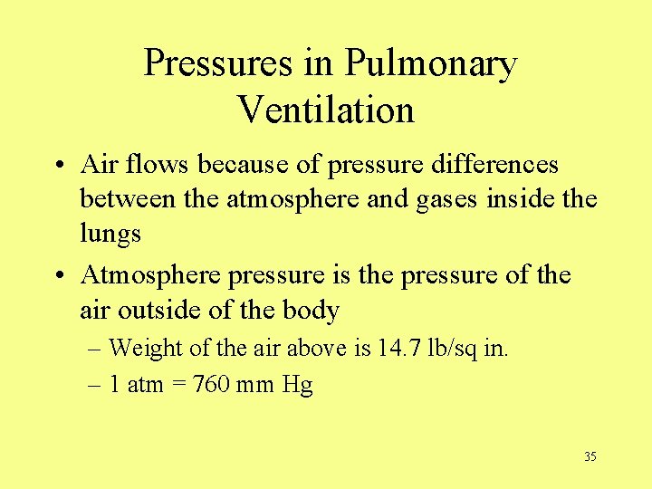
Pressures in Pulmonary Ventilation • Air flows because of pressure differences between the atmosphere and gases inside the lungs • Atmosphere pressure is the pressure of the air outside of the body – Weight of the air above is 14. 7 lb/sq in. – 1 atm = 760 mm Hg 35
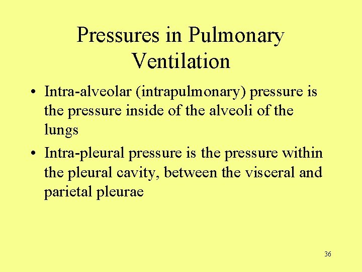
Pressures in Pulmonary Ventilation • Intra-alveolar (intrapulmonary) pressure is the pressure inside of the alveoli of the lungs • Intra-pleural pressure is the pressure within the pleural cavity, between the visceral and parietal pleurae 36
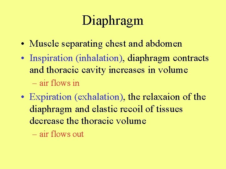
Diaphragm • Muscle separating chest and abdomen • Inspiration (inhalation), diaphragm contracts and thoracic cavity increases in volume – air flows in • Expiration (exhalation), the relaxaion of the diaphragm and elastic recoil of tissues decrease thoracic volume – air flows out
Boyles Law • The pressure of a given quantity of gas is inversely proportional to its volume assuming constant temperature • (very important when diving to release compressed air on the way back up) 38
Sequence of events in inspiration and expiration • Inspiration – During inspiration, the diaphragm contracts and the thoracic cavity increases in volume – An increase in thoracic volume decreases the pressure below atmospheric pressure so that air flows into the lungs 39
Sequence of events in inspiration and expiration • Expiration – During expiration, the relaxation of the diaphragm and elastic recoil of tissues decrease thoracic volume – The decrease in thoracic volume increases the intra-alveolar pressure so the air flows out of the lungs 40
Sufactant Importance • Sufactant reduces the surface tension inside of the walls of the alveoli so that they do not adhere to each other and collapse 41
Collapsed Lung When the negative pressure inside of the pleural cavity is interrupted by damage in the thoracic walls, the lungs may collapse. 42
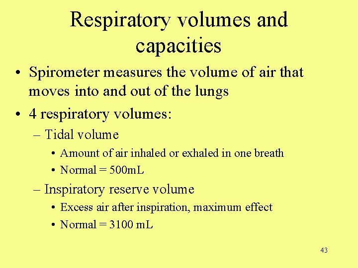
Respiratory volumes and capacities • Spirometer measures the volume of air that moves into and out of the lungs • 4 respiratory volumes: – Tidal volume • Amount of air inhaled or exhaled in one breath • Normal = 500 m. L – Inspiratory reserve volume • Excess air after inspiration, maximum effect • Normal = 3100 m. L 43
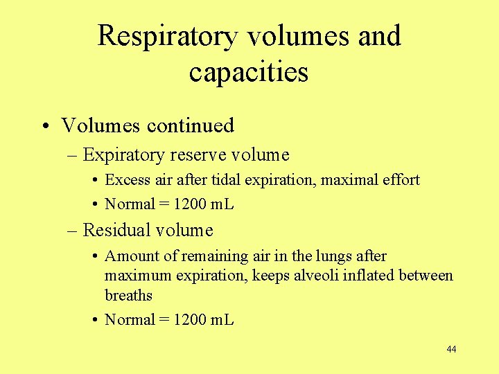
Respiratory volumes and capacities • Volumes continued – Expiratory reserve volume • Excess air after tidal expiration, maximal effort • Normal = 1200 m. L – Residual volume • Amount of remaining air in the lungs after maximum expiration, keeps alveoli inflated between breaths • Normal = 1200 m. L 44
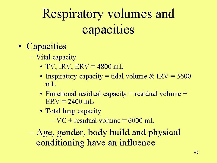
Respiratory volumes and capacities • Capacities – Vital capacity • TV, IRV, ERV = 4800 m. L • Inspiratory capacity = tidal volume & IRV = 3600 m. L • Functional residual capacity = residual volume + ERV = 2400 m. L • Total lung capacity – VC + residual volume = 6000 m. L – Age, gender, body build and physical conditioning have an influence 45
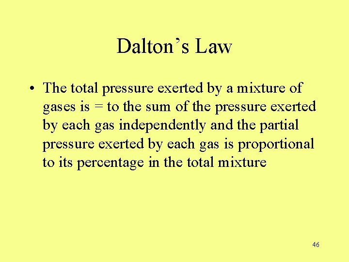
Dalton’s Law • The total pressure exerted by a mixture of gases is = to the sum of the pressure exerted by each gas independently and the partial pressure exerted by each gas is proportional to its percentage in the total mixture 46
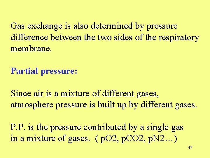
Gas exchange is also determined by pressure difference between the two sides of the respiratory membrane. Partial pressure: Since air is a mixture of different gases, atmosphere pressure is built up by different gases. P. P. is the pressure contributed by a single gas in a mixture of gases. ( p. O 2, p. CO 2, p. N 2…) 47
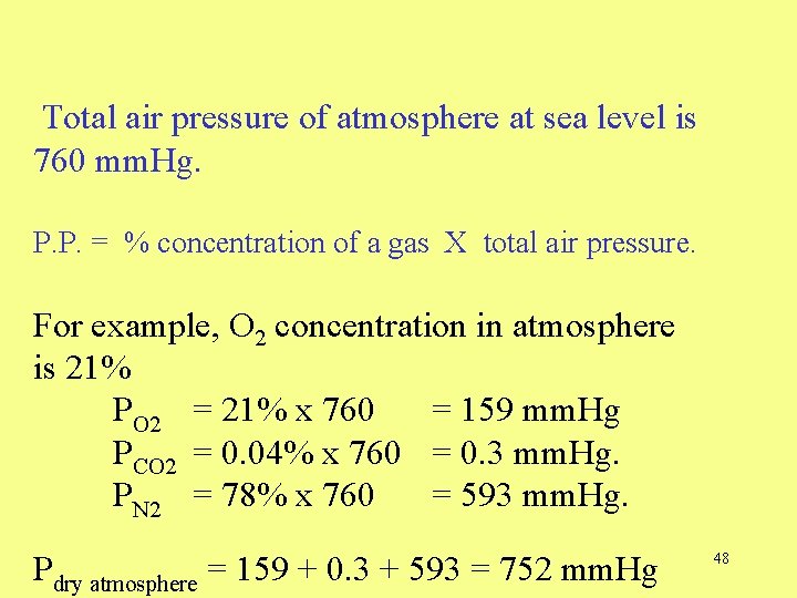
Total air pressure of atmosphere at sea level is 760 mm. Hg. P. P. = % concentration of a gas X total air pressure. For example, O 2 concentration in atmosphere is 21% PO 2 = 21% x 760 = 159 mm. Hg PCO 2 = 0. 04% x 760 = 0. 3 mm. Hg. PN 2 = 78% x 760 = 593 mm. Hg. Pdry atmosphere = 159 + 0. 3 + 593 = 752 mm. Hg 48
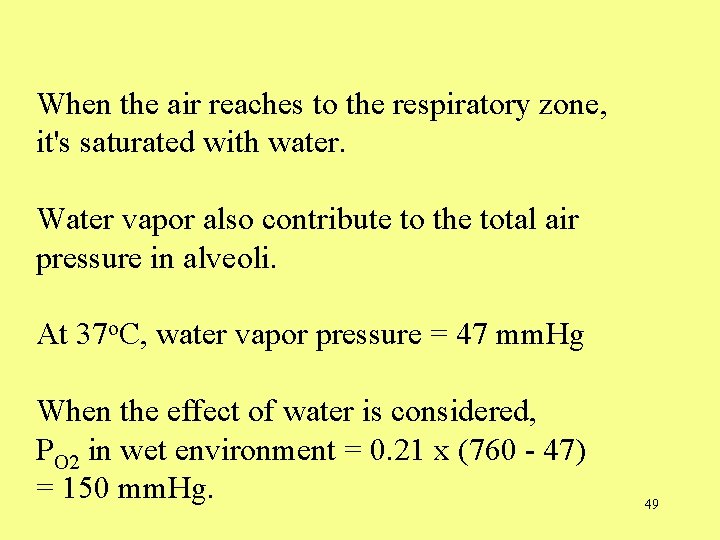
When the air reaches to the respiratory zone, it's saturated with water. Water vapor also contribute to the total air pressure in alveoli. At 37 o. C, water vapor pressure = 47 mm. Hg When the effect of water is considered, PO 2 in wet environment = 0. 21 x (760 - 47) = 150 mm. Hg. 49
After mixing with the residual air, PO 2 of alveolar air is further decreased to 105 mm. Hg (Fig. 16. 20). 50
Henry’s Law • States that when a mixture of gases is in contact with a liquid, each gas dissolves in the liquid in proportion to its own solubility and partial pressure 51
When air is dissolved in solution (blood), the concentration of each gas is proportional to its partial pressure in the air. PO 2 is 100 mm. Hg in pulmonary vein and systemic artery, and 40 mm. Hg in pulmonary artery and systemic vein. The gas diffusion across the respiratory membrane follows their concentration gradients. 52
External and internal respiration • Internal respiration is the exchange of gases between the tissue cells and the blood in the tissue capillaries • Oxygen diffuses from the blood into the tissue cells and carbon dioxide diffuses from the tissue cells into the blood during internal respiration 53
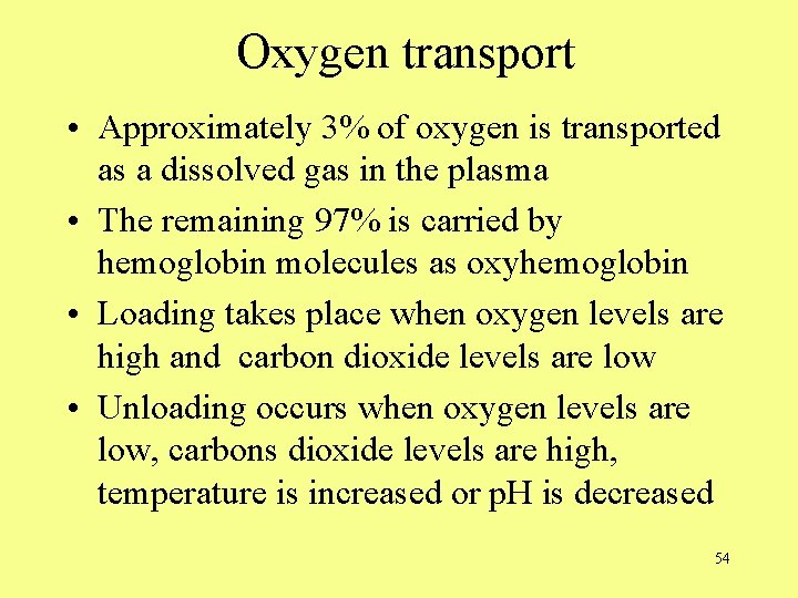
Oxygen transport • Approximately 3% of oxygen is transported as a dissolved gas in the plasma • The remaining 97% is carried by hemoglobin molecules as oxyhemoglobin • Loading takes place when oxygen levels are high and carbon dioxide levels are low • Unloading occurs when oxygen levels are low, carbons dioxide levels are high, temperature is increased or p. H is decreased 54
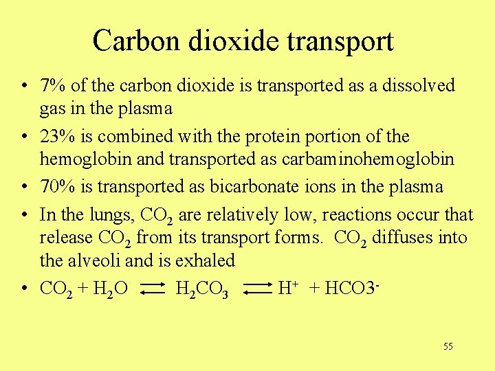
Carbon dioxide transport • 7% of the carbon dioxide is transported as a dissolved gas in the plasma • 23% is combined with the protein portion of the hemoglobin and transported as carbaminohemoglobin • 70% is transported as bicarbonate ions in the plasma • In the lungs, CO 2 are relatively low, reactions occur that release CO 2 from its transport forms. CO 2 diffuses into the alveoli and is exhaled • CO 2 + H 2 O H 2 CO 3 H+ + HCO 355
Brain and respiration • Respiratory center of the brain – Medulla oblongata – pons 56
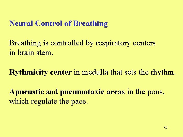
Neural Control of Breathing is controlled by respiratory centers in brain stem. Rythmicity center in medulla that sets the rhythm. Apneustic and pneumotaxic areas in the pons, which regulate the pace. 57
Respiratory Nerves • Phrenic nerve – Carries the impulses to the diaphragm and along the intercostals to the external intercostal muscles • Intercostal nerves – When a more forceful contraction is needed, the expiratory center sends impulses along the intercostal nerves to the internal intercostal muscles 58
Breathing regulation • Central chemoreceptors in the medulla oblongata are sensitive to increases in CO 2, H+ and O 2 levels • Peripheral chemoreceptors in the aortic and carotid bodies detect decreases in O 2 levels- not a strong stimulus for breathing • Stretch receptors in the lungs initiate the Hering-Breuer reflex that prevents over inflation of the lungs 59
Breathing Regulation • Voluntary and involuntary impulses from the higher brain centers may override the respiration center temporarily but after a limited time, the respiratory center resumes control • Increase in temperature increases the breathing rate 60
Factors that change breathing rate and depth: The autonomic control of breathing is influenced by input from the chemoreceptors that monitor the chemical composition of the blood. Blood levels of CO 2, p. H or O 2 will be sensed and fed back to the control centers. 61
There are central chemoreceptors in medulla and peripheral chemoreceptors in aorta and carotid arteries. If either CO 2 or H+ levels rises, the rate and depth of breathing will increase. ventilation increases, CO 2 is released, blood acidity - breathing rate will return to normal. Fig. 16. 29 62
This reflex control system is more sensitive to CO 2 levels. PCO 2 provides most potent stimulus to the reflex control of ventilation. Ventilation is adjusted to maintain a constant PCO 2. Proper PO 2 of blood is achieved as a side product of this reflex. 63