CORNEA AND RETINA Friends or Foes DR AJAY















- Slides: 76
CORNEA AND RETINA Friends…. or Foes…. ? DR. AJAY I. DUDANI M. S. , DNB, FCPS, DOMS, Vitreoretinal surgery & Laser Specialist, Consulting eye surgeon K. J. Somaiya Hospital, Bombay Hospital.
CORNEA & RETINA FRIENDS …. OR FOES …. . ?
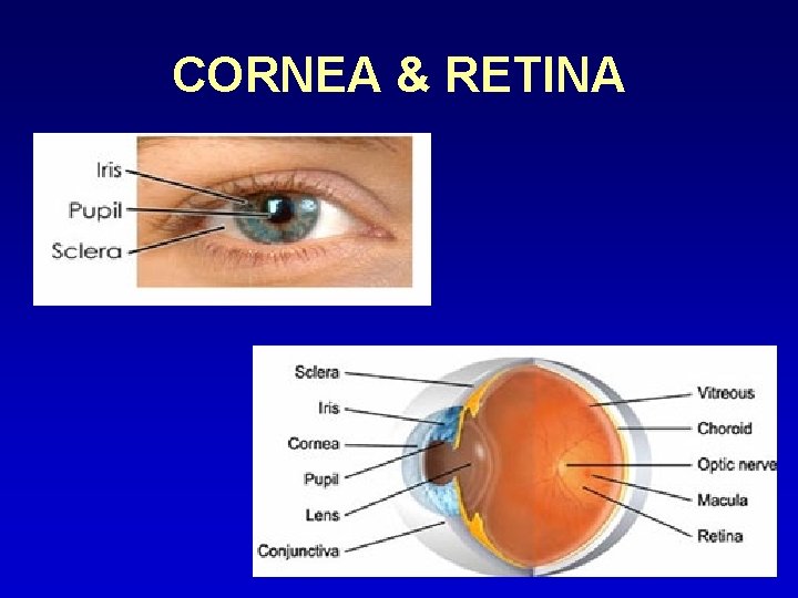
CORNEA & RETINA
ROLE OF NORMAL HEALTHY TRANSPARENT CORNEA • Clear visualisation of retina; normal or diseased ( DO, I/O, Slit lamp Biomicroscopy) • Investigations for diagnosis of retinal conditions ( FFA, ERG) • Diagnosing complications of retinal conditions; neovascular glaucoma ( Gonioscopy) • Easy & adequate treatment of retinal disorders (Laser or even Vitreoretinal Surgeries)
CONDITIONS AFFECTING CORNEA & RETINA ( INDEPENDENT OF EACH OTHER) • Congenital anomalies – Micro or megalocornea ; medullated nerve fibres in retina • Degenerations or Dystrophies – Lattice or Granular corneal dystrophies or Spheroidal degenerations ; Retinitis Pigmentosa or ARMD
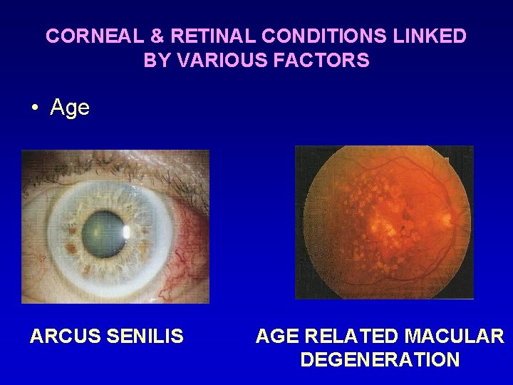
CORNEAL & RETINAL CONDITIONS LINKED BY VARIOUS FACTORS • Age ARCUS SENILIS AGE RELATED MACULAR DEGENERATION
CONGENITAL • Congenital syphilis – -Interstitial keratitis -Chorioretinitis • Congenital rubella syndrome – -microcornea, corneal clouding -rubella keratitis resembling CHED (congenitalhereditary endothelial dysfunction) -speckled retinitis of posterior pole
INFECTIONS • HIV • Syphilis ( interstitial keratitis & salt pepper fundus) • Lyme disease ( bilateral keratitis, papillodema, retinal haemorrhages, exudative RD’s, panophthalmitis) • Infectious mononucleosis ( nummular keratitis, retinal periphlebitis) • Onchocerciasis (superficial & deep keratitis, chorioretinitis)
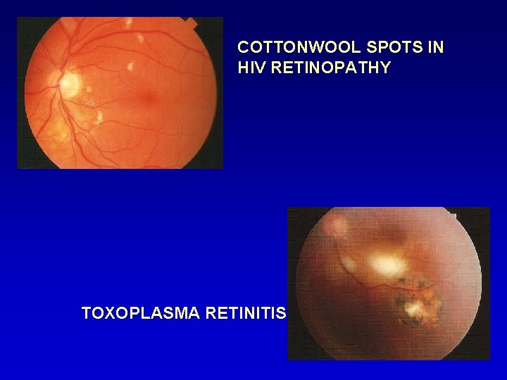
SYSTEMIC INFECTIONS AFFECTING BOTH CORNEA & RETINA Acquired immunodeficiency syndrome • CORNEA • RETINA -Herpes zoster keratitis -Herpes simplex keratitis -Keratoconjunctivitis sicca -CMV retinitis VZV- PORN -(progressive outer retinal necrosis) -Toxoplasmosis
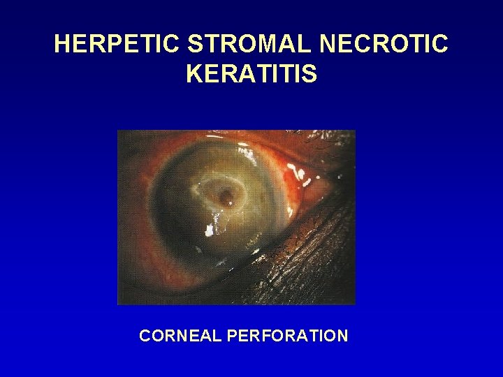
HERPETIC STROMAL NECROTIC KERATITIS CORNEAL PERFORATION
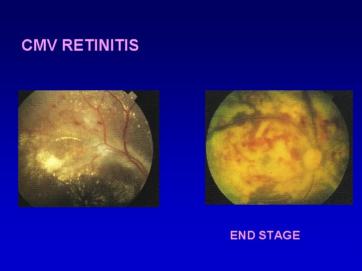
CMV RETINITIS END STAGE
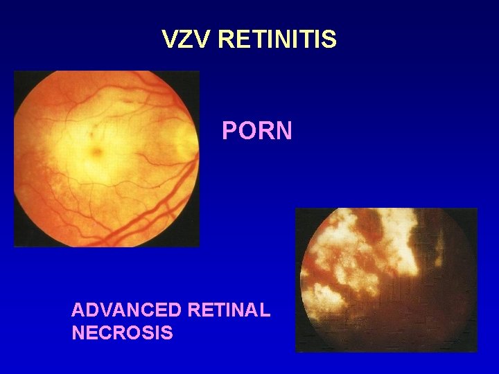
VZV RETINITIS PORN ADVANCED RETINAL NECROSIS
COTTONWOOL SPOTS IN HIV RETINOPATHY TOXOPLASMA RETINITIS
COLLAGEN DISEASES Commonly associated with keratitis (peripheral ulcerative) , keratoconjunctivitis sicca (dry eye syndromes)& retinal odema, vascular changes, hemorrhages… • • Periarteritis nodosa Scleroderma Systemic lupus erythematosus Wegeners granulomatosis Chronic granulomatous disease (sarcoidosis) associated with KCS & Fundal Granulomas
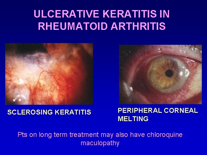
ULCERATIVE KERATITIS IN RHEUMATOID ARTHRITIS SCLEROSING KERATITIS PERIPHERAL CORNEAL MELTING Pts on long term treatment may also have chloroquine maculopathy
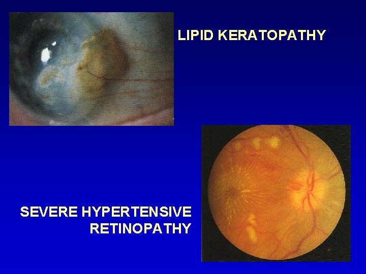
CARDIOVASCULAR DISEASES • Arcus senilis • Lipid keratopathy • Retinal odema, haemorrhages, hard exudates • Vessel tortuosity, copper silver wire vascular changes • Papilledema • Star maculopathy
LIPID KERATOPATHY SEVERE HYPERTENSIVE RETINOPATHY
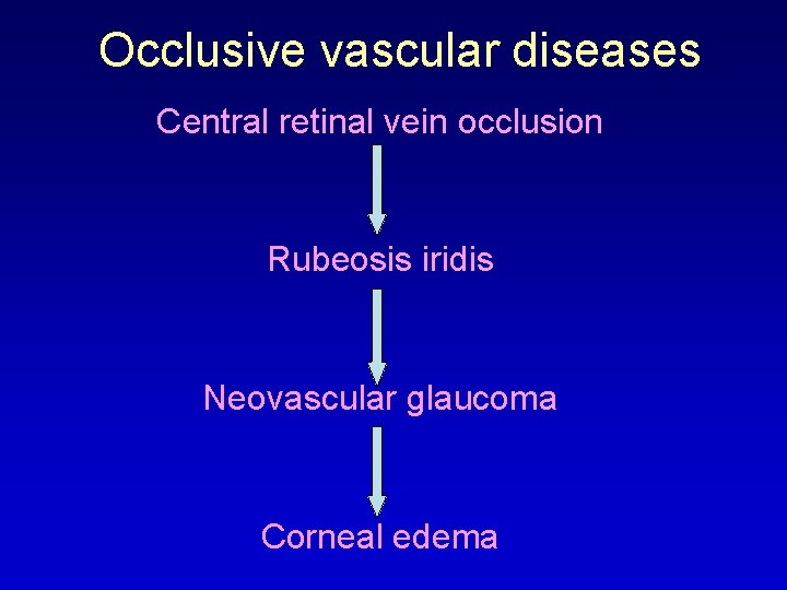
Occlusive vascular diseases Central retinal vein occlusion Rubeosis iridis Neovascular glaucoma Corneal edema
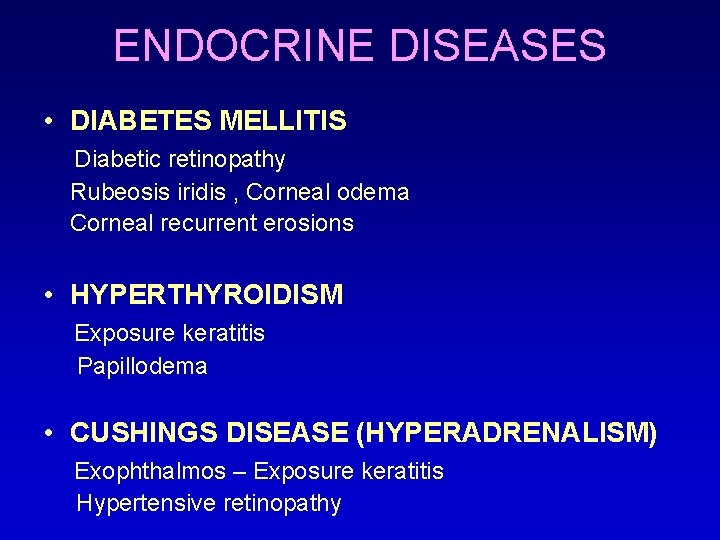
ENDOCRINE DISEASES • DIABETES MELLITIS Diabetic retinopathy Rubeosis iridis , Corneal odema Corneal recurrent erosions • HYPERTHYROIDISM Exposure keratitis Papillodema • CUSHINGS DISEASE (HYPERADRENALISM) Exophthalmos – Exposure keratitis Hypertensive retinopathy
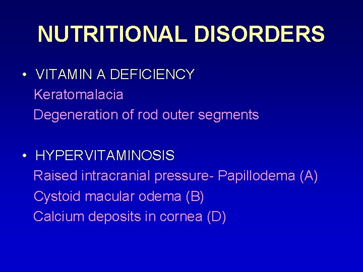
NUTRITIONAL DISORDERS • VITAMIN A DEFICIENCY Keratomalacia Degeneration of rod outer segments • HYPERVITAMINOSIS Raised intracranial pressure- Papillodema (A) Cystoid macular odema (B) Calcium deposits in cornea (D)
METABOLIC DISEASES • AMYLOIDOSIS Amyloid nodules in corneal stroma Vitreous opacities, retinal haemorrhages • CYSTINOSIS Corneal crystals in anterior stroma Retinal peripheral pigment clumping • FABRY’S DISEASE Vortex pattern corneal epithelial opacities Retinal haemorrhages, macular edema • LIPIDOSIS Lipid keratopathy Lipaemia retinalis
Pigmentary retinopathy occurs in all Mucopoly-Saccharidoses except Morquio & Maroteaux Lamy CORNEAL CLOUDING IN HURLER SYNDROME (in all MPS except Hunter & Sanfilippo)
CORNEAL SURGERIES AFFECTING RETINA • Refractive corneal surgeries ( leading to RRD, endophthalmitis) • Penetrating keratoplasty ( leading to endophthamitis, RD)
RRD FOLLOWING LASIK • Is infrequent • A study reported 0. 05% incidence at mean of 24 mths after lasik • Occurred 1 – 36 mths (mean 12. 6 mths) after lasik • Occurred in eyes with mean -6. 96 D of myopia before lasik
• If managed promptly, can result in good vision • No cause effect relationship between lasik & RRD was proven • However it is recommended that all pts scheduled for lasik undergo a thorough dilated fundus examination with scleral depression & also treatment of retinal lesions predisposing to RRD before the refractive surgery
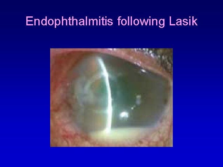
ENDOPHTHALMITIS FOLLOWING LASIK • Incidence of sight threatening complications after lasik still remains low. • Reports of endophthalmitis after incisional refractive surgeries – RK, hexagonal keratotomy, Ruiz procedure exist
Endophthalmitis following Lasik
Causes Corneal stroma may come in contact with infectious agents from • • Patients own body Contaminants present on instruments Surgeon or operating room Breaks in epithelial barrier & excessive surgical manipulation • Post op delayed epithelialisation of cornea, topical steroids, therapeutic CL’s, decreased corneal sensitivity & dry eye state
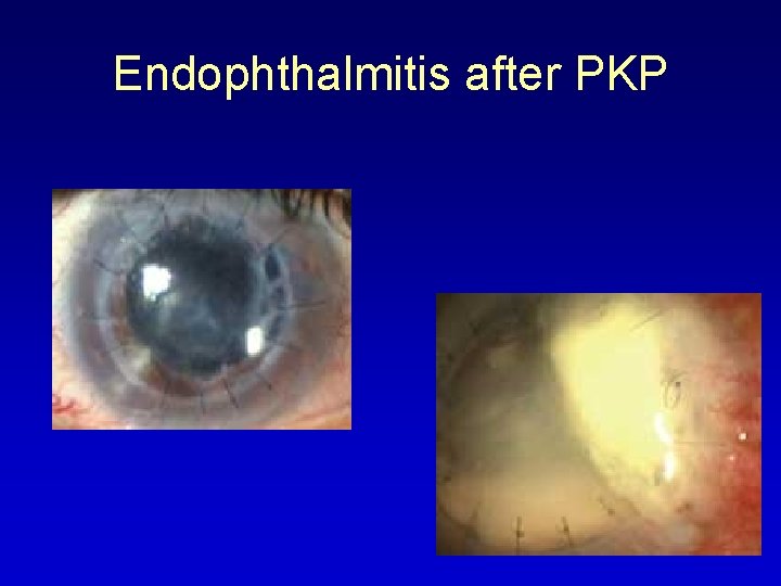
ENDOPHTHALMITIS AFTER PKP • Reported incidence of 0. 2%, has decreased in the last decade • Infections developed within 72 hrs & in majority the donor rim culture grew the same organism as was obtained from the AC or vitreous • Both bacterial & fungal • Fungal enophthalmitis transmitted by K-sol stored corneas • Torulopsis glabrata endophthalmitis after keratoplasty with organ cultured corneas
Endophthalmitis after PKP
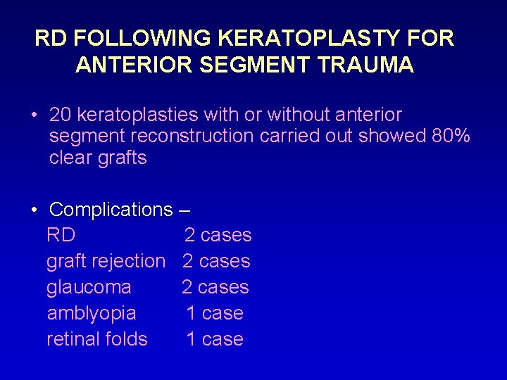
RD FOLLOWING KERATOPLASTY FOR ANTERIOR SEGMENT TRAUMA • 20 keratoplasties with or without anterior segment reconstruction carried out showed 80% clear grafts • Complications – RD 2 cases graft rejection 2 cases glaucoma 2 cases amblyopia 1 case retinal folds 1 case
RETNAL SURGERIES AFFECTING CORNEA • Retinal Detachment Surgeries • Vitreoretinal surgeries (VRS)
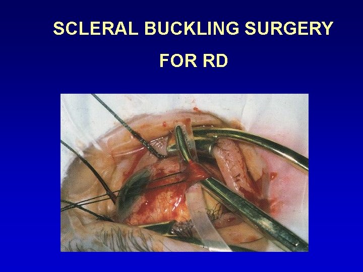
CORNEAL ODEMA FROM IOP RISE AFTER RD SURGERY Scleral buckling procedures alter anatomical configuration of globe & affect rise in IOP Factors affecting IOP include • Degree of shortening of encirclage if SRF drainage done (2 -3 mm) if SRF drainage not done (3 -6 mm) • Tightness with which scleral fixation sutures are tied (1 tight suture raises IOP by 10 mm. Hg immediately)
SCLERAL BUCKLING SURGERY FOR RD
CORNEAL ODEMA FROM GLAUCOMA AFTER VRS • Erythroclastic glaucoma – secondary to inadequate removal of intraocular haemorrhage • Inflammatory glaucoma – trabeculitis • Expanding gas bubble – mixing error (confusing cubic cm in syringe for %, pupillary block or unwise decision to use expanding gas in a total fill surgical situation)
• Emulsification glaucoma – uncommon delayed complication of silicone oil use • Steroid glaucoma • Hyperoxygenation of vitreous cavity & secondarily acqueous humour occur after vitrectomy which is responsible for trabecular damage (Sanley Chang)
SILICON OIL IN AC EMULSIFIED SILICON OIL IN AC
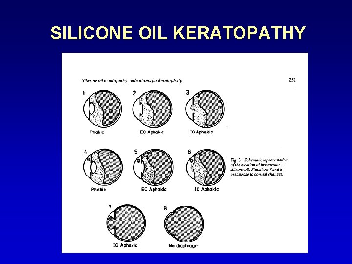
SILICONE OIL KERATOPATHY
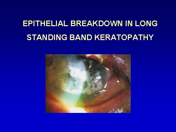
EPITHELIAL BREAKDOWN IN LONG STANDING BAND KERATOPATHY
VITREO- RETINAL SURGERIES • Self retaining corneal contact lens system
CORNEAL CONTACT LENS SYSTEM FOR VITREOUS SURGERY • They neutralise the refractive power of cornea • They afford excellent visualisation of fundus, vitreoretinal pathologies • Allow corneal contact on rotation of globe & eliminate accumulation of blood or bubbles between lens & cornea
CORNEAL OPACITIES – HINDRANCE IN RETINAL SURGERIES • Cornea may become cloudy, opacified due to injury, infection or scar tissue • Scar tissue prevents light from passing through cornea resulting not only in vision loss but also in difficult visualisation & treatment of retinal (or other posterior segment) lesions.
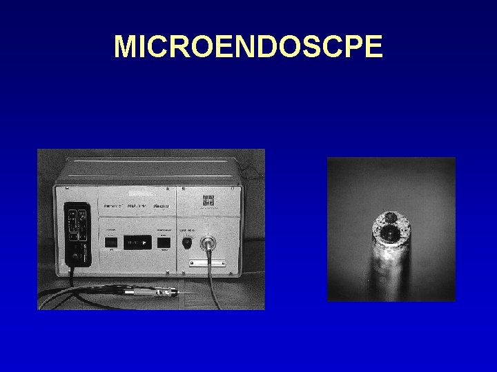
• Epithelial scrapping done to improve visualisation • Use of new ophthalmic microendoscopes • Endoscopic laser photocoagulation of ischaemic retina against opacity of anterior eye • Use of temporary keratoprosthesis followed later by keratolpasty
MICROENDOSCPE
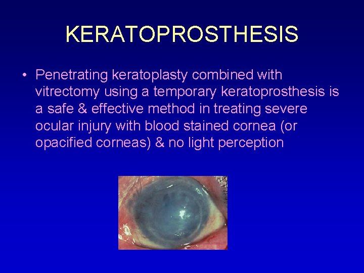
KERATOPROSTHESIS • Penetrating keratoplasty combined with vitrectomy using a temporary keratoprosthesis is a safe & effective method in treating severe ocular injury with blood stained cornea (or opacified corneas) & no light perception
KERATOPROSTHESIS
LASIK AFTER RD SURGERY • Myopic refractive errors are common in eyes that develop RD • Myopic changes may also be induced by RD surgeries because of changes in axial length, anterior chamber depth or position of the lens • A study has shown improvement in UCVA in all eyes & no decline in BCVA in any of them No retinal complication in post-op period although F/U is required • Only problem found was that of extensive conjunctival scarring which hampers the function of suction ring of microkeratome
OCULAR TRAUMA • • • Blunt Penetrating Intraocular foreign bodies Sympathetic ophthalmitis Radiation
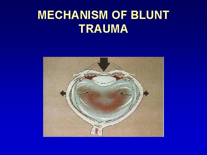
MECHANISM OF BLUNT TRAUMA
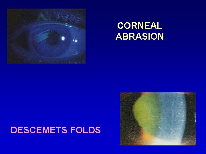
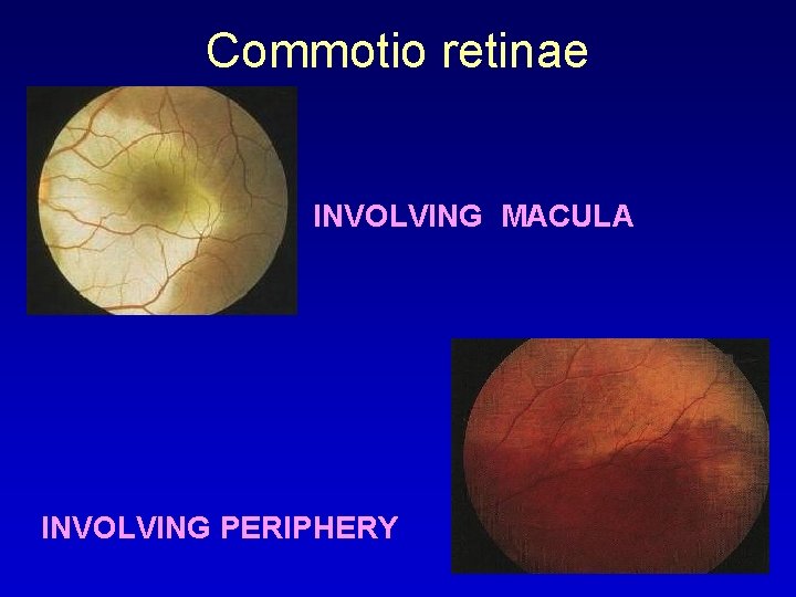
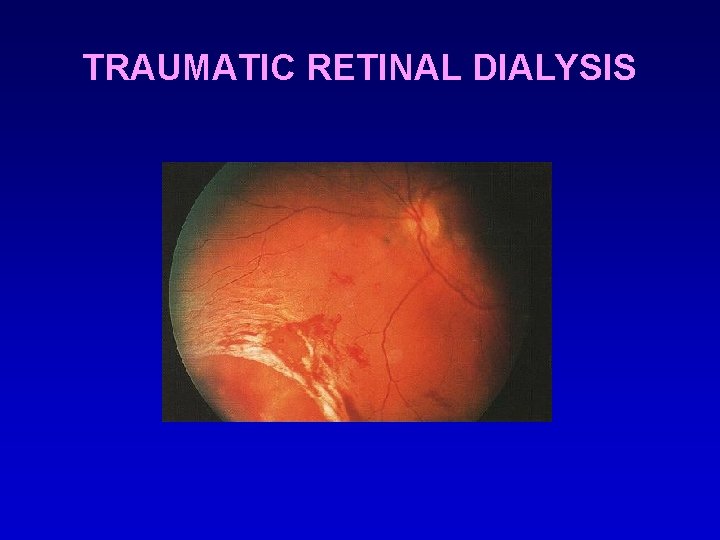
• Corneal abrasion – which stains with fluorescein • Acute corneal edema – due to focal or diffuse dysfunction of corneal endothelium may be associated with folds in descemet membrane. • Commotio retinae – gives grey appearance to fundus, frequently temporal occasionally may involve macula causing cherry red spot at fovea. Subsequent progressive pigmentary degeneration & macular hole formation may occur. • Retinal breaks leading to RD may occur in the form of retinal dialysis, equatorial tears or macular holes.
CORNEAL ABRASION DESCEMETS FOLDS
Commotio retinae INVOLVING MACULA INVOLVING PERIPHERY
TRAUMATIC RETINAL DIALYSIS
PENETRATING OCULAR TRAUMA • Assault, domestic accidents, sports injuries may cause corneal lacerations with or without iris prolapse. • Tractional RD may occur secondary to vitreous incarceration in the wound & intragel vitreous haemorrhage which stimulates fibroplastic proliferation. Subsequent contraction of membranes leads to tractional RD.
PENETRATING INJURY CORNEAL WOUND WITH IRIS PROLAPSE VITREOUS PROLAPSE , INTRAOCULAR HAEMMORHAGE, TRD
INTRAOCULAR FOREIGN BODIES • May traumatize the eye mechanically • Introduce infection – endophthalmitis or panophthalmitis • Exert other toxic effects on intraocular structures depending on their type stone, organic FB’s – infection iron, copper - sideosis, chalcosis respectively glass, plastics, gold, silver - inert
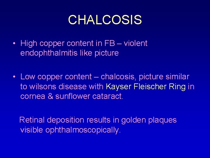
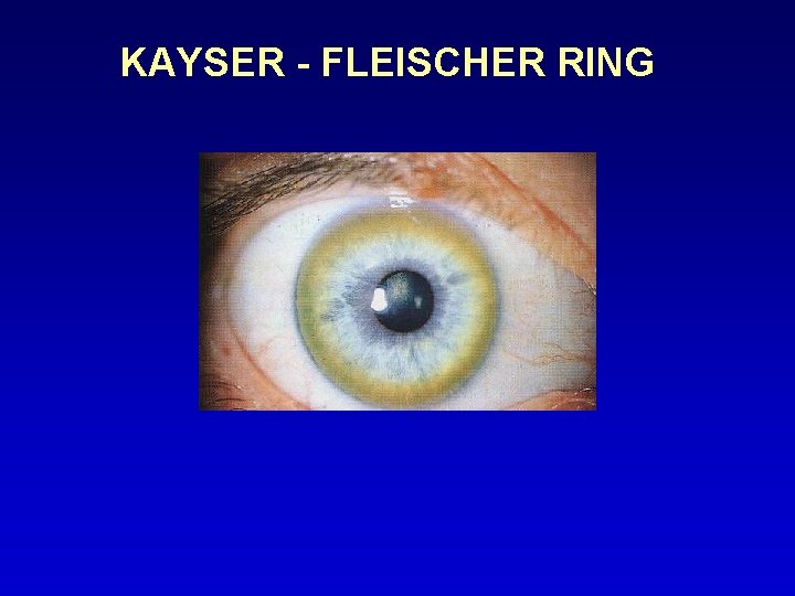
CHALCOSIS • High copper content in FB – violent endophthalmitis like picture • Low copper content – chalcosis, picture similar to wilsons disease with Kayser Fleischer Ring in cornea & sunflower cataract. Retinal deposition results in golden plaques visible ophthalmoscopically.
KAYSER - FLEISCHER RING
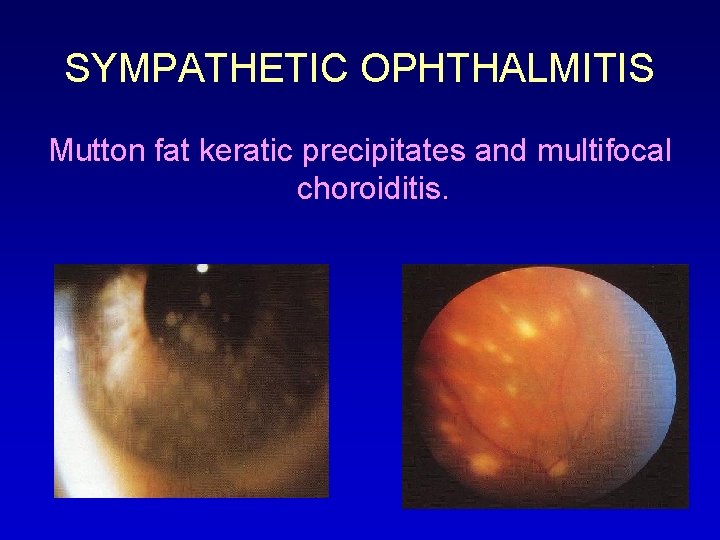
SYMPATHETIC OPHTHALMITIS Mutton fat keratic precipitates and multifocal choroiditis.
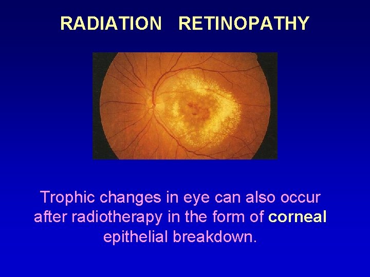
RADIATION RETINOPATHY Trophic changes in eye can also occur after radiotherapy in the form of corneal epithelial breakdown.
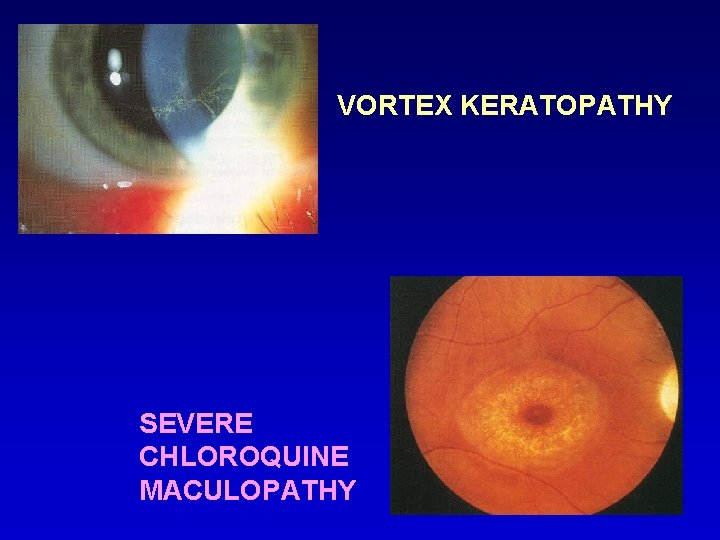
DRUGS AFFECTING CORNEA & RETINA Chloroquine & hydroxychloroquine • Vortex keratopathy – whorl like fine greyish to golden brown corneal epithelial deposits in form of arborizing horizontal lines resembling cats whiskers. • Unlike retinopathy, it bears no relationship to dosage, duration or treatment.
VORTEX KERATOPATHY SEVERE CHLOROQUINE MACULOPATHY
CHLOROQUINE MACULOPATHY • Risk increases when cumulative dose exceeds 300 g. (250 mg daily for 3 yrs) • Loss of foveal reflex • Central foveal pigmentation surrounded by depigmented zone of RPE atrophy which is again surrounded by hyperpigmented ring • Bull’s eye macular lesion • Unmasking of larger choroidal blood vessels & development of pigment clumps in retinal periphery.
Tamoxifen crystalline maculopathy & vortex keratopathy • Specific anti estrogen used in treatment of selected patients with breast carcinoma • Multiple yellow crystalline ring like deposits at the maculae • Persist on cessation of treatment • Maculopathy rare, routine screening not warranted
TAMOXIFEN CRYSTALLINE MACULOPATHY
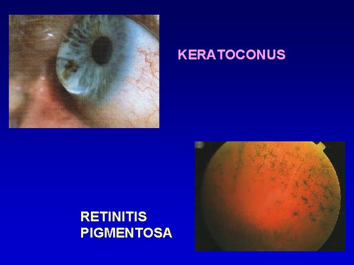
SYNDROMES AFFECTING CORNEA & RETINA • Ehlers danlos syndrome type 6 • Alports syndrome • Marfans syndrome Other ocular associations include: • Keratoconus associated with retinitis pigmentosa & ROP
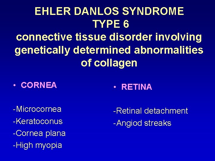
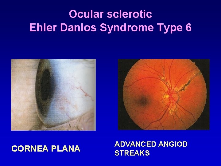
EHLER DANLOS SYNDROME TYPE 6 connective tissue disorder involving genetically determined abnormalities of collagen • CORNEA • RETINA -Microcornea -Keratoconus -Cornea plana -High myopia -Retinal detachment -Angiod streaks
Ocular sclerotic Ehler Danlos Syndrome Type 6 CORNEA PLANA ADVANCED ANGIOD STREAKS
MICROCORNEA
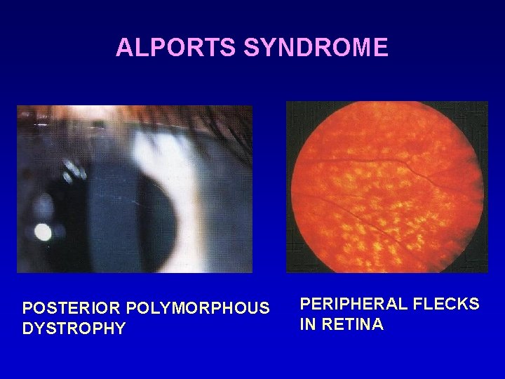
ALPORTS SYNDROME • Rare abnormality of glomerular basement membrane caused by mutations in genes encoding particular forms of type 4 collagen • Yellow punctate flecks in premacular area sparing fovea • Larger flecks become confluent in periphery • Associated with anterior lenticonus & occasionally posterior polymorphous corneal dystrophy
ALPORTS SYNDROME POSTERIOR POLYMORPHOUS DYSTROPHY PERIPHERAL FLECKS IN RETINA
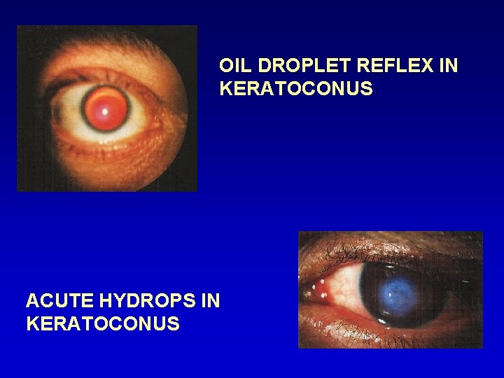
KERATOCONUS WITH RETINITIS PIGMENTOSA • Non-infective, progressive, bilateral thinning of cornea with ectasia of conical shape • Direct ophthalmoscopy from a distance of 1 foot shows an oil droplet reflex • Retinoscopy shows an irregular scissor reflex • Slit lamp biomicroscopy shows very fine vertical deep stromal striae (vogt lines) which disappear with external pressure on the globe
KERATOCONUS RETINITIS PIGMENTOSA
OIL DROPLET REFLEX IN KERATOCONUS ACUTE HYDROPS IN KERATOCONUS
THUS, NO PART OF THE HUMAN EYE CAN BE STUDIED IN ISOLATION THE CORNEA & RETINA INFLUENCE EACH OTHER IN HEALTH , FUNCTION & DISEASE THE INFLUENCE OF ONE ON THE OTHER IS OF CLINICAL SIGNIFICANCE TO THE PRACTISING CLINICIAN