
Eye VIBS 443602 192 Eye conjuctiva on white
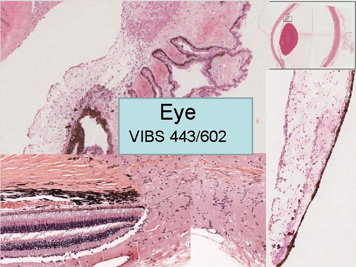
Eye VIBS 443/602
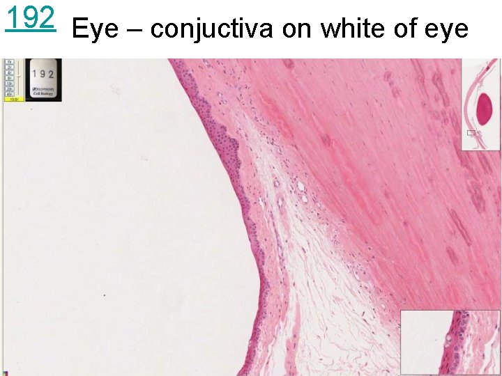
192 Eye – conjuctiva on white of eye
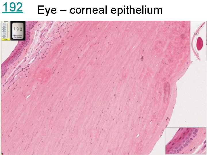
192 Eye – corneal epithelium
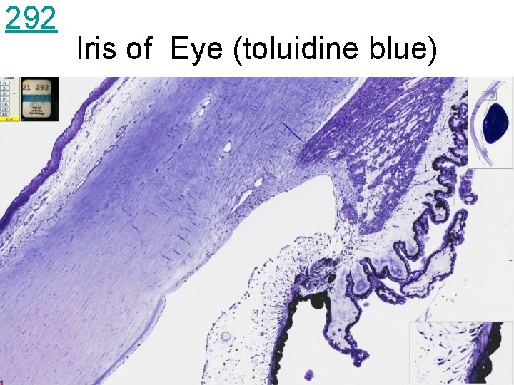
292 Iris of Eye (toluidine blue)
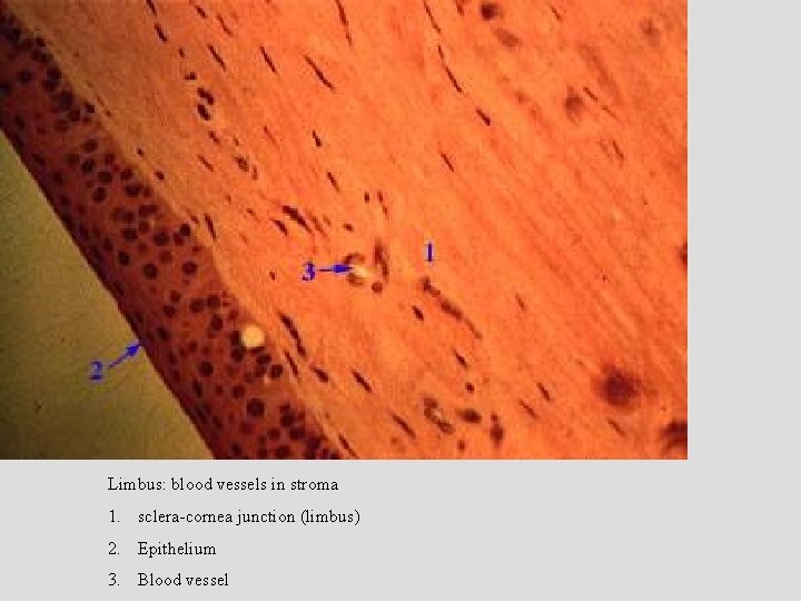
Limbus: blood vessels in stroma 1. sclera-cornea junction (limbus) 2. Epithelium 3. Blood vessel
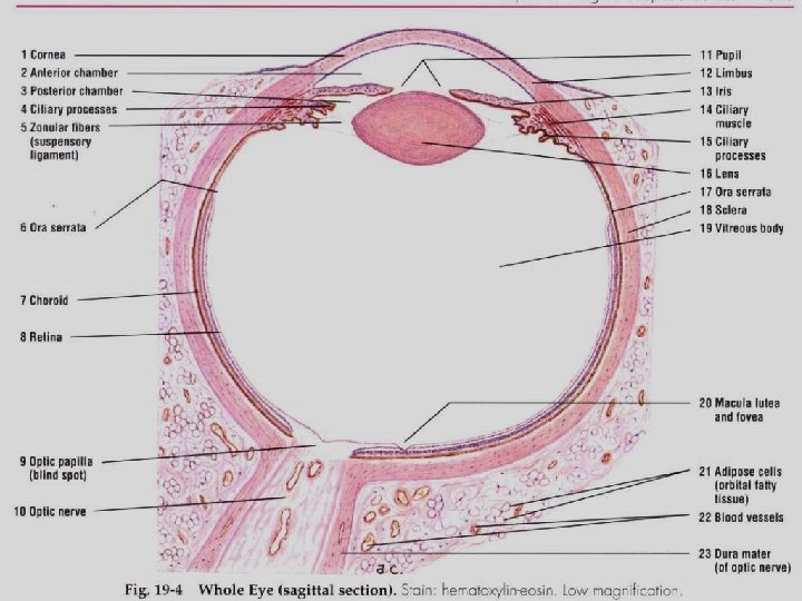
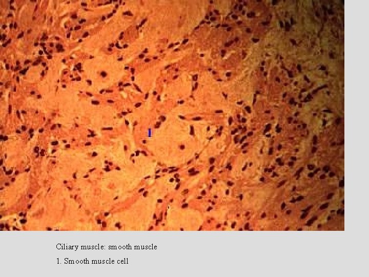
Ciliary muscle: smooth muscle 1. Smooth muscle cell
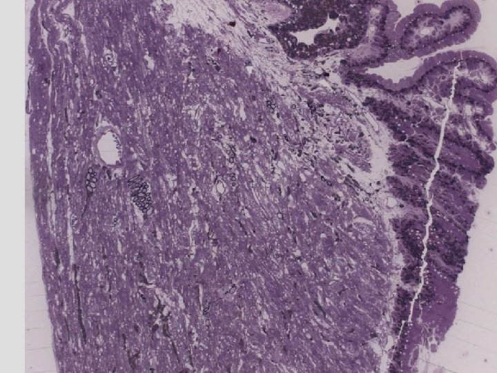
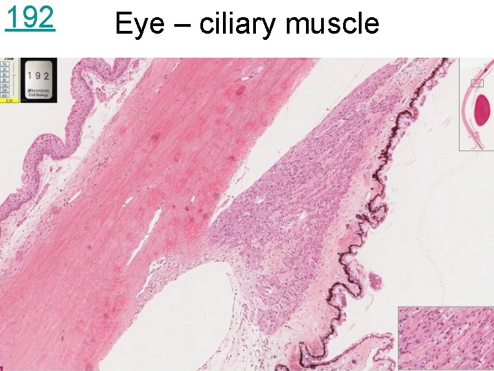
192 Eye – ciliary muscle
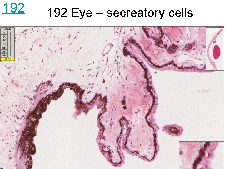
192 Eye – secreatory cells
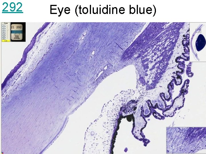
292 Eye (toluidine blue)
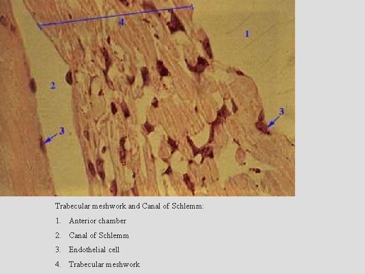
Trabecular meshwork and Canal of Schlemm: 1. Anterior chamber 2. Canal of Schlemm 3. Endothelial cell 4. Trabecular meshwork
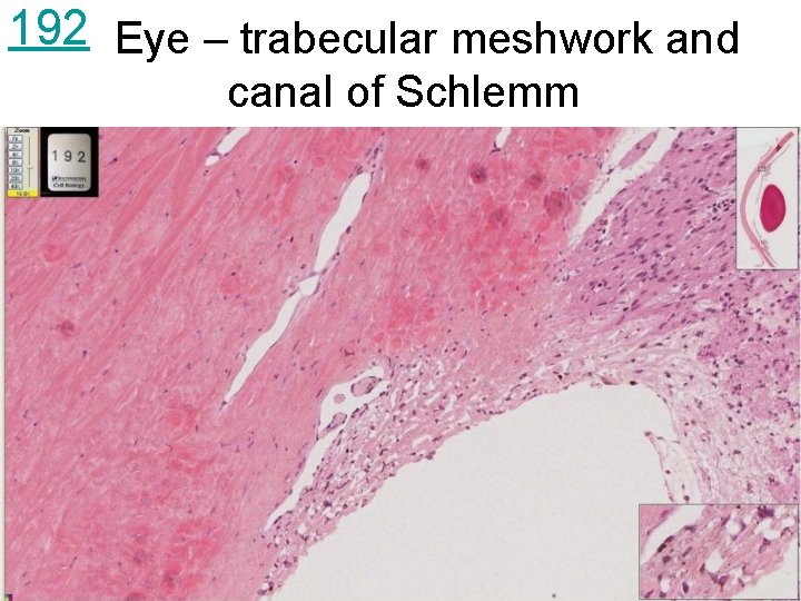
192 Eye – trabecular meshwork and canal of Schlemm
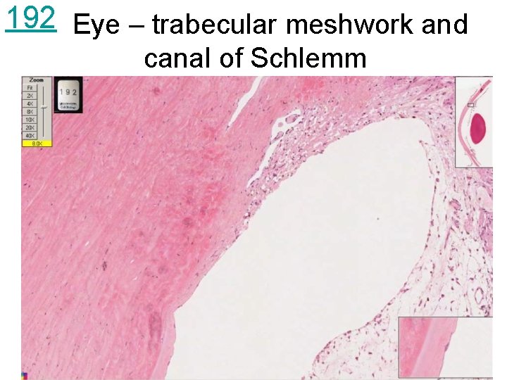
192 Eye – trabecular meshwork and canal of Schlemm
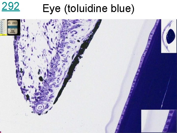
292 Eye (toluidine blue)
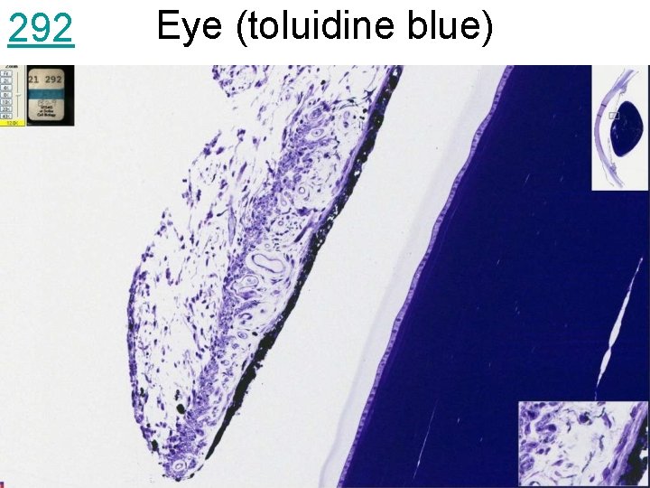
292 Eye (toluidine blue)
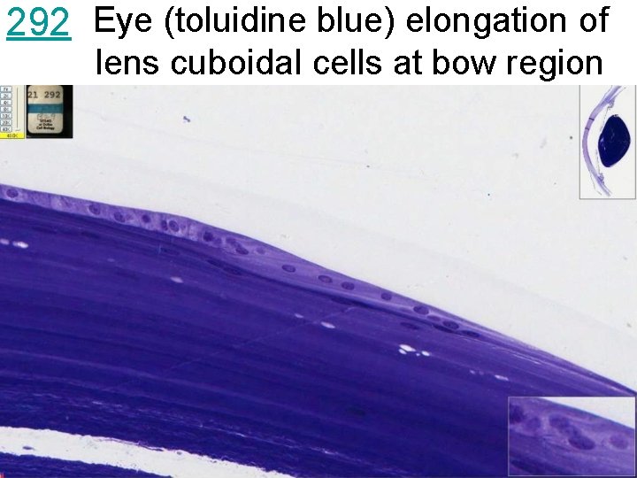
292 Eye (toluidine blue) elongation of lens cuboidal cells at bow region
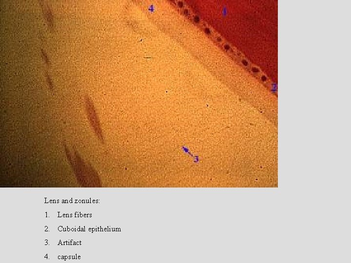
Lens and zonules: 1. Lens fibers 2. Cuboidal epithelium 3. Artifact 4. capsule
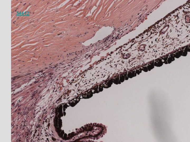
34412
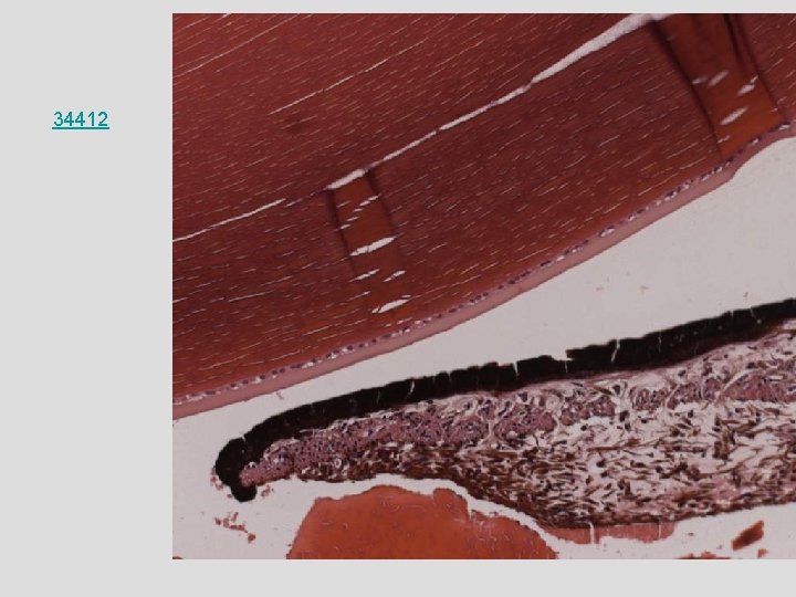
34412
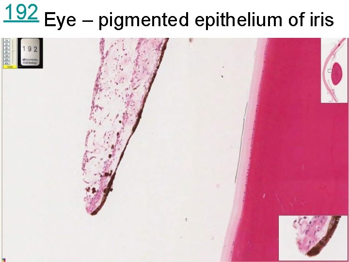
192 Eye – pigmented epithelium of iris
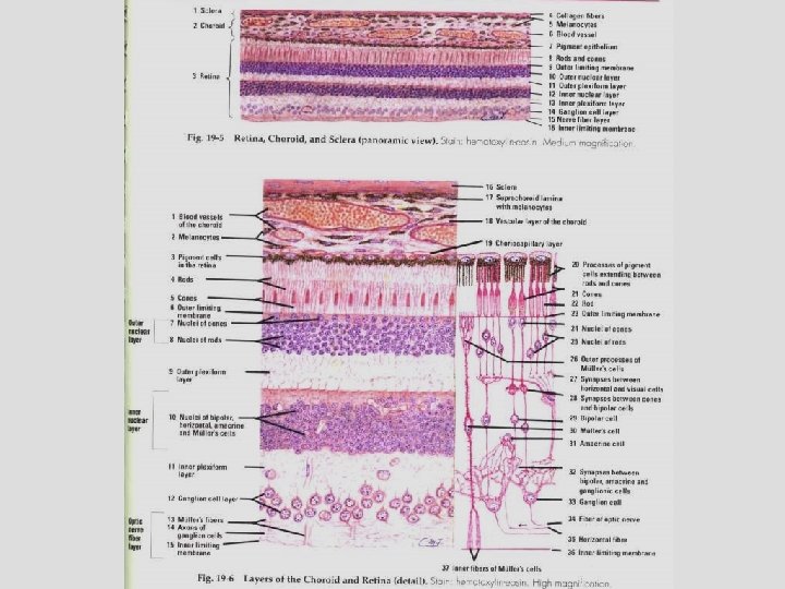
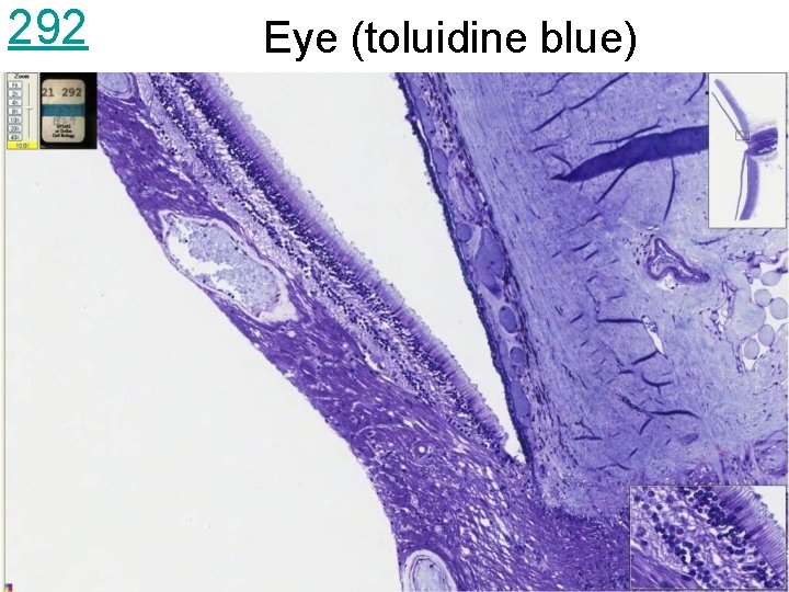
292 Eye (toluidine blue)
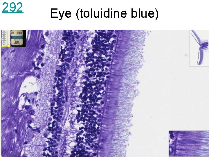
292 Eye (toluidine blue)
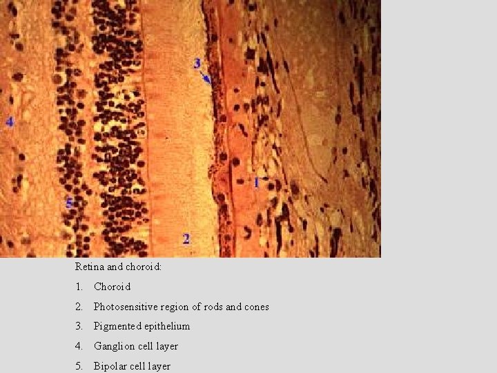
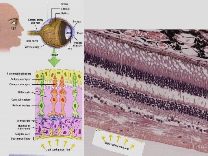
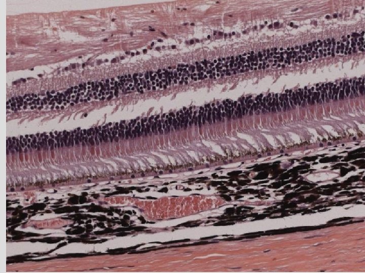
Retina and choroid: 1. Choroid 2. Photosensitive region of rods and cones 3. Pigmented epithelium 4. Ganglion cell layer 5. Bipolar cell layer
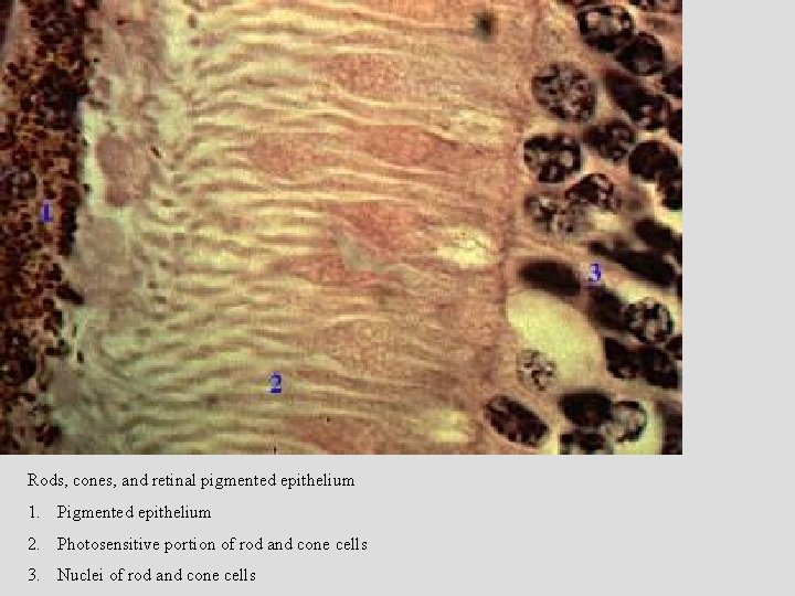
Rods, cones, and retinal pigmented epithelium 1. Pigmented epithelium 2. Photosensitive portion of rod and cone cells 3. Nuclei of rod and cone cells
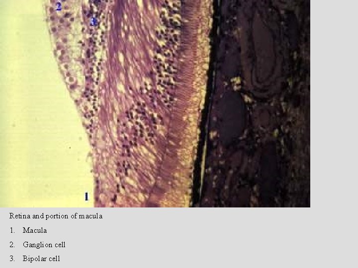
Retina and portion of macula 1. Macula 2. Ganglion cell 3. Bipolar cell
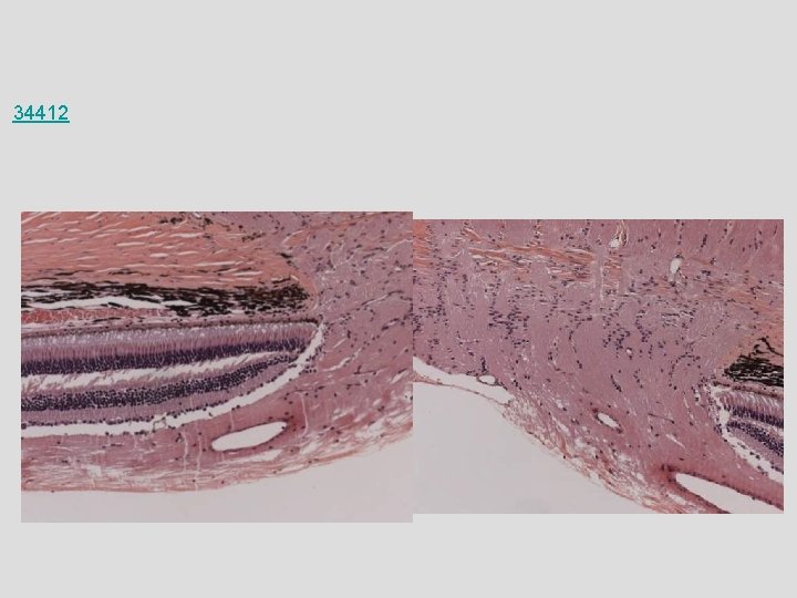
34412
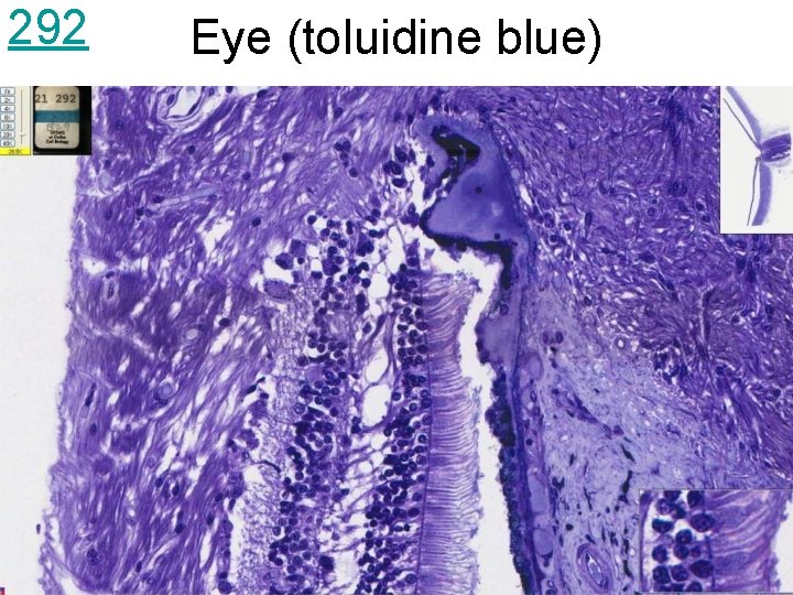
292 Eye (toluidine blue)
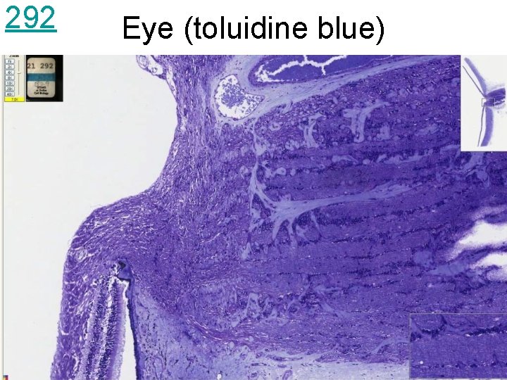
292 Eye (toluidine blue)
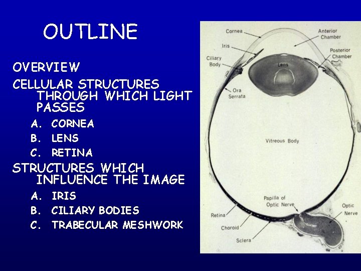
OUTLINE OVERVIEW CELLULAR STRUCTURES THROUGH WHICH LIGHT PASSES A. CORNEA B. LENS C. RETINA STRUCTURES WHICH INFLUENCE THE IMAGE A. IRIS B. CILIARY BODIES C. TRABECULAR MESHWORK
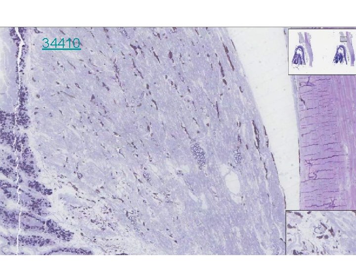
34410
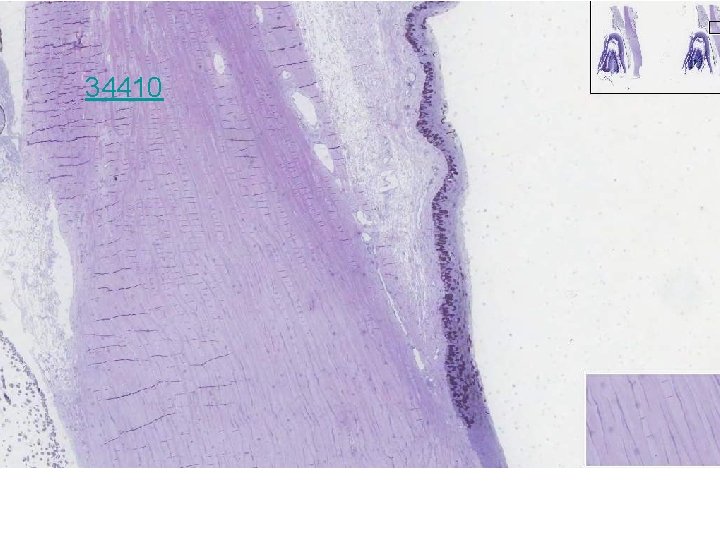
34410
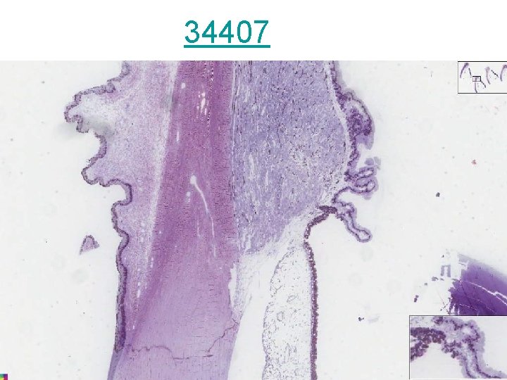
34407
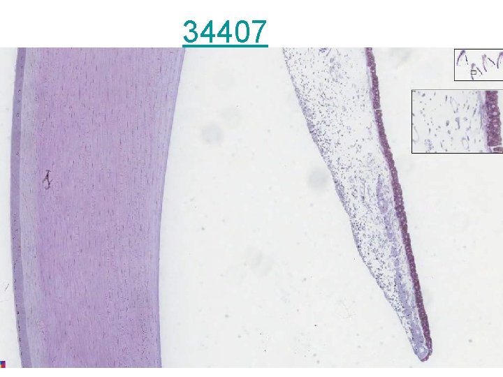
34407
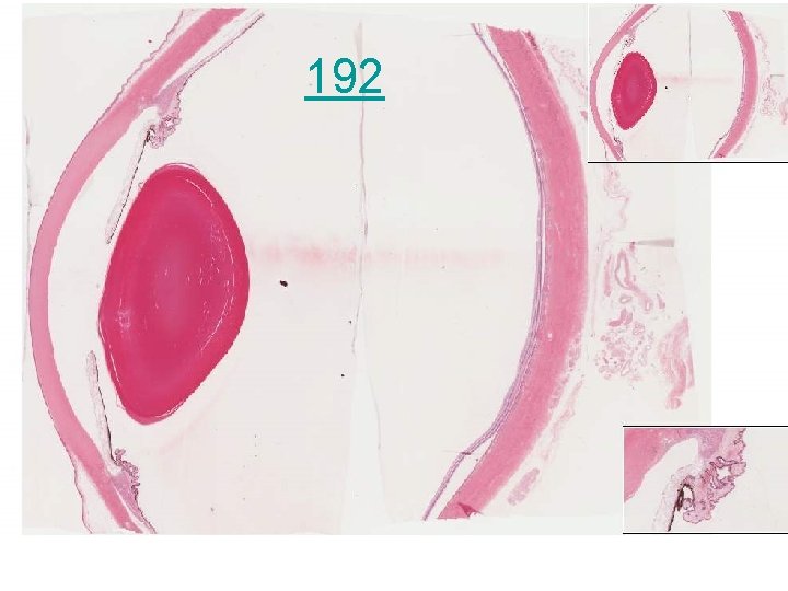
192
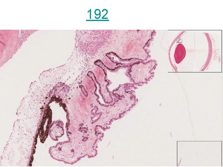
192
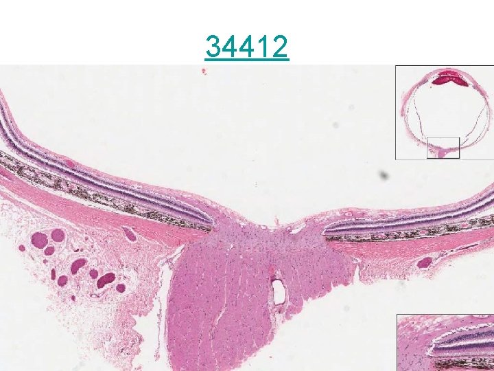
34412
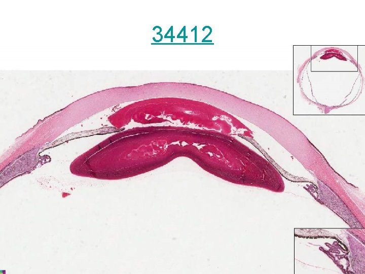
34412
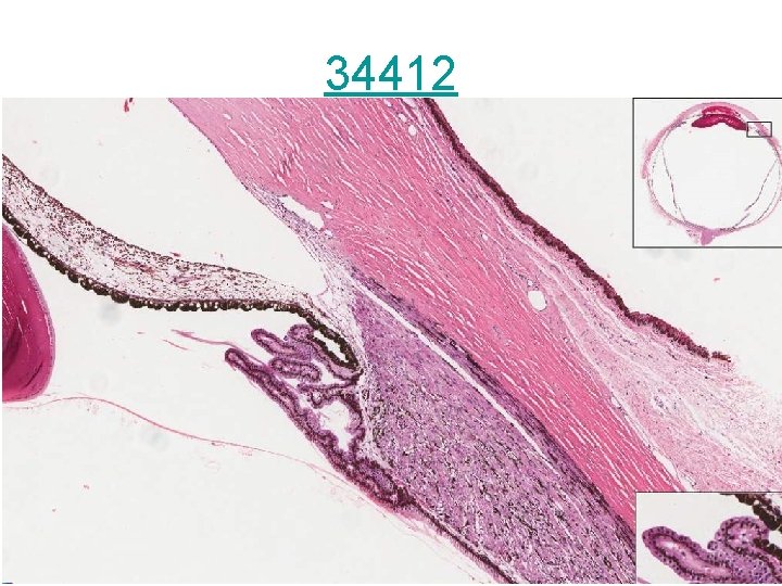
34412
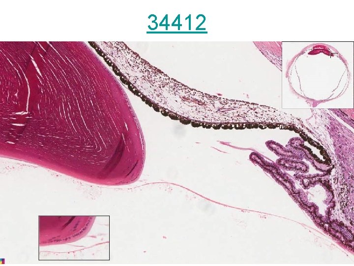
34412
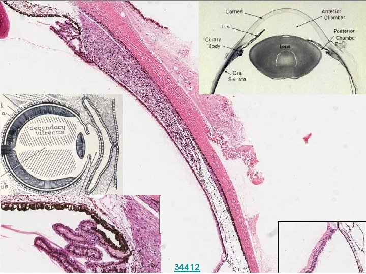
34412
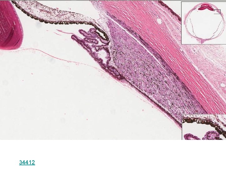
34412
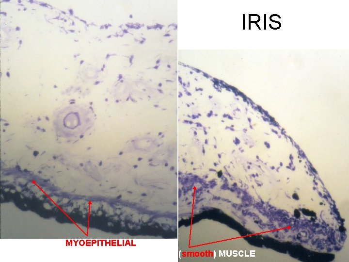
IRIS DILATOR (MYOEPITHELIAL) AND CONSTRICTOR (smooth) MUSCLE
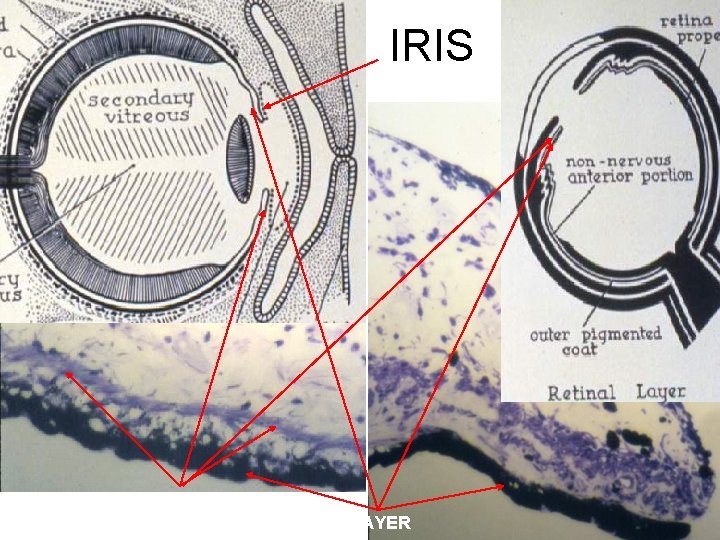
IRIS MYOEPITHELIAL PIGMENTED LAYER
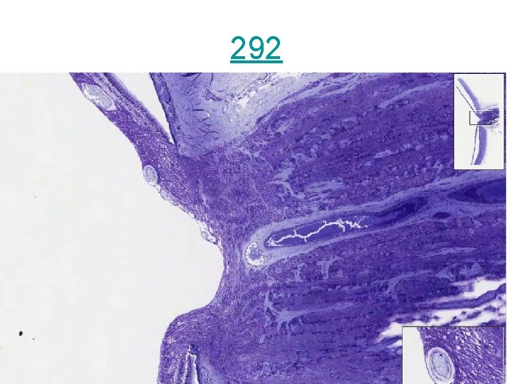
292
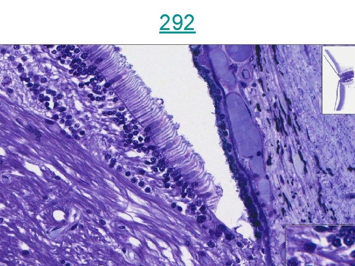
292
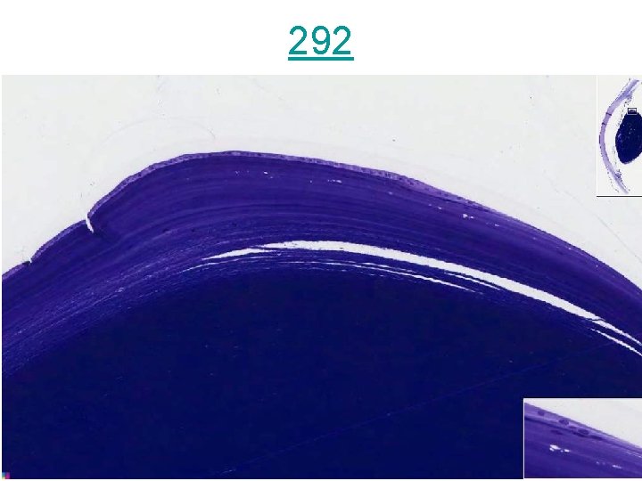
292
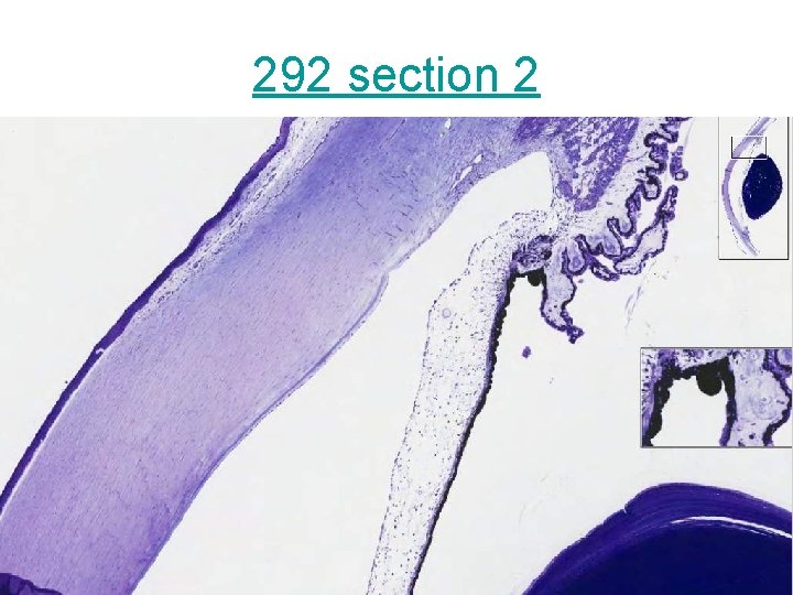
292 section 2
- Slides: 54