RETINA It has 10 layers interiortoExterior 1 Inner
 1. Inner limiting membrane. 2. Nerve fiber layer")


 1. Inner limiting membrane. 2. Nerve fiber layer")
- Slides: 15
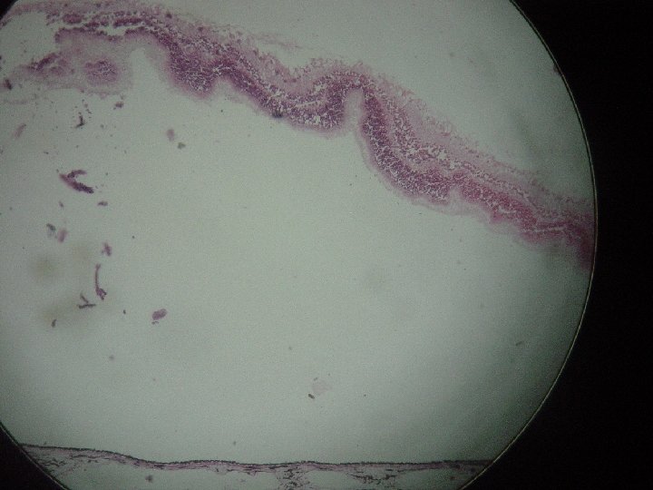
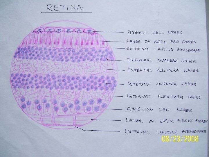
RETINA It has 10 layers (interior-to-Exterior) 1. Inner limiting membrane. 2. Nerve fiber layer 3. Layer of ganglion cells/optic nerve cells. 4. Inner plexiform layer. 5. Inner nuclear layer. 6. Outer plexiform layer. 7. Outer nuclear layer. 8. External limiting membrane. 9. Layers of rodes & cones 10. Pigment layer.
1. INNER LIMITING MEMBRANE. Neuroglial cells in retina are called retinal gliocytes. Protoplasmic processes of adjoining retinal gliocytes meet to form a thin internal-limiting membrane, internally on the inner surface of retina. It separates retina from vitreous humor.
2. NERVE FIBER LAYER It contains optic nerve fibres made of axons of ganglion cells. Nerve fibres converge on optic-disc, passes through foramina of lamina cribrosa to enter the optic nerve.
3. LAYER OF GANGLION CELLS/OPTIC NERVE CELLS. It contains cell bodies of ganglion cells. Dendrites of ganglion cells enter internal plexiform layer, synapse with processes of- bipolar cells & amacrine cells. Axons of bipolar cells form fibres of optic nerve.
4. INNER PLEXIFORM LAYER. Contains-processes of optic nerve cells and cells of inner nuclear layer which form synapse.
5. INNER NUCLEAR LAYER. It is composed of neurons of 3 types: Bipolar neurons. dendrites enter external layer to synapse axons of rod cells & cone-cells. Axons enter internal plexiform layer & synapse with dendrites of ganglion cell. Horizontal neurons. its processes runs parallel to to retinal surface. It enters outer plexiform layer. synapse with rods, cones, dendrites of bipolar cells. Amacrine neurons. lies horizontally in retina. Its processes enter inner plexiform layer. They synapse with axon of bipolar cells & dendrites of ganglion cells.
6. OUTER PLEXIFORM LAYER. It is also called- synaptic zone. It contains axons of rods & cones which synapse with dendrites of bipolar neuron (in the outer nuclear membrane) & horizontal cells.
7. OUTER NUCLEAR LAYER. It contains cell body and nuclei of rod cells & cone cells. They are- photo-receptos. Rods and cone cells-are modified neurons This neuron contains a)central process. b)peripheral process is rod-shaped in rod cells. peripheral process is cone-shaped in cone cells central process of rod & cone-cell forms axons. It extends into external plexiform layer.
8. EXTERNAL LIMITING MEMBRANE. Protoplasmic processes of adjoining retinal gliocytes meet to form thin external limiting membrane. It is formed at the junction of layer of rods & cones and external nuclear layer.
9. LAYERS OF RODS & CONES Processes of rod cells & cone cells are called rods & cones. Tip of processes is surrounded by processes of pigment cells.
10. PIGMENT LAYER It contains single layer of cells containing pigment. Processes from pigment cells extend into the next layer.
Pigment layer. Layers of rodes & cones. External limiting membrane. Outer nuclear layer Outer plexiform layer Inner plexiform layer. Layer of ganglion cells/ optic nerve cells. Nerve fiber layer Inner limiting membrane Layers of RETINA
RETINA It has 10 layers (interior-to-Exterior) 1. Inner limiting membrane. 2. Nerve fiber layer 3. Layer of ganglion cells/optic nerve cells. 4. Inner plexiform layer. 5. Inner nuclear layer. 6. Outer plexiform layer. 7. Outer nuclear layer. 8. External limiting membrane. 9. Layers of rodes & cones 10. Pigment layer.