INFECTIVE UVEITIS DR P PRADEEP PROFESSOR DEPARTMENT OF


, which is")


 Macular lesion; (B) multiple peripheral scars on wide-field")


 Typical ‘satellite’ lesion adjacent to an old")
 and")

 peripheral exudation and vitreoretinal traction bands (C) ultrasonography shows a")


 The fovea shows")


 Early macular involvement (B) established disease resulting from confluence of")


- Slides: 45
INFECTIVE UVEITIS DR. P. PRADEEP, PROFESSOR, DEPARTMENT OF OPHTHALMOLOGY ASRAM, ELURU
CLASSIFICATION § UVEITIS ASSOCIATED WITH BACTERIAL INFECTIONS: - Tubercular uveitis - Syphilitic uveitis - Leprotic uveitis § PARASITIC UVEITIS - Toxoplasmosis - Toxocariasis - Onchocerciasis - Amoebiasis § FUNGAL UVEITIS - Presumed Ocular Histoplasmosis Syndrome - Candidiasis § VIRAL UVEITIS -Herpes Simplex - Herpes Zoster - Acquired Cytomegalovirus Uveitis - Uveitis In AIDS
TUBERCULAR UVEITIS § Tb is a chronic granulomatous infection caused by bovine or human tubercle bacilli. § Can cause both anterior and posterior uveitis CLINICAL PRESENTATIONS: § Tubercular Anterior Uveitis § Tubercular Posterior Uveitis § Vasculitis
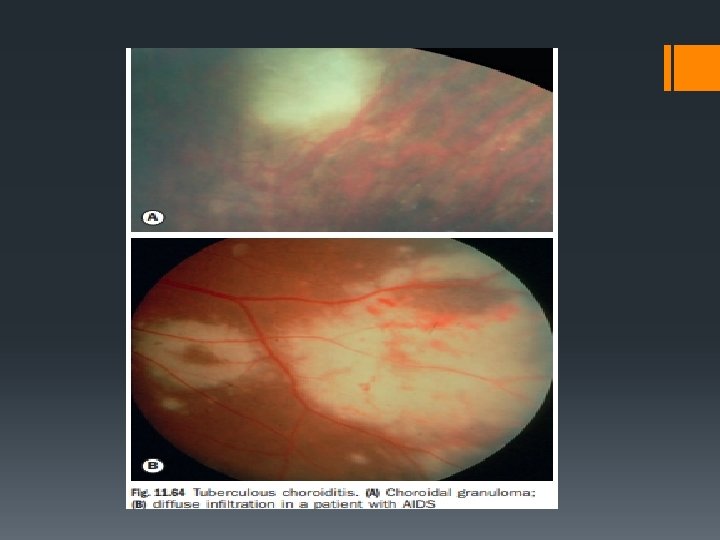
TUBERCULAR ANTERIOR UVEITIS: § May occur as acute non granulomatous iridocyclitis or granulomatous anterior uveitis which in turn may be in the form of miliary tubercular iritis or conglomerate granuloma. TUBERCULAR POSTERIOR UVEITIS: may occur as § Multiple miliary tubercles in the choroid which appear as round yellow white nodules usually associated with TB menigitis. § Diffuse or disseminated choroiditis in chronic TB. § Choroidal granuloma may occur rarely as a solitary lesion.
DIAGNOSIS: § Positive skin test § Associated findings of systemic TB. § Intractable uveitis unresponsive to steroid therapy § Positive response to Isoniazid(response of iritis to isoniazid 300 m OD for 3 weeks). TREATMENT: § Chemotherapy with rifampicin and isoniazid for 12 months along with usual treatment of uveitis.
ACQUIRED SYPHILITIC UVEITIS § It is a chronic venereal infection caused by Treponema pallidum which affects both anterior and posterior uvea. SYPHILITIC ANTERIOR UVEITIS: § Occur as acute plastic iritis or granulomatous iritis. § Acute plastic iritis occurs in secondary stage of syphilis and as Herxheimer reaction 24 -48 hours after therapeutic dose of penicillin. § Gummatous anterior uveitis occurs in secondary or rarely tertiary stage of syphilis. § It is characterised by formation of yellowish red highly vascularised miltiple nodules arranged near the pupillary border or ciliary border of iris. § SYPHILITIC POSTERIOR UVEITIS: § May occur as disseminated, peripheral or diffuse choroiditis.
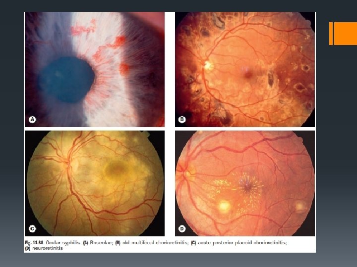
VARIOUS OCULAR MANIFESTATIONS IN ACQUIRED SYPHILIS § Chorioretinitis often multifocal , bilateral and associated with vitritis may develop. § Roseolae are dilated iris capillaries that may develop into yellowish nodules may be present. § Acute syphilitic posterior placoid chorioretinopathy (ASPPC) is characterized by large pale-yellowish subretinal lesions in the posterior pole. § Retinitis has a ‘ground glass’ appearance § Optic neuritis and neuroretinitis § Other features include conjunctivitis, episcleritis, intermediate uveitis, glaucoma, cataract and Argyll Robertson pupils
DIAGNOSIS: § Clinical detection § Confirmed by FTS-ABS test(fluoroscent treponemal antibody absorption), which is specific and sensitive than TPI and VDRL. TREATMENT: § Local therapy for uveitis § Systemic penicillin
LEPROTIC UVEITIS § Caused by Mycobacterium leprae, an acid-fast bacillus. § Occurs in 2 forms: § Lepromatous form: predominantly involves anterior uveitis. § Tuberculoid form § Clinically it may occur as acute iritis(nongranulomatous) or chronic iritis(granulomatous).
Acute iritis: § caused by antigen- antibody deposition. § Characterised by severe exudative reaction. Chronic granulomatous iritis: § Occurs due to direct organismal invasion. § Characterised by small glistening iris pearls near pupillary margin in a necklace form. § Small pearls enlarge and coalesce to form large pearls. TREATMENT: § Anti leprotic treatment with Dapsone 50 -100 mg daily. § Local therapy of uveitis.
PARASITIC UVEITIS
TOXOPLASMOSIS § Protozoan infestation caused by Toxoplama gondii. § Definitive host: cats § Intermediate host: humans, cattle, sheep, pigs § Disease primarily affects CNS(brain and retina). § Systemic lesions are more marked in immunocompromised patients. § MC cause of posterior uveitis. § Accounts for 90% of focal necrotizing retinitis. § Occurs in 3 forms: - congenital toxoplasmosis - acquired toxoplasmosis - recurrent toxoplasmic retinochoroiditis
LIFE CYCLE OF TOXOPLASMA
CONGENITAL TOXOPLASMOSIS: § More common than acquired form. § Acquired by foetus through transplacental route from mother contracting acute infestaton during pregnancy. § Triad of congenital toxoplasmosis include: - Convulsions - Chorioretinitis - Intracranial calcification § In active stage the typical lesion is necrotic granulomatous retinochoroiditis involving macular region with vitritis. § inactive stage is characterised by b/l healed punched out heavily pigmented chorioretinal scars in macular area.
Retinochoroidal scars in congenital toxoplasmosis. (A) Macular lesion; (B) multiple peripheral scars on wide-field imaging;
ACQUIRED TOXOPLAMOSIS: § acquired by eating under cooked meat of intermediate host containing cyst form of the parasite. § Most cases are subclinical § Lesions similar to congenital toxoplamosis. § Other lesions include punctate outer retinal toxoplasmosis. § More common in HIV+ patients
§ RECURRENT TOXOPLASMIC RETINOCHOROIDITIS § pathogenesis: parasite reach foetus through placenta § involve brain and retina § excite antibodies formation § healing of active retinal lesion § retinal cysts rupture after 10 -40 yrs § release hundreds of parasites § fresh lesion of focal necrotizing retinochoriditis
CLINICAL FEATURES: § Characterised by whitish yellow , slightly raised area of infiltration located near the margin of old punched out scarred lesion in the macular region associated with severe vitritis. § There may be associated non granulomatous type of mild anterior uveitis. DIAGNOSIS: § Confirmed by indirect fluorescein antibody test, haemagglutination test, ELISA test. TREATMENT: § Active lesions treated by topical and sytemic steroids. § Antitoxoplasmic drugs like spiramycin, clindamycin, sulfadiazine or pyremethamine can be given.
Other ocular features Active Toxoplasma retinitis. (A) Typical ‘satellite’ lesion adjacent to an old scar (B) two small foci severe vitreous haze and ‘headlight in the fog’ appearance of lesion
TOXOCARIASIS: § It is an infestation caused by intestinal roundworm of dogs(toxocara canis) and cats(Toxocara catis). § young children who play with dogs and cats are infested by ova of these worms. § Ova develop into larva in the human gut and produce condition called visceral larva migrans. OCULAR TOXOCARIASIS: § It is ocular infestation by the larva. § Always U/L
Clinical presentation: § Toxocara chronic endophthalmitis: § Presents with leucocoria due to marked vitreous clouding. § Seen in children between 2 -10 yrs of age. § Mimics retinoblastoma. Posterior pole granuloma: § Presents as yellow, round, solitary, raised nodule about 1 -2 disc diameter in size, located either at macula or centrocaecal area. § Seen in children between 5 and 15 yrs of age. § Presents with U/L loss of vision.
Chronic Toxocara endophthalmitis. (B) peripheral exudation and vitreoretinal traction bands (C) ultrasonography shows a vitreoretinal traction band Juxtapapillary granuloma
Peripheral granuloma: § Situated anterior to equator and associated with vitreous band formation. § Presents from 6 -40 yrs of age. DIAGNOSIS: § Clinical picture § ELISA TREATMENT: § Consists of periocular injection of steroids and systemic steroids. § Pars plana vitrectomy in unresponsive patients with endophthalmitis and with vitreous band formation. peripheral granuloma
FUNGAL UVEITIS
PRESUMED OCULAR HISTOPLASMOSIS SYNDROME § Causative agent: Histoplasma capsulatum CLINICAL FEATURES: Histospots: § Atrophic spots scattered in mid retinal periphery. § Rounded yellowish white lesions measuring 0. 2 to 0. 7 DD. § Appear in childhood and represent scars of disseminated histoplasma choroiditis. Macular lesion: § Starts as atrophic macular scar hole in Brucs membrane subretinal choroidal neovascularisation. § Leakage of fluid from neovascular membrane causes serous detachment. § Repeated haemorrhages leads to haemorrhagic detachment. § Fibrous disciform scar develops associated with marked permanent visual loss.
Peripheral ‘histo’ spots Choroidal neovascularization in presumed ocular histoplasmosis. (A) The fovea shows a focal area of oedema and a few small haemorrhages, and a small histo spot temporally
DIAGNOSIS: § Fluorescein angiography reveals subretinal neovascular membrane. TREATMENT: § Active lesions at macula treated with systemic corticosteroids. § Early laser photocoagulation of subretinal neovascular membrane prevent marked permanent visual loss. § For subfoveal membrane - PDT should be considered.
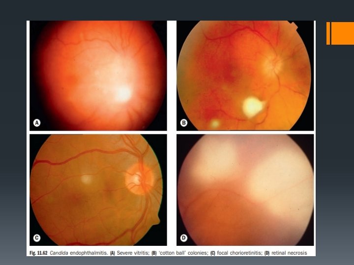
§ CANDIDIASIS § It is an opportunistic infection caused by Candida albicans. § Occurs in immunocompromised patients. § Ocular candidiasis: manifestations include§ Anterior uveitis associated with hypopyon. § Multifocal chorioretinitis is a more common lesion characterised by multiple, small, round, whitish areas associated with areas of haemorrhages with pale centre(Roth spots). § Candida endophthalmitis characterised by areas of severe retinal necrosis associated with vitreoretinal abscess. § Vitreous exudates present as puff ball or cotton ball colonies which when joined by exudative strands form string of pearls.
TREATMENT: § Topical cyclopegics , antifungal drugs § Systemic antifungal drugs like ketoconazole, flucytosine, amphotericin – B can be given. § Pars plana vitrectomy.
VIRAL UVEITIS
UVEITIS IN HERPES ZOSTER OPHTHALMICUS: § It is involvement of ophthalmic division of fifth nerve by varicella zoster. § Anterior uvetis develops in 40 -50% cases with HZO within 2 weeks of onset of skin rashes. § Mild iritis present in patients with vesicular eruption on tip of nose. § Iridocyclitis is non-granulomatous characterised by presence of small KPs, mild aqueous flare and ocassional haemorrhage hypopyon. § COMPLICATIONS: § Iris atrophy § Secondary glaucoma § Complicated cataract § TREATMENT: topical steroids, cyclopegics for several months. systemic acyclovir for early lesions.
HERPES SIMPLEX UVEITIS § Associated with keratitis, geographical or dendritic corneal ulceration. § Anterior uveitis may occur even without keratitis. § It is a mild grade non-granulomatous iridocyclitis excited by hypersensitivity reaction. TREATMENT: § Antivirals § Cyclopegics § Steroids C/I in case of active viral ulcers
CYTOMEGALOVIRUS RETINITIS § Occurs in immunocompromised patients. § Most frequent ocular opportunistic infection in patients with AIDS. § Occurs only when CD 4+ counts < 50 cells/mm³. § CLINICAL FEATURES: § Often asymptomatic , patients may present with scotoma or decreased vision. § Anterior segment signs are absent. § Posterior segment signs include: Haemorrhagic retinitis § - characterised by areas of necrosis - areas of perivasculitis(perivascular sheathing) - retinal haemorrhages § - lesions present posteriorly appear alongnretinal vessels as large areas of thick white infilrate with retinal haemorrhages(pizza pie appearance or cheese pie) atypical presentation in fulminant form. Granular retinitis characterised by peripheral granular opacities with haemorrhage.
Pizza pie appearance White granular retinitis with intraretinal hemorrhage
Early necrosis at periphery Attenuation of vessels in area affected by retinitis Retinitis starts in mid periphery and progress in bush fire pattern
COMPLICATIONS: § Retinal detachment § Retinal atrophy § Optic nerve disease TREATMENT: § HAART therapy to reduce retroviral load and to increase CD 4 count. § Specific anti CMV drugs include valaganciclovir, foscarnet, iv injections of cidifovir
ACUTE RETINAL NECROSIS § It is a rare clinical syndrome caused by any of the following viruses: § Varicella zoster virus: it is commonest cause of ARN § Herpes simplex 1 & 2 in younger patients. § CMV – rarely CLINICAL FEATURES: § Patient c/o decreased visual acuity, floaters, ocular discomfort, pain, photophobia. § Signs: anterior segment may or may not show markers of inflammation. § Vitritis § Retinal lesions include areas of focal , well demarcated peripheral necrotising retinitis, full-thickness retinal necrosis
COMPLICATIONS: § Retinal detachment § Ischemic optic neuropathy. TREATMENT: § Systemic antiviral drugs include acyclovir i. v. 10 mg/kg body weight TID for 2 weeks then PO dose for 6 -12 weeks. § Systemic steroids § Aspirin to prevent arterial occlusion § Barrier laser photocoagulation for retinal breaks § Vitrectomy with silicon oil injection for retinal detachment
PROGRESSIVE OUTER RETINAL NECROSIS: § Rare devastating necrotizing retinitis caused by VZV infection in immunocompromised patients. C/F: § Painless rapid loss of vision § Retinal lesions include rapidly coalescing white areas of outer retinal necrosis. § TREATMENT: § iv injection of ganciclovir or foscarnet combined with intravitreal ganciclovir.
Progressive retinal necrosis. (A) Early macular involvement (B) established disease resulting from confluence of multiple foci – there is little or no haemorrhage
THANKQ