POSTERIOR UVEITIS Dr Anupam Associate Professor AIIMS Rishikesh


 International Uveitis Study Group 1) Anterior Uveitis – Inflammation")
 Acute – sudden symptomatic onset. Persists for 3 weeks")
 Exogenous. Introduction of organism into the eye through a")
 Granulomatous 2) Non-granulomatous")
. • Since")
 • • II.")


endophthalmitis • Sterile endophthalmitis refers to inflammation of inner structures of eyeball")


 SYNDROME • It is an idiopathic multisystem disorder which includes cutaneous, neurological")


 eye. It shows clinical features of persistent low")


 • It denotes inflammation of pars plana part of ciliary")


- Slides: 60
POSTERIOR UVEITIS Dr Anupam Associate Professor AIIMS Rishikesh
Definition & Classification of Uveitis • Inflammation of uveal tract is uveitis. • CLASSIFICATION: • • I. ANATOMICAL CLASSIFICATION II. CLINICAL CLASSIFICATION III. ETIOLOGICAL CLASSIFICATION IV. PATHOLOGICAL CLASSIFICATION
A. Anatomical Classification – (IUSG) International Uveitis Study Group 1) Anterior Uveitis – Inflammation of iris and anterior part of ciliary body. 2) Intermediate Uveitis – Involvement of posterior part of ciliary body and extreme periphery of retina. (Pars planitis) 3) Posterior uveitis – Retinochoroiditis, retinitis, chorioretinitis 4) Diffuse or pan uveitis – Involvement of entire uveal tract
B. Clinical Classification • 1) Acute – sudden symptomatic onset. Persists for 3 weeks or less. • 2) Chronic – Frequently insidious and asymptomatic. Persists for months or years. • 3) Recurrent
C. Etiological Classification • 1) Exogenous. Introduction of organism into the eye through a perforating wound or ulcer. Acute iridocyclitis of suppurative type, panophthalmitis. • 2) Secondary infection- Due to direct spread from adjoining structures— Cornea — Sclera — Retina
D. Pathological Classification 1) Granulomatous 2) Non-granulomatous
POSTERIOR UVEITIS • Posterior uveitis refers to inflammation of the choroid (choroiditis). • Since the outer layers of retina are in close contact with the choroid and also depend on it for the nourishment, the choroidal inflammation almost always involves the adjoining retina and the resultant lesion is called chorioretinitis.
Clinical types • I. Suppurative choroiditis (Purulent inflammation of the choroid) • • II. Non-suppurative choroiditis 1. Diffuse choroiditis. 2. Disseminated choroiditis. 3. Circumscribed/localised/focal choroiditis. i. Central choroiditis. ii. Juxtacaecal or juxtapapillary choroiditis. iii. Anterior peripheral choroiditis. iv. Equatorial choroiditis.
Symptoms • • 1. Defective vision. 2. Photopsia. 3. Black spots floating in front of the eyes. 4. Metamorphopsia. 5. Micropsia 6. Macropsia 7. Positive scotoma
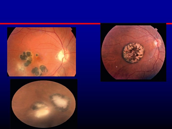
Signs • 1. Vitreous opacities • 2. Features of a patch of choroiditis. i. In active stage it looks as a pale-yellow or dirty white raised area with ill-defined edges. This results due to exudation and cellular infiltration of the choroid which hide the choroidal vessels. The lesion is typically deeper to the retinal vessels. The overlying retina is often cloudy and oedematous.
Signs • ii. In atrophic stage or healed stage, when active inflammation subsides, the affected area becomes more sharply defined and delineated from the rest of the normal area. The involved area shows white sclera below the atrophic choroid and black pigmented clumps at the periphery of the lesion.
Complications These include extension of the inflammation to anterior uvea, complicated cataract, vitreous degeneration, macular oedema, secondary periphlebitis retinae and retinal detachment.
Treatment • It is broadly on the lines of anterior uveitis. • 1. Non-specific therapy: In the form of topical and systemic steroids. • 2. Specific treatment is required for the causative disease.
PURULENT UVEITIS • Purulent uveitis is suppurative inflammation of the uveal tract occurring as a result of direct invasion by the pyogenic organisms. • • Purulent iridocylitis Purulent choroiditis Endophthalmitis Panophthalmitis
ENDOPHTHALMITIS • Endophthalmitis is defined as an inflammation of the inner structures of the eyeball i. e. , uveal tissue and retina associated with pouring of exudates in the vitreous cavity, anterior chamber and posterior chamber. • Etiologically endophthalmitis may be infectious or non-infectious (sterile).
A. Infective endophthalmitis • Exogenous infections. • Endogenous or metastatic endophthalmitis. • Secondary infections from surrounding structures.
Causative organisms • 1. Bacterial endophthalmitis. S. epidermidis and S. aureus, Streptococci, Pseudomonas, Pneumococci, Cornebacterium.
Propionio bacterium acnes and actinomyces are gram-positive organisms capable of producing slow grade endophthalmitis. • 2. Fungal endophthalmitis is caused by aspergillus, fusarium, candida etc.
B. Non-infective (sterile)endophthalmitis • Sterile endophthalmitis refers to inflammation of inner structures of eyeball caused by certain toxins/toxic substances. It occurs in following situations. • 1. Postoperative sterile endophthalmitis • 2. Post-traumatic sterile endophthalmitis • 3. Intraocular tumour • 4. Phacoanaphylactic endophthalmitis
Symptoms. • Acute bacterial endophthalmitis usually occurs within 7 days of operation and is characterized by severe ocular pain, redness, lacrimation, photophobia and marked loss of vision.
Signs • Lid swelling • Conjunctival chemosis and congestion • Corneal edema • Wound gape & necrosis • Hypopyon • Iris becomes edematous and muddy • Yellow reflex • IOP may or may not be raised
Treatment • Intravitreal antibiotics , S/C injections, Topical fortified antibiotic drops & oral antibiotics. • Cycloplegics • Antiglaucoma therapy • Vitrectomy
PANOPHTHALMITIS • It is an intense purulent inflammation of the whole eyeball including the Tenon’s capsule. • • • Symptoms. Severe ocular pain and headache Complete loss of vision Watering & discharge Marked redness and swelling of the eyes
Signs • Lids swelling. • Eyeball is slightly proptosed, ocular movements are limited & painful. • Ciliary as well as conjunctival congestion. • Cornea is cloudy and oedematous. • Anterior chamber is full of pus. • Vision is completely lost • Intraocular pressure is markedly raised.
• Complications include • Orbital cellulitis • Cavernous sinus thrombosis • Meningitis or encephalitis
Treatment • 1. Anti-inflammatory and analgesics • 2. Broad spectrum antibiotics • 3. Evisceration
Non-suppurative Uveitis • I. UVEITIS ASSOCIATED WITH CHRONIC SYSTEMIC BACTERIAL INFECTIONS: 1. Tubercular uveitis 2. Syphilitic uveitis 3. Leprotic uveitis • UVEITIS ASSOCIATED WITH NONINFECTIOUS SYSTEMIC DISEASES 1. Uveitis in sarcoidosis 2. Behcet’s disease.
Non-suppurative Uveitis • III. UVEITIS ASSOCIATED WITH ARTHRITIS 1. Uveitis with Ankylosing spondylitis 2. Reiter’s syndrome 3. Still’s disease • IV. PARASITIC UVEITIS 1. Toxoplasmosis 2. Toxocariasis 3. Onchocerciasis 4. Amoebiasis
Non-suppurative Uveitis • V. FUNGAL UVEITIS 1. Presumed ocular histoplasmosis syndrome 2. Candidiasis • VI. VIRAL UVEITIS 1. Herpes simplex uveitis 2. Herpes zoster uveitis 3. Acquired cytomegalovirus uveitis 4. Uveitis in acquired immune deficiency syndrome (AIDS)
Non-suppurative Uveitis • VII. LENS INDUCED UVEITIS 1. Phacotoxic uveitis 2. Phacoanaphylactic endophthalmitis • VIII. TRAUMATIC UVEITIS • IX. UVEITIS ASSOCIATED WITH MALIGNANT INTRAOCULAR TUMOURS
Non-suppurative Uveitis • IDIOPATHIC SPECIFIC UVEITIS SYNDROMES 1. Fuchs’ uveitis syndrome 2. Intermediate uveitis (pars planitis) 3. Sympathetic ophthalmitis 4. Glaucomatocyclitic crisis. 5. Vogt-Koyanagi-Harada’s syndrome. 6. Bird shot retinochoroidopathy. 7. Acute multifocal placoid pigment epitheliopathy (AMPPE) 8. Serpiginous choroidopathy.
VOGT-KOYANAGI-HARADA (VKH) SYNDROME • It is an idiopathic multisystem disorder which includes cutaneous, neurological and ocular lesions, more common in Japanese who are usually positive for HLADR 4 and DW 15. • Clinical features • 1. Cutaneous lesions include: alopecia, poliosis and vitiligo. • 2. Neurological lesions are in the form of meningism, encephalopathy, tinnitus, vertigo and deafness. • 3. Ocular features are bilateral chronic granulomatous panuveitis and exudative retinal detachment. • Treatment. It comprises steroids administered topically, periocularly and systemically.
SYMPATHETIC OPHTHALMITIS • It is a rare bilateral granulomatous panuveitis. • Occurs following penetrating ocular trauma associated with incarceration of uveal tissue in the wound. • The injured eye is called ‘exciting eye’ and the fellow eye which also develops uveitis is called ‘sympathizing eye’.
• A. Predisposing factors • 1. It almost always follows a penetrating wound. • 2. Wounds in the ciliary region • 3. Wounds with incarceration of the iris, ciliary body or lens capsule are more vulnerable. • 4. It is more common in children than in adults. • 5. It does not occur when actual suppuration develops in the injured eye.
• Dalen-Fuchs’ nodules are formed due to proliferation of the pigment epithelium (of the iris, ciliary body and choroid) associated with invasion by the lymphocytes and epitheloid cells.
Clinical picture • I. Exciting (injured) eye. It shows clinical features of persistent low grade plastic uveitis, which include ciliary congestion, lacrimation and tenderness. • 2. Sympathizing (sound) eye. It is usually involved after 4 -8 weeks of injury in the other eye. Can occur upto 1 year after injury.
• Retrolental flare is first clinical sign. • If not prevented loss of sight is inevitable • Prophylaxis • Treatment.
Ocular Toxoplasmosis Clinical features Unilateral focal chorio-retinitis adjacent to healed chorioretinal scar a. Healed scars may be multiple, but usually only one reactivates at a time. b. Atypical forms of extensive chorio-retinitis can occur in immunocompromised individuals. c. Active chorio-retinitis is yellow-white, slightly elevated, with a relatively well-defined border.
Clinical features: Intraocular inflammation a. Iritis. Often granulomatous & may be associated with ocular hypertension b. Vitritis. Often intensified over the lesion c. Vasculitis. Variably present; Often arteritis, but also periphlebitis and the vasculitis can be remote from the chorio-retinitis d. Optic neuritis or neuro-retinitis.
Appropriate Laboratory Testing 1. Conformation of exposure to toxoplasmosis by serum ab. titer; high sensitivity and low specificity because of high prevalence of positive antibody titers in general population 2. Determination of toxoplasmosis Ig. G or Ig. A titers in aqueous humor useful in cases with atypical features 3. PCR of aqueous humor for toxoplasmosis DNA useful in older patients with large lesions or in immunocompromised patients
Risk of congenital infection Acquired toxoplasmosis infection in a pregnant woman a. Most severe effects on fetus if acquired during first trimester b. Risk of transmission greatest if acquired during third trimester Seroconversion treated with antibiotic therapy Prenatal treatment reduces fetal effects.
Differential Diagnosis 1. Toxocariasis 2. Cytomegalovirus retinitis 3. Necrotizing herpetic retinitis 4. Syphilis 5. Focal fungal or bacterial infections 6. Intraocular lymphoma
Treatment Decision to treat based on proximity to macula and optic nerve, amount of inflammation, and vision 1. Sight-threatening infections almost always treated. 2. Small, peripheral lesions often observed. 3. Infection is self-limited in most cases in healthy patients.
Treatment 1. Pyrimethamine is most common agent combined with sulfadiazine or triple-sulfa, azithromycin, or clindamycin Usually given with leukovorin to mitigate hematologic toxicity 2. Trimethoprim-sulfamethoxazole increasing in use combined with clindamycin for increased efficacy 3. Monotherapy-generally reserved for non-sight threatening disease; Doxycycline or minocycline
Anti-inflammatory Treatment Topical corticosteroids Oral corticosteroids Indicated for vision threatening inflammation Low to moderate doses for 2 to 3 weeks Not given alone because of risk of worsened infection without antibiotic coverage Periocular steroids felt to be contraindicated because of reports of uncontrolled infection after injection Generally not used in immunocompromised patients
INTERMEDIATE UVEITIS (PARS PLANITIS) • It denotes inflammation of pars plana part of ciliary body and most peripheral part of the retina. • Etiology. It is an idiopathic disease usually affecting both eyes of young adults. • Symptoms. Most of the patients present with history of floaters. Some patients may come with defective vision due to associated cystoid macular oedema.
Signs • Fundus examination with indirect ophthalmoscope reveals the whitish exudates present near the ora serrata in the inferior quadrant. These typical exudates are referred as snow ball opacities. These may coalesce to form a grey white plaque called snow banking.
Complications • Cystoid macular oedema, • Complicated cataract and • Tractional retinal detachment • Treatment 1. Corticosteroids ( Topical or systemic) 2. Immunosuppressive drugs may be helpful in steroid resistant cases. 3. Peripheral cryotherapy is also reported to be effective.
MCQ 1. A 60 yr old female having h/o cataract surgery 1 yr back. She had complain of sudden onset painful diminution of vision and was diagnosed as a case of late onset endophthalmitis. Organism most commonly implicated is? • A. Pseudomonas aeruginosa • B. Staphylococcus epidermidis • C. Candida albicans • D. Propionibacterium acnes
MCQ 2. All of the following are involved in endophthalmitis except? • A. Retina • B. Vitreous • C. Sclera • D. Uvea
MCQ • 3. A 30 yr old female had complains of floaters with blurring of vision. On indirect ophthalmoscopy pt had clear central fundus but there was inflammation of the pheripheral part. What is your diagnosis? • • A. Panuveitis B. Posterior uveitis C. Endophthalmitis D. Pars planitis
MCQ • 4. A 50 yr old female having h/o cataract surgery 1 week back. She had complains of sudden onset painful diminution of vision, redness & watering. On clinical examination she had hypopyon, AC cells and flare with vitreous exudates on USG. What is your diagnosis? • A. Panuveitis • B. Posterior uveitis • C. Endophthalmitis • D. Panophthalmitis •
MCQ • 5. A 30 yrs old male had history of trauma followed by loss of vision, severe pain, redness and watering. On clinical examination he had inflammation of all ocular structures including tenon’s capsule. What is your diagnosis? • A. Panuveitis • B. Posterior uveitis • C. Endophthalmitis • D. Panophthalmitis
MCQ • 6. A 50 yr old female having h/o cataract surgery 1 week back. She had complains of sudden onset painful diminution of vision, redness & watering. On clinical examination she had hypopyon, AC cells and flare with vitreous exudates on USG. Which is the most common organism responsible for the condition? • • A. Pseudomonas aeruginosa B. Staphylococcus epidermidis C. Candida albicans D. Propionibacterium acnes
MCQ • 7. A 15 yrs old male had history of penetrating trauma to right eye followed by loss of vision, severe pain, redness and watering in the left eye 3 months after trauma. On clinical examination he retrolental flare with signs of panuveitis. What is your diagnosis? • A. Sympathetic ophthalmitis • B. Posterior uveitis • C. Endophthalmitis • D. Panophthalmitis
MCQ • 8. What is the most common cause of loss of vision in pars planitis? • • A. Panuveitis B. Posterior uveitis C. Cystoid macular edema D. Floaters
MCQ 9. Snow banking is seen in: A. Panuveitis B. Posterior uveitis C. Endophthalmitis D. Pars planitis
MCQ • 10. Dalen-Fuchs’ nodules are seen in: • • A. Sympathetic ophthalmitis B. Posterior uveitis C. Endophthalmitis D. Panophthalmitis
THANK YOU