
Infective endocarditis Olcay ZVEREN M D Definition Infective


- Slides: 85
Infective endocarditis Olcay ÖZVEREN, M. D.
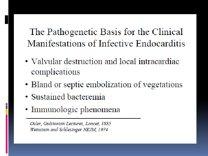
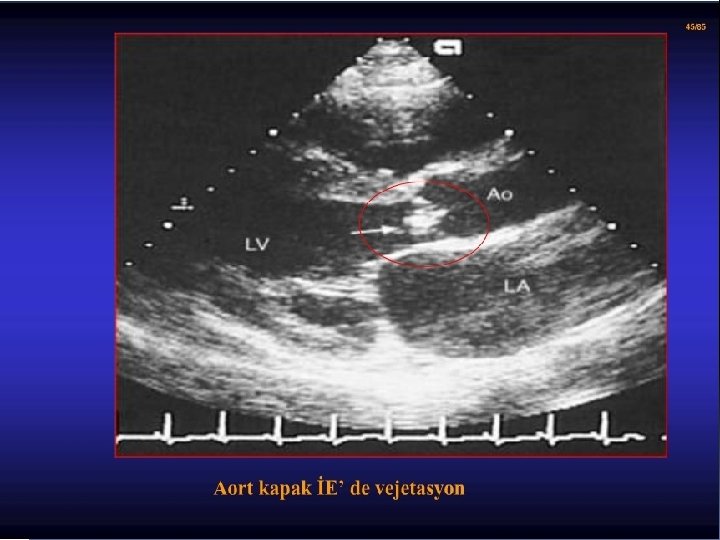
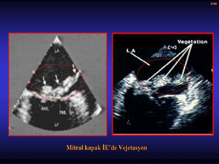
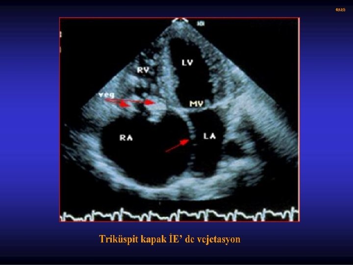
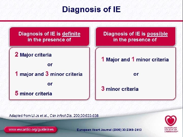
Definition Infective endocarditis is characterized by colonization or invasion of the heart valves or the mural endocardium by a microbe, leading to the formation of bulky, friable vegetations composed of thrombotic debris and organisms, often associated with destruction of the underlying cardiac tissues
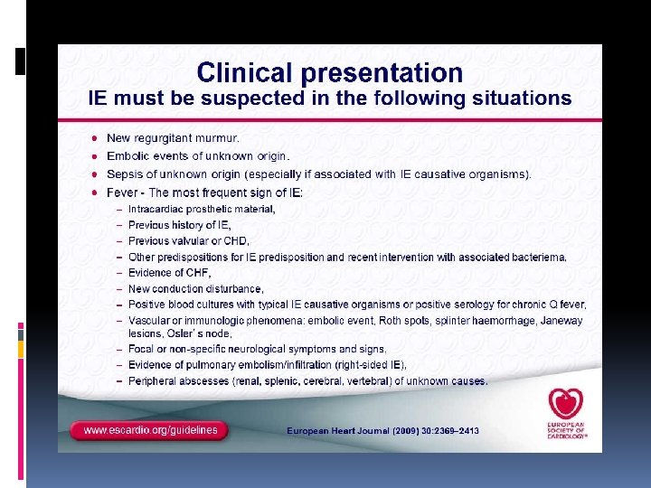
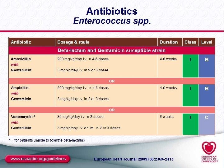
A changing epidemiology: Once a disease affecting young adults with previously well-identified (mostly rheumatic) valve disease, IE is now affecting older patients who more often develop IE as the result of health care-associated procedures, either in patients with no previously known valve disease or in patients with prosthetic valves. Trials showed an increasing incidence of IE associated with a prosthetic valve, an increase in cases with underlying mitral valve prolapse, and a decrease in those with underlying rheumatic heart disease. Newer predisposing factors have emerged—valve prostheses, degenerative valve sclerosis, intravenous drug abuse—associated with increased use of invasive procedures at risk for bacteraemia, resulting in health care-associated IE.
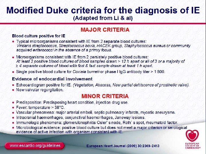
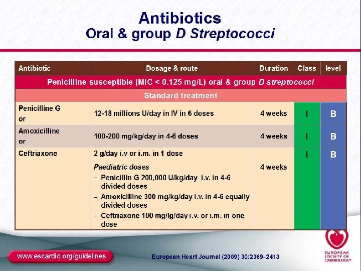
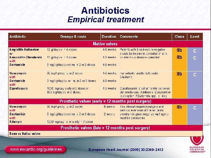
3– 10 episodes/100 000 person-years. increased dramatically with age. The male: female ratio is 2: 1. Positive blood cultures representing 85% of all IE. Causative microorganisms are most often staphylococci, streptococci, and enterococci. S. Aureus is the most frequent cause not only of IE but also of prosthetic valve IE. Conversely, coagulasenegative staphylococci can also cause native valve IE. Especially S. lugdunensis, which frequently has an aggressive clinical course. Oral (formerly viridans) streptococci species such as S. sanguis, S. mitis, S. salivarius, S. mutans, and Gemella morbillorum. Microorganisms of this group are almost always susceptible to penicillin G. Infective endocarditis with negative blood cultures because of prior antibiotic treatment.
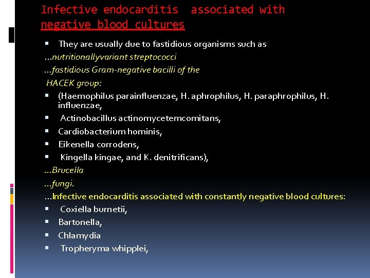
Infective endocarditis associated with negative blood cultures They are usually due to fastidious organisms such as …nutritionallyvariant streptococci …fastidious Gram-negative bacilli of the HACEK group: (Haemophilus parainfluenzae, H. aphrophilus, H. paraphrophilus, H. influenzae, Actinobacillus actinomycetemcomitans, Cardiobacterium hominis, Eikenella corrodens, Kingella kingae, and K. denitrificans), …Brucella …fungi. …Infective endocarditis associated with constantly negative blood cultures: Coxiella burnetii, Bartonella, Chlamydia Tropheryma whipplei,
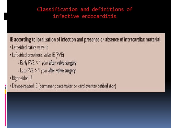
Classification and definitions of infective endocarditis
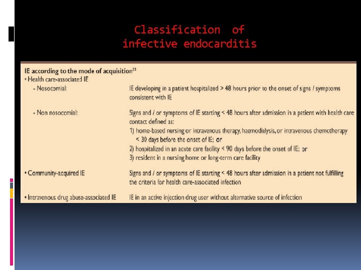
Classification of infective endocarditis
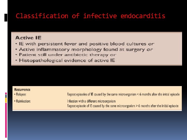
Classification of infective endocarditis
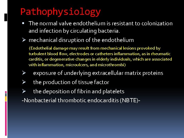
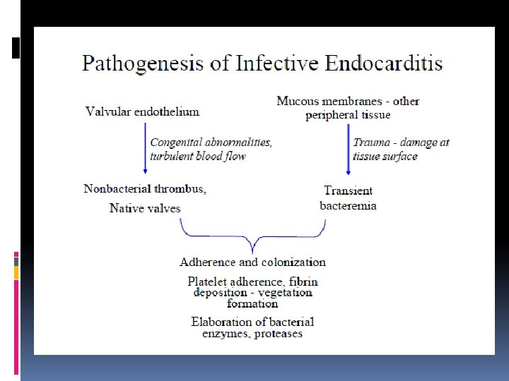
Pathophysiology The normal valve endothelium is resistant to colonization and infection by circulating bacteria. Ø mechanical disruption of the endothelium (Endothelial damage may result from mechanical lesions provoked by turbulent blood flow, electrodes or catheters inflammation, as in rheumatic carditis, or degenerative changes in elderly individuals, which are associated with inflammation, microulcers, and microthrombi) exposure of underlying extracellular matrix proteins Ø the production of tissue factor Ø the deposition of fibrin and platelets -Nonbacterial thrombotic endocarditis (NBTE)Ø
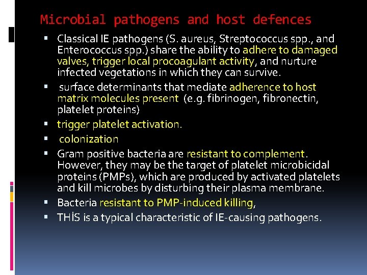
Microbial pathogens and host defences Classical IE pathogens (S. aureus, Streptococcus spp. , and Enterococcus spp. ) share the ability to adhere to damaged valves, trigger local procoagulant activity, and nurture infected vegetations in which they can survive. surface determinants that mediate adherence to host matrix molecules present (e. g. fibrinogen, fibronectin, platelet proteins) trigger platelet activation. colonization Gram positive bacteria are resistant to complement. However, they may be the target of platelet microbicidal proteins (PMPs), which are produced by activated platelets and kill microbes by disturbing their plasma membrane. Bacteria resistant to PMP-induced killing, THİS is a typical characteristic of IE-causing pathogens.
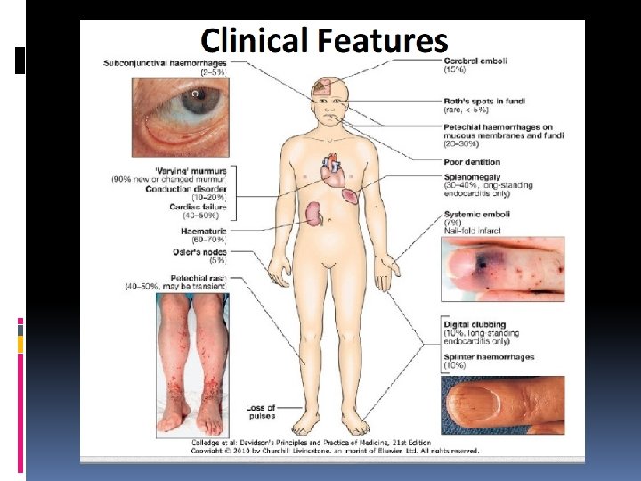
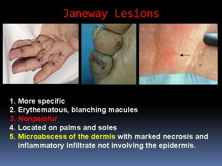
Janeway Lesions 1. More specific 2. Erythematous, blanching macules 3. Nonpainful 4. Located on palms and soles 5. Microabscess of the dermis with marked necrosis and inflammatory infiltrate not involving the epidermis.
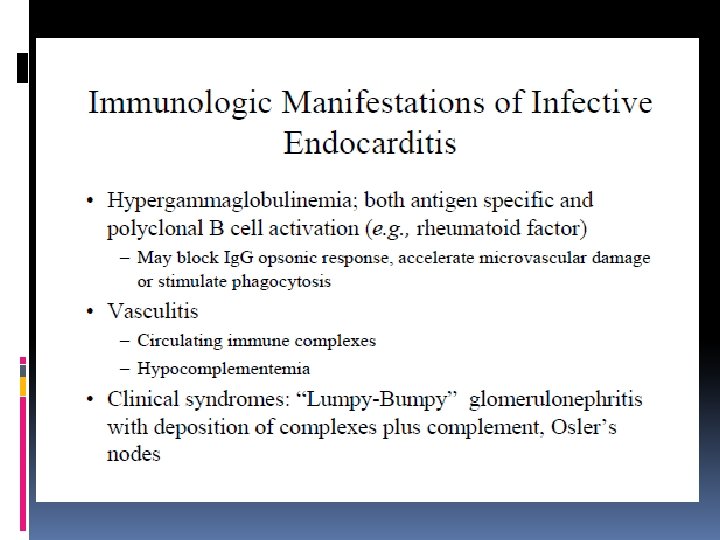
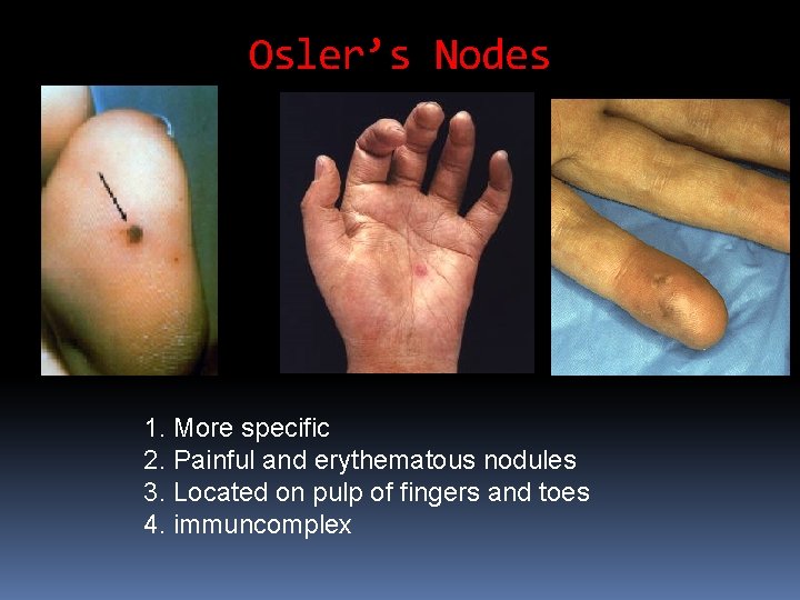
Osler’s Nodes 1. More specific 2. Painful and erythematous nodules 3. Located on pulp of fingers and toes 4. immuncomplex
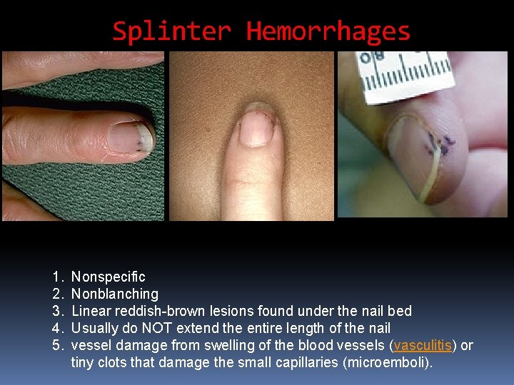
Splinter Hemorrhages 1. 2. 3. 4. 5. Nonspecific Nonblanching Linear reddish-brown lesions found under the nail bed Usually do NOT extend the entire length of the nail vessel damage from swelling of the blood vessels (vasculitis) or tiny clots that damage the small capillaries (microemboli).
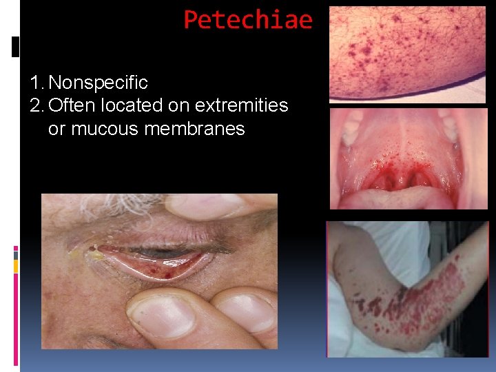
Petechiae 1. Nonspecific 2. Often located on extremities or mucous membranes
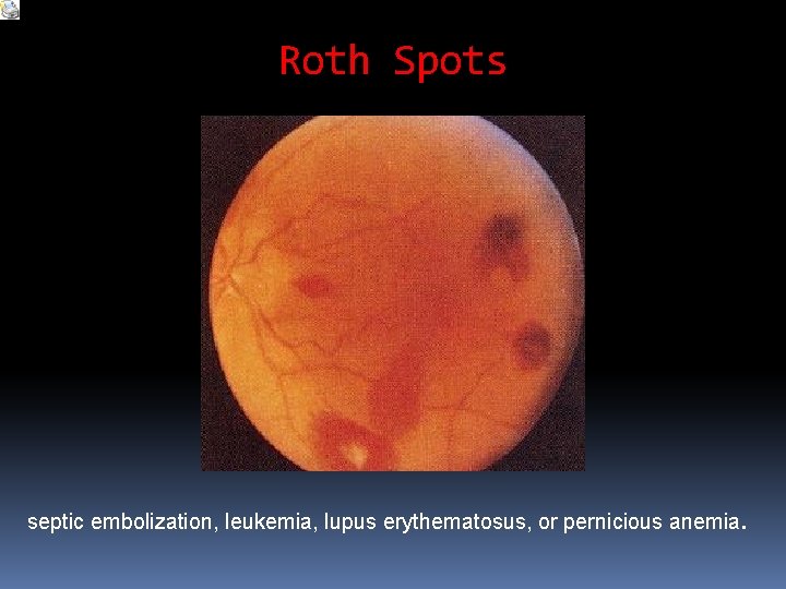
Roth Spots septic embolization, leukemia, lupus erythematosus, or pernicious anemia.
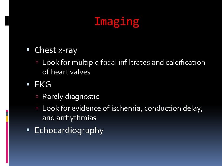
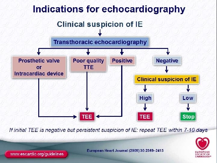
Imaging Chest x-ray Look for multiple focal infiltrates and calcification of heart valves EKG Rarely diagnostic Look for evidence of ischemia, conduction delay, and arrhythmias Echocardiography
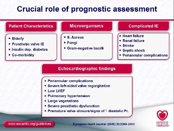
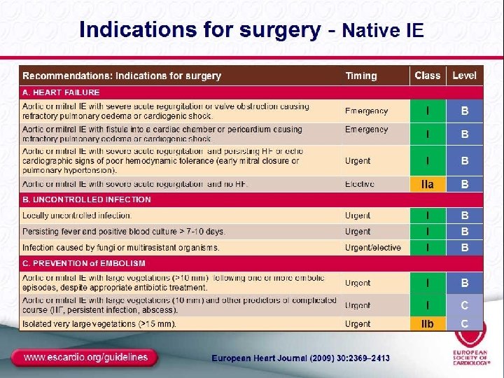
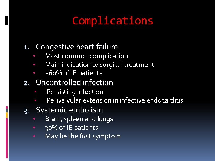
Complications 1. Congestive heart failure • • • Most common complication Main indication to surgical treatment ~60% of IE patients 2. Uncontrolled infection • • Persisting infection Perivalvular extension in infective endocarditis 3. Systemic embolism • • • Brain, spleen and lungs 30% of IE patients May be the first symptom
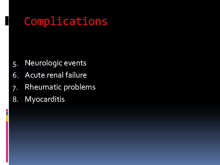
Complications 5. Neurologic events 6. Acute renal failure 7. Rheumatic problems 8. Myocarditis
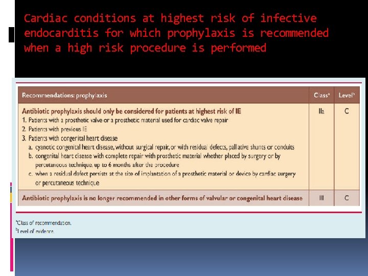
Cardiac conditions at highest risk of infective endocarditis for which prophylaxis is recommended when a high risk procedure is performed
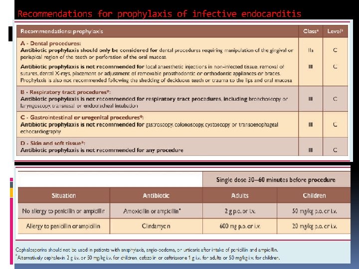
Recommendations for prophylaxis of infective endocarditis
ACUTE RHEUMATIC FEVER
Definition Current Diagnosis 07 An acute systemic immune disease that may develop after an infection with Group A betahemolytic Streptococcal infection of the pharynx. This disease can affect the HEART, JOINTS, SKIN, SUBCUTANEOUS TISSUE, BRAIN, RESPIRATORY SYSTEM, VESSELS, SEROSAL MEMBRANES, TENDONS AND FASCIAL SHEATHS
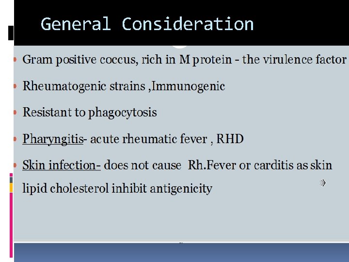
General Consideration
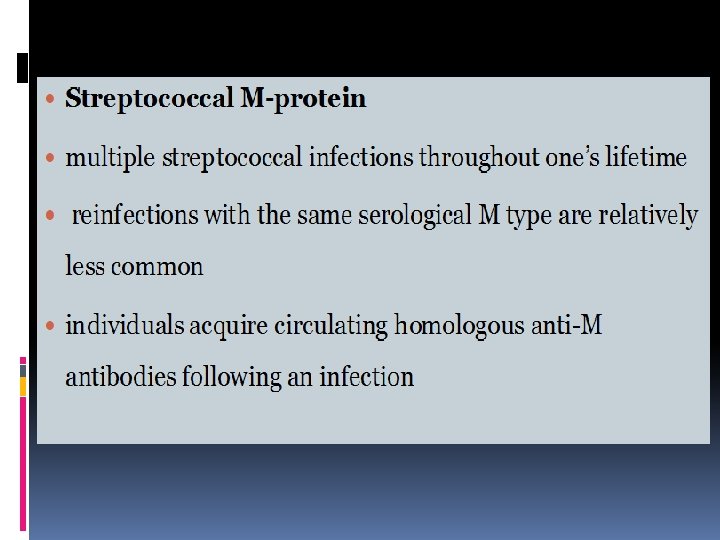
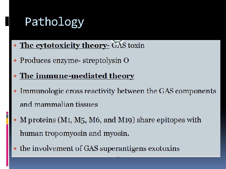
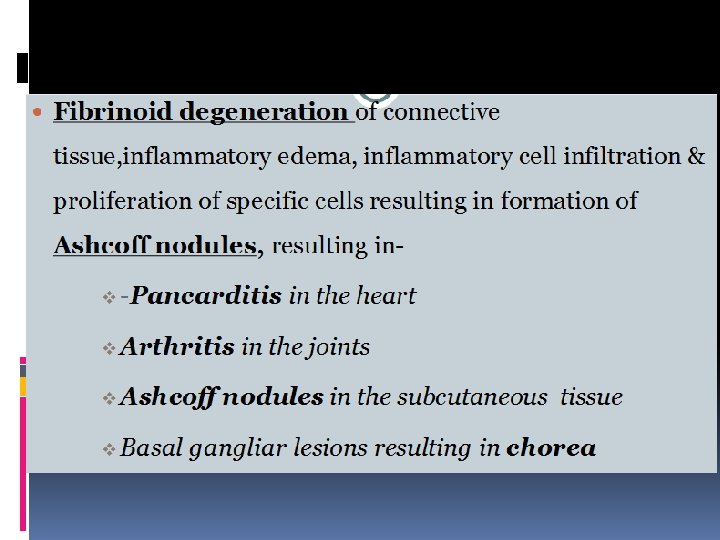
Pathology
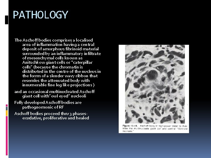
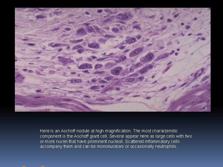
PATHOLOGY The Aschoff bodies comprises a localised area of inflammation having a central deposit of amorphous fibrinoid material surrounded by an inflammatory infiltrate of mesenchymal cells known as Anitschkow giant cells or “caterpillar cells” (because the chromatin is distributed in the centre of the nucleus in the forrm of a slender wavy ribbon that resemles the attenuated body with innumerable fine leg like projections ) and an occasional multinucleated Aschoff giant cell with”owl eyed” nucleoli Fully developed Aschoff bodies are pathognomonic of RF Aschoff bodies proceed thru 3 phasesexudative, proliferative and healed
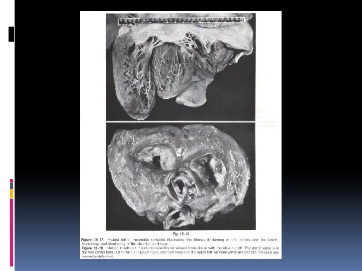
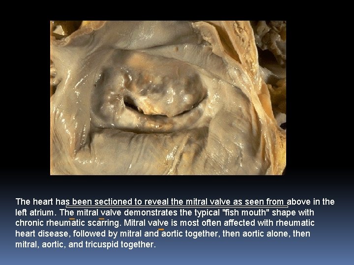
The heart has been sectioned to reveal the mitral valve as seen from above in the left atrium. The mitral valve demonstrates the typical "fish mouth" shape with chronic rheumatic scarring. Mitral valve is most often affected with rheumatic heart disease, followed by mitral and aortic together, then aortic alone, then mitral, aortic, and tricuspid together.
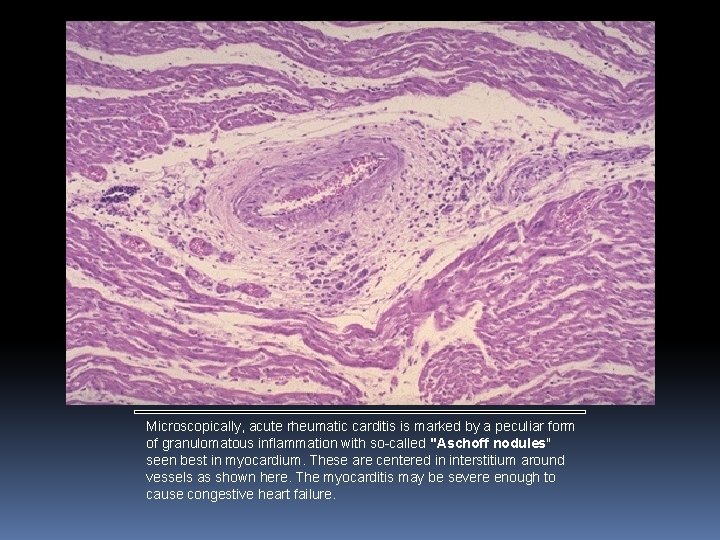
Microscopically, acute rheumatic carditis is marked by a peculiar form of granulomatous inflammation with so-called "Aschoff nodules" seen best in myocardium. These are centered in interstitium around vessels as shown here. The myocarditis may be severe enough to cause congestive heart failure.
Here is an Aschoff nodule at high magnification. The most characteristic component is the Aschoff giant cell. Several appear here as large cells with two or more nuclei that have prominent nucleoli. Scattered inflammatory cells accompany them and can be mononuclears or occasionally neutrophils.
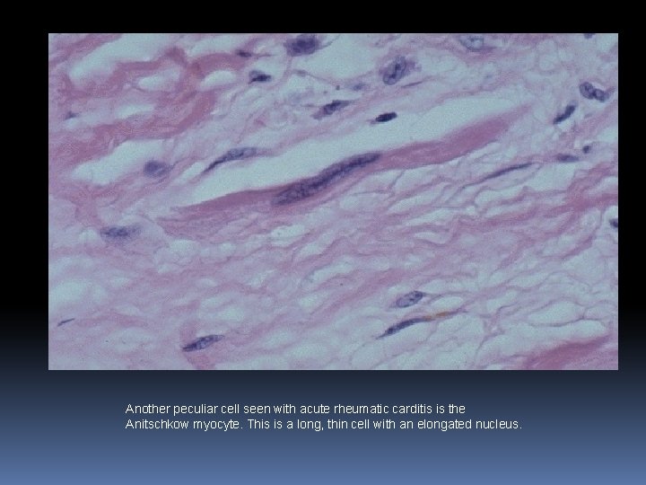
Another peculiar cell seen with acute rheumatic carditis is the Anitschkow myocyte. This is a long, thin cell with an elongated nucleus.
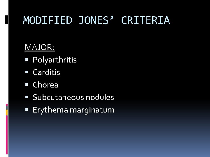
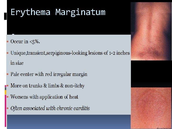
MODIFIED JONES’ CRITERIA MAJOR: Polyarthritis Carditis Chorea Subcutaneous nodules Erythema marginatum
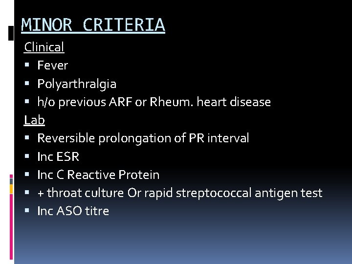
MINOR CRITERIA Clinical Fever Polyarthralgia h/o previous ARF or Rheum. heart disease Lab Reversible prolongation of PR interval Inc ESR Inc C Reactive Protein + throat culture Or rapid streptococcal antigen test Inc ASO titre
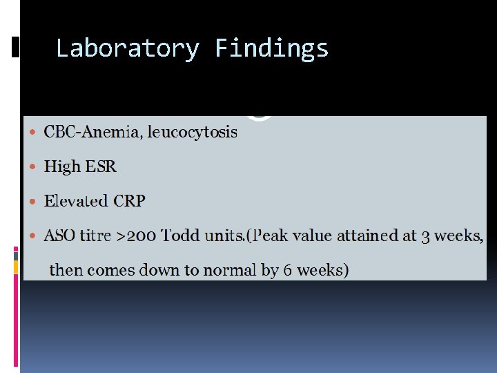
Laboratory Findings
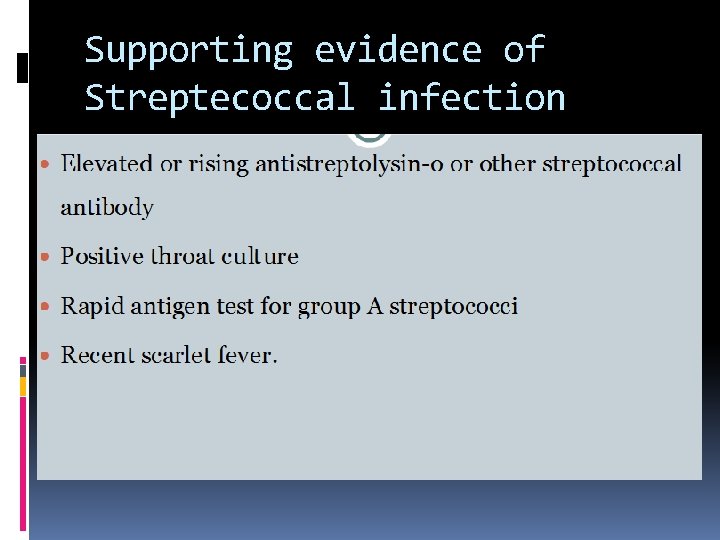
Supporting evidence of Streptecoccal infection
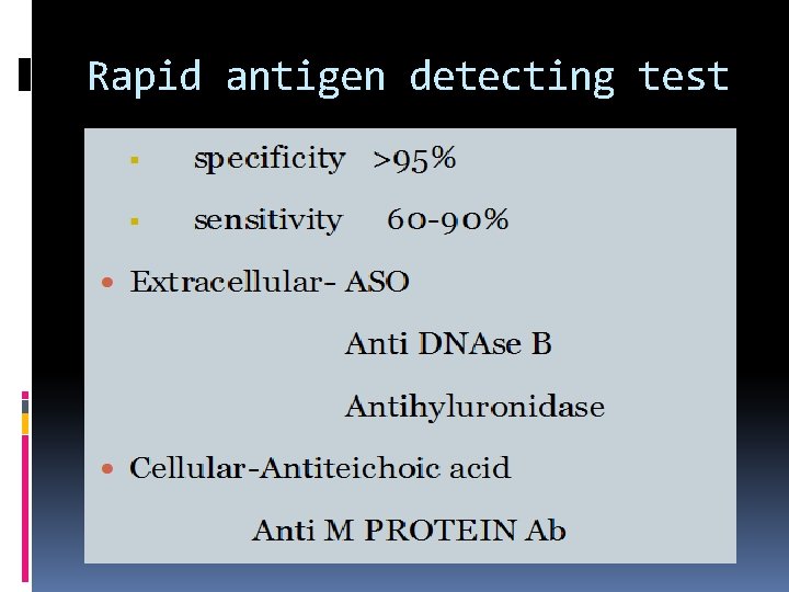
Rapid antigen detecting test
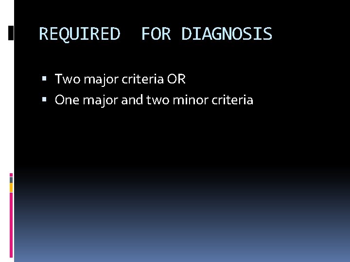
REQUIRED FOR DIAGNOSIS Two major criteria OR One major and two minor criteria
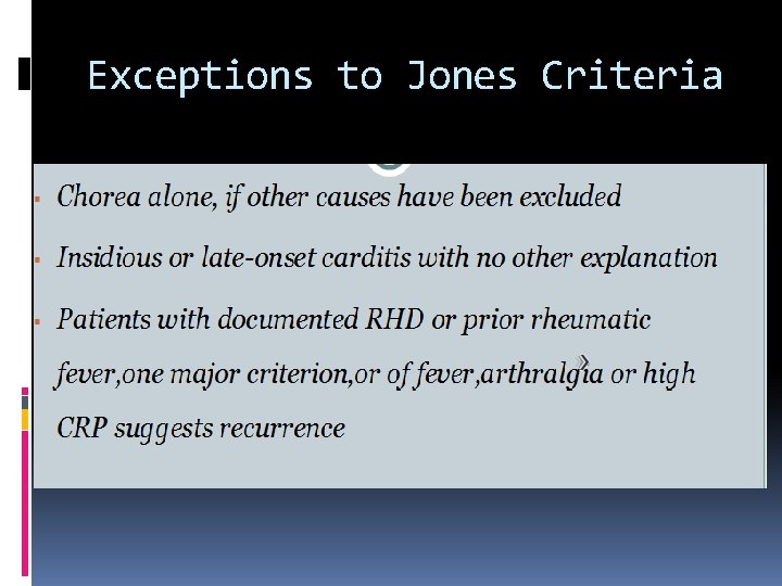
Exceptions to Jones Criteria
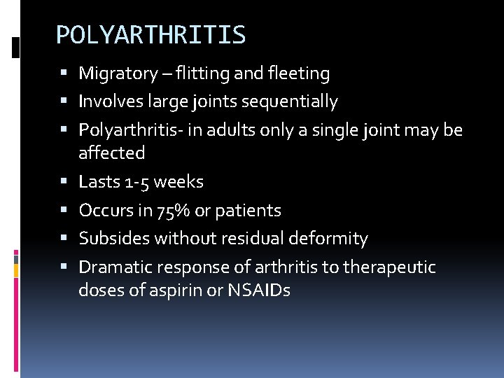
POLYARTHRITIS Migratory – flitting and fleeting Involves large joints sequentially Polyarthritis- in adults only a single joint may be affected Lasts 1 -5 weeks Occurs in 75% or patients Subsides without residual deformity Dramatic response of arthritis to therapeutic doses of aspirin or NSAIDs
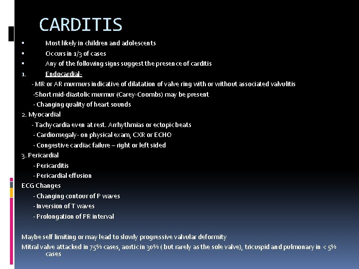
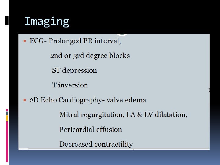
CARDITIS Most likely in children and adolescents Occurs in 1/3 of cases Any of the following signs suggest the presence of carditis Endocardial- MR or AR murmurs indicative of dilatation of valve ring with or without associated valvulitis -Short mid-diastolic murmur (Carey-Coombs) may be present - Changing quality of heart sounds 2. Myocardial - Tachycardia even at rest. Arrhythmias or ectopic beats - Cardiomegaly- on physical exam, CXR or ECHO - Congestive cardiac failure – right or left sided 3. Pericardial - Pericarditis - Pericardial effusion ECG Changes - Changing contour of P waves - Inversion of T waves - Prolongation of PR interval 1. Maybe self limiting or may lead to slowly progressive valvular deformity Mitral valve attacked in 75% cases, aortic in 30% ( but rarely as the sole valve), tricuspid and pulmonary in < 5% cases
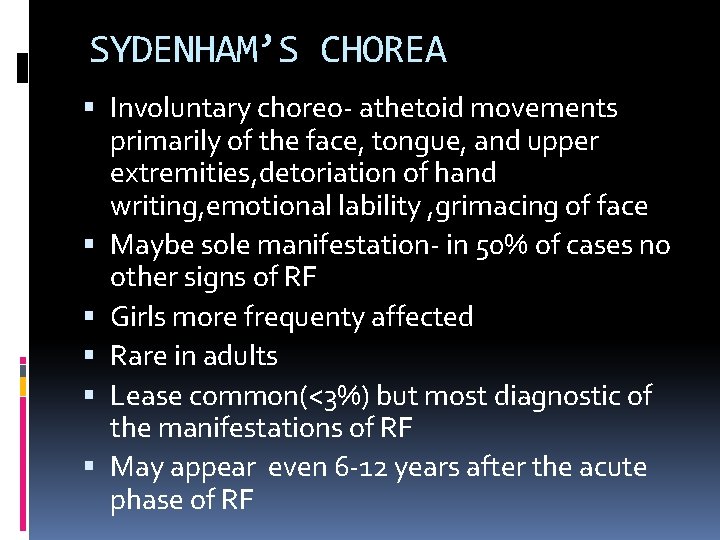
SYDENHAM’S CHOREA Involuntary choreo- athetoid movements primarily of the face, tongue, and upper extremities, detoriation of hand writing, emotional lability , grimacing of face Maybe sole manifestation- in 50% of cases no other signs of RF Girls more frequenty affected Rare in adults Lease common(<3%) but most diagnostic of the manifestations of RF May appear even 6 -12 years after the acute phase of RF
Erythema Marginatum .
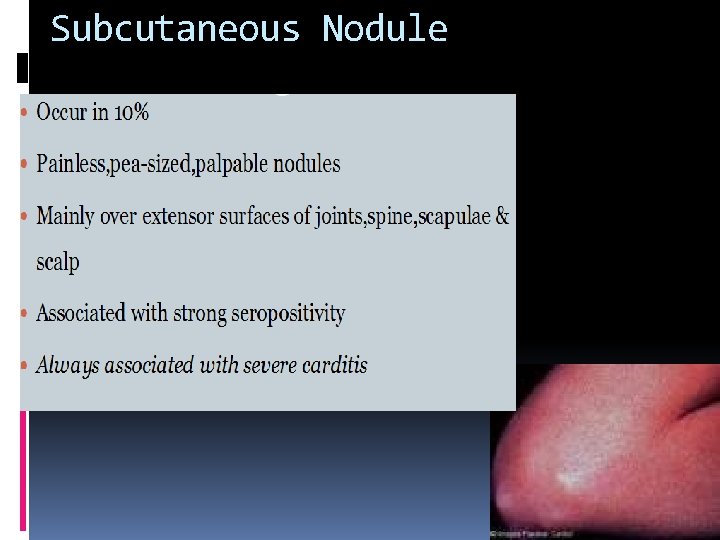
Subcutaneous Nodule
Imaging
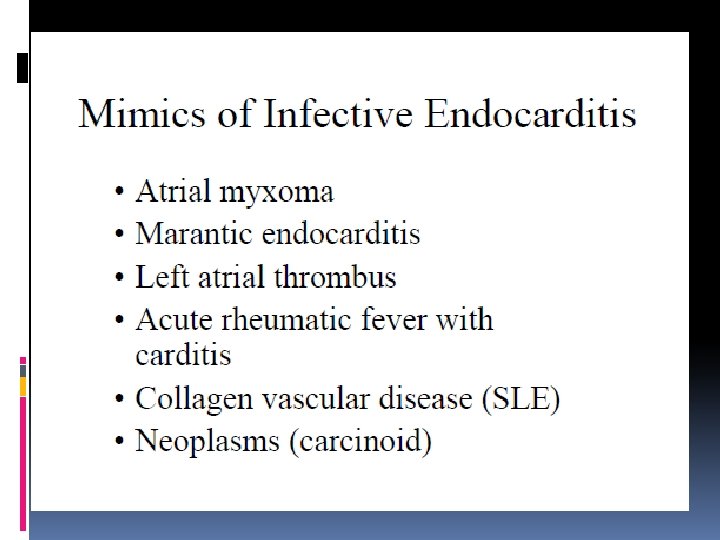
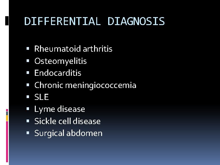
DIFFERENTIAL DIAGNOSIS Rheumatoid arthritis Osteomyelitis Endocarditis Chronic meningiococcemia SLE Lyme disease Sickle cell disease Surgical abdomen
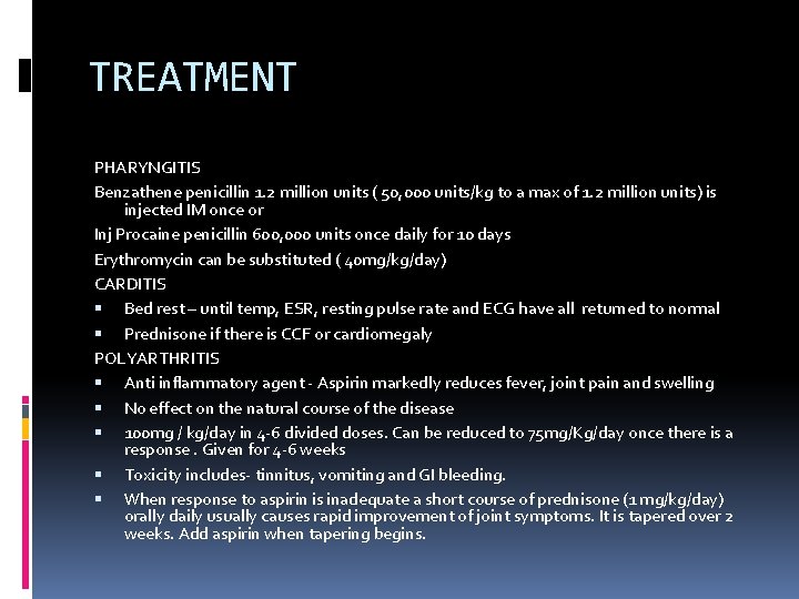
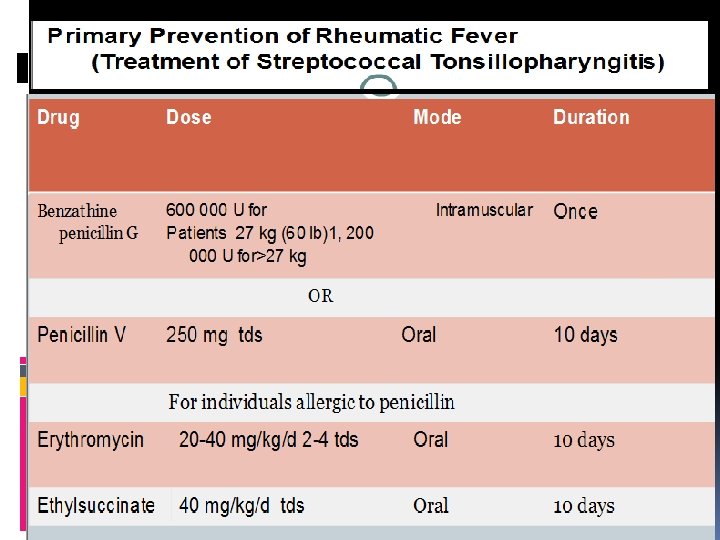
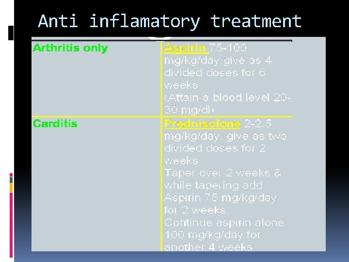
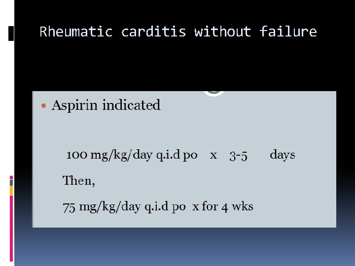
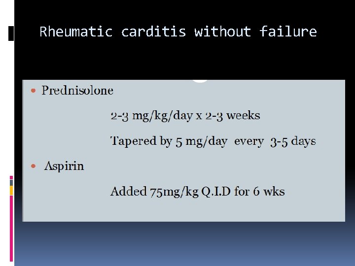
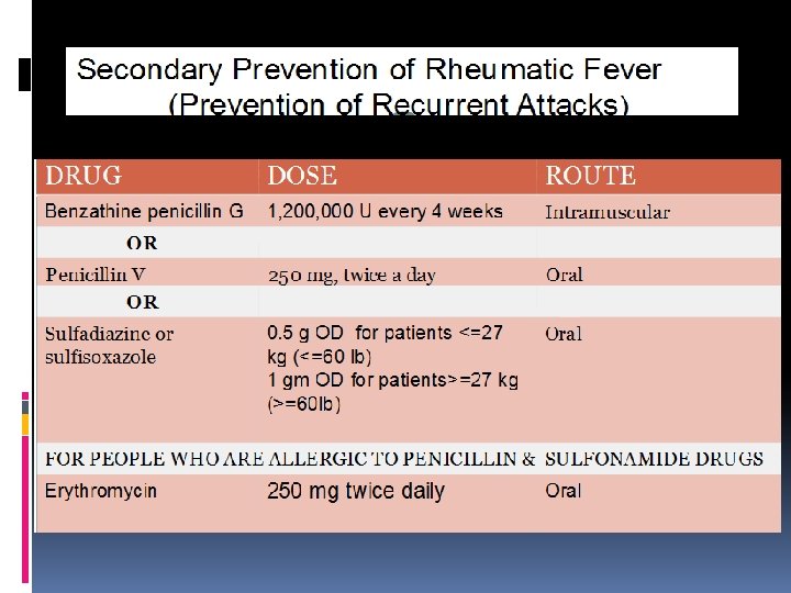
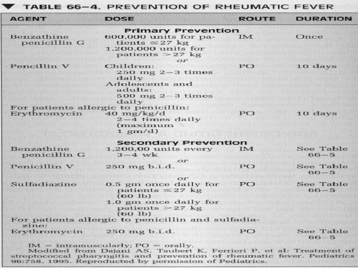
TREATMENT PHARYNGITIS Benzathene penicillin 1. 2 million units ( 50, 000 units/kg to a max of 1. 2 million units) is injected IM once or Inj Procaine penicillin 600, 000 units once daily for 10 days Erythromycin can be substituted ( 40 mg/kg/day) CARDITIS Bed rest – until temp, ESR, resting pulse rate and ECG have all returned to normal Prednisone if there is CCF or cardiomegaly POLYARTHRITIS Anti inflammatory agent - Aspirin markedly reduces fever, joint pain and swelling No effect on the natural course of the disease 100 mg / kg/day in 4 -6 divided doses. Can be reduced to 75 mg/Kg/day once there is a response. Given for 4 -6 weeks Toxicity includes- tinnitus, vomiting and GI bleeding. When response to aspirin is inadequate a short course of prednisone (1 mg/kg/day) orally daily usually causes rapid improvement of joint symptoms. It is tapered over 2 weeks. Add aspirin when tapering begins.
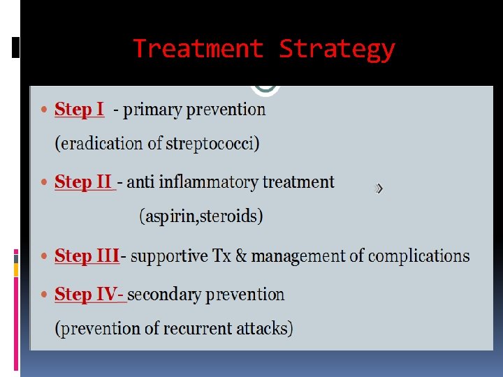
Treatment Strategy
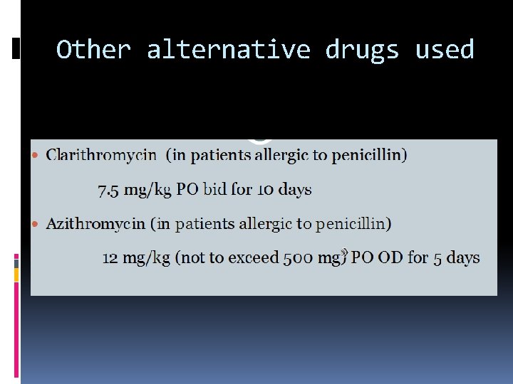
Other alternative drugs used
Anti inflamatory treatment
Rheumatic carditis without failure
Rheumatic carditis without failure
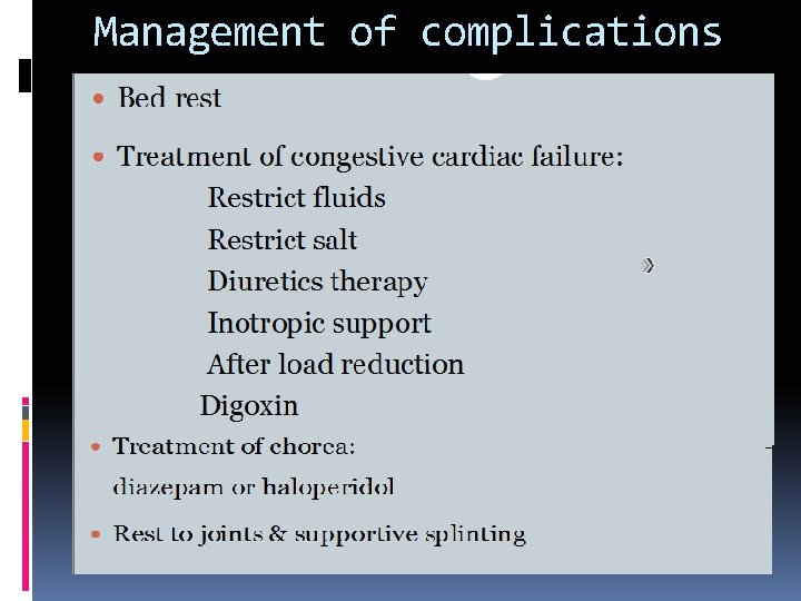
Management of complications