ED SlitLamp Examination Andrew Shannon MD MPH Department


 2) 3)")

 Seidel’s")


 SLE lateral view showing intracorneal lens anterior")

")


")


- Slides: 23
ED Slit-Lamp Examination Andrew Shannon, MD MPH Department of Emergency Medicine Jacobi Medical Center
Why bother? • ED Ophthalmology goals: – • R/O or R/I ruptured globe, retained foreign body, corneal abrasion, diagnose HSV corneal ulcer The Slit-lamp Exam: – – – makes you look like you know what you’re doing provides superior magnification & stabilizes pt’s head for removal of corneal FB tangential illumination aids in dx of uveitis/iritis • – • “cells and flare” billing? Eye exam interactive tutorial at – http: //www. kellogg. umich. edu/theeyeshaveit/index. html
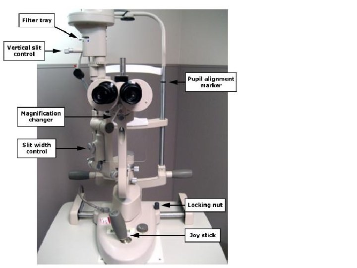
The Slit Lamp • Low- to medium-powered binocular horizontally mounted microscope 1) 2) 3) 4) 5) 6) Magnification Elevation Joystick for R/L movement & to focus Slit width &/or height Slit centration or offset Slit height & Intensity
– Slit width • Wide- survey globe/cornea • Narrow- depth, width & position of small abnormalities – beam as wide as cornea is thick – forms a parallelepiped volume: a box of illuminated tissue is seen • Thin (slit)- narrowest beam forms an optical section – so thin it's just discernible – valuating small changes in clarity & pinpointing depth of pathology – Light-source intensity • Medium to high: most purposes • High: optical section – Filters • neutral, cobalt blue (for fluorescein), red-free – Magnification • low power (~10 x) is used for survey • medium to high (16 -40 x) for optic section & parallelepiped • high (40 x) for specular reflection – normally, light is focused at same point as microscope (“parfocal”)
“Welder’s keratitis”-- diffuse punctate lesions of the cornea caused by UV radiation (+) Seidel’s test: ruptured globe dendritic appearance of HSV keratitis linear corneal abrasion
Slit lamp technique • Start w/ 10 x eyepieces & lower powered objective – (“ 1 x” or “ 12” on JMC scopes) • Use lowest voltage setting on transformer – ensure open aperture • Select the longest slit length • Adjust chin rest – Pt's eyes approx level w/ marker on head rest • Slit arm in line w/ microscope • Lamp height w/ slit beam centered vertically on Pt's medial canthus • Focus by moving joystick
Ocular focus to 0 adjust beam height for tall, narrow vertical beam locking nut: loose for free movement adjust width for narrow beam w/ good illumination
slit width adjustment
magnification adjustment beam height slit-width adjustment filter rheostat slit-arm locking knob focus & lamp height locking nut (horizontal) intensity locking bar (vertical)
• Magnification adjustment can be found in various locations, including btwn the eyepieces • The filter rheostat can be used to decrease Pt discomfort under exam w/ the lamp (neutral density filter)
Position of the Light – Slit-arm pivots 1800 around microscope mount • 450 angle, directed temporal to nasal is standard • In-line w/ scope for initial survey of lids, lashes, lacrimals, conjunctiva and sclera • Methods of viewing – Direct illumination: beam directly pointed at specimen • gross pathology – Retro-illumination: beam de-centered to illuminate behind area of interest while it is still in focus • may bring out subtle optical changes – thin vascularization, small incisions, endothelial abnormalities – Sclerotic scatter: light spreads by total internal reflection • uses a beam ~1 mm wide, ½ height of cornea & pointed at limbus • subtle abnormalities as light through the cornea scatters off of any pathology
Forehead should be in contact w/ restraint Eyeline should be at level of indicator Angle of slit-arm ~ 600
Direct/focal illumination • Most common; focused slit; magnification 10 x 40 x – wide beam for surface study; narrow beam for sections • Broad beam (parallelepiped) section of cornea • 2 mm slit: corneal surface & stroma • to ascertain depth (FB, abrasion) • Narrow beam (optic section): easier to determine precise depth • resolution improved by reducing slit width; clarity improved w/ increasing mag – angle btwn slit-arm & scope ~ 450 - 600 • increasing angle up to 900 will increase amnt of cross-section – 4 layers of corneal section: • • tears (outer) epithelium (& Bowman’s membrane) stroma: seen as central gray granular area endothelium (& Descemet’s membrane): fainter back line – lens: • opacities scatter & reflect more light - appear white (or pigmented) against gray background
SLE w/ wide slit on a post-op IOL Pt dx’d w/ Propionobacterium acnes endophthalmitis iris on tangential illumination light source outer epithelium of cornea
SLE thru dilated pupil: anterior chamber (AC) SLE lateral view showing intracorneal lens anterior to native crystalline lens light source cornea crystalline lens
• van Herick’s Technique: to assess anterior chamber angle – – low mag (6 x or 10 x) set beam 600 to side of scope place narrow slit as close to limbus as possible & perpendicular to the cornea compare width of cornea seen by optical section w/ the dark section seen btwn front surface of iris & back of cornea • • GRADE 4: ratio of aqueous to cornea is 1: 1 - open angle GRADE 3: ratio of aqueous to cornea is 1: 2 - open angle GRADE 2: ratio of aqueous to cornea is 1: 4 - narrow angle GRADE 1: ratio is < 1: 4 - dangerously narrow angle • Conical beam: used to detect aqueous flare – inflammatory cells in AC (eg acute anterior uveitis) • • room illumination must be completely dark can only be seen using conical beam of light set slit-arm angle btwn 450 -- 600 focus onto front surface of cornea w/ high magnification – W/ pupil as dark background, flare may be seen btwn focused beam of light on cornea & the out-of-focus beam on lens • in normal eye this space will be clear
keratic precipitates iris cornea “flare” in anterior chamber (AC)
iris cells & flare cornea no cells or flare cornea iris light source
Indirect Illumination • Evaluate tissue outside directly illuminated area – reduced glare; easier to view opacities, corneal nerves & limbal vessels • focus on feature directly & then swing lamp to one side • Retro-illumination (rarely useful in ED!) – light reflected off deeper structures (iris or retina) w/ microscope focused on ant. structures • study cornea in light reflected from iris; lens in light reflected from retina – light-opaque features are dark agnst light backgrnd (scars, pigment, vessels containing blood) • light-scattering features appear lighter than background (e. g. corneal precipitates) – useful for examining size / density of opacities (not location) – 1) use a parallelepiped beam, focus on retina – 2) direct retro-illumination: observed corneal feature viewed in direct pathway of reflected light • angle btwn microscope & illuminating arm ~ 60° – 3) indirect retro-illumination: angle is greatly reduced /increased so feature on cornea is viewed against dark bckgrnd
keratic precipitates (direct & retro-illumination )
Hypopyon layering in AC “Rust ring” residual from metallic FB
References: • • • My Hanh Nguyen. Ophthalmology Grand Rounds. Tufts Unviersity. http: //ocw. tufts. edu/Content/37/topics/487903/488024. Content accessed 9/23/08. Introduction to Slit Lamp Technique. CYBER-SIGHT: Copyright © 2003 Project ORBIS International Inc. http: //www. cybersight. org/bins/content_page. asp? cid=1 -1581 -1604. Content accessed 9/23/08. G Papaliodis. Propionibacterium acnes Endophthalmitis. Ocular Immunology and Uveitis Foundation. Massachusetts Eeye Research and Surgery Institution. Copyright © 1996 -2008 C. Stephen Foster M. D. http: //www. uveitis. org/medical/articles/case/P_acnes. html. Content accessed 9/23/08. Vance Thompson. Postoperative Care for Phakic Intraocular Lens Implants. In: Phakic Intraocular Lenses: Principles and Practice by Hardten, Lindstrom, and Davis. Slack, Inc. www. slackbooks. com/excerpts/66402. asp. Content accessed 9/23/08. Jared Schultz. One intracorneal segment treats keratoconus better than two. Copyright 2008 SLACK Inc. , www. osnsupersite. com/view. asp? r. ID=23456. Content accessed 9/23/08. Craig Blackwell. Narrated Eye Exam: Copyright 2008. www. blackwelleyesight. com/narrated-eye-exam/. Content accessed 9/23/08. Patient Glossary. Ocular Immunology and Uveitis Foundation. Massachusetts Eeye Research and Surgery Institution. Copyright © 1996 -2008 C. Stephen Foster M. D. http: //www. uveitis. org/patient/glossary/a_f. html. Content accessed 9/23/08. JG O'Shea, DA Infeld, RB Harvey. Uveitis- a photoessay. http: //medweb. bham. ac. uk/easdec/eyetextbook/Uveitis/uveitis. htm. Content accessed on 9/23/08. Second Year 99/00: Clinical Optometry 3. Slit lamp examination: Practical. http: //www. academy. org. uk/lectures/eperjesi 5. htm. Content accessed 9/23/08. KJ Knoop. Slit-lamp exam. Uptodate. com. Last updated: February 12, 2008. http: //www. uptodateonline. com/online/content/topic. do? topic. Key=ad_proc/2391&selected. Title=1~61&sou rce=search_result. Content accessed 9/23/08.